 Perspective


Affiliation:
1Dept. Biochemistry and Molecular Biomedicine, University of Barcelona, 08028 Barcelona, Spain
2Centro de Investigación Biomédica en Red Enfermedades Neurodegenerativas (CiberNed), National Institute of Health Carlos III, 28029 Madrid, Spain
3Institut de Química Teòrica i Computacional (IQTCUB), University of Barcelona, 08028 Barcelona, Spain
Email: rfranco123@gmail.com; rfranco@ub.edu
ORCID: https://orcid.org/0000-0003-2549-4919


Explor Neuroprot Ther. 2026;6:1004170 DOI: https://doi.org/10.37349/ent.2026.1004170
Received: August 11, 2026 Accepted: October 21, 2026 Published: November 04, 2026
Academic Editor: Elliott Mufson, Barrow Neurological Institute, USA
Dopamine presents a striking postsynaptic paradox: it is stored in synaptic vesicles and released in quantal events, yet every established mammalian dopamine receptor (D1–D5) is a GPCR. Whether—and by what mechanism—dopamine can generate rapid postsynaptic ion flow in the mammalian brain therefore remains unresolved. This Perspective shifts attention from release to the receiving cell and reassesses evidence for fast dopamine-evoked changes in membrane conductance. Although quantal dopamine release can generate relatively rapid D2R–GIRK-mediated postsynaptic currents, this indirect metabotropic mechanism may not account for all reported rapid dopamine-dependent changes in membrane conductance. Electrophysiological studies show millisecond-scale dopaminergic responses and direct dopamine-gated channels in invertebrates, whereas a vertebrate-derived hybrid cell line and chick neurons display rapid dopamine-dependent conductance changes or ion-channel regulation. These findings do not identify a mammalian ionotropic dopamine receptor, but they establish electrophysiological phenomena that remain mechanistically unexplained. A recent preprint presents evidence that the delta-type glutamate receptors GluD1 and GluD2 (previously known as delta1 and delta2 glutamate receptors) may act as dopamine-responsive ion channels tuned by G-protein subunits. Independent studies must now determine whether physiological dopamine concentrations evoke rapid GluD currents in native neurons and whether these currents influence defined dopaminergic circuits in vivo. Other experimentally tractable hypotheses include an unconventional dopamine-gated channel, direct coupling between a dopamine GPCR and an ion channel, and receptor homomers or heteromers with emergent ion-handling properties. Molecular-dynamics simulations showing spontaneous Na+ entry deep into D2R support intrareceptor ion access, but not complete transmembrane permeation. By separating established observations, recent preprint data, and mechanistic speculation, this Perspective defines a testable framework for identifying how dopamine binding is converted into postsynaptic ion flow and for determining whether dopamine can support fast neurotransmission in mammals.
The classical operational criteria for identifying a molecule as a neurotransmitter include its synthesis or presence in the presynaptic neuron, accumulation within the nerve terminal, storage in synaptic vesicles, and release in quantal units when depolarization triggers Ca2+-dependent vesicle fusion. Once released, the molecule should bind to specific receptors on a postsynaptic or target cell and elicit a measurable physiological response; its action must then be terminated by reuptake, enzymatic degradation, or diffusion. Some signalling molecules, notably endocannabinoids and nitric oxide, both involved in retrograde neurotransmission, do not conform to these criteria, and the boundaries of what should count as a neurotransmitter remain debated. For conventional synaptic neurotransmission, however, vesicular storage, quantal release, and the postsynaptic response form a continuous sequence. This Perspective is centered on the final step: the molecular and electrical events initiated in the postsynaptic cell by dopamine.
Table 1 provides a representative overview of mammalian neurotransmitters and the ionotropic and metabotropic receptors that mediate their effects; all metabotropic receptors listed here are G protein-coupled receptors (GPCRs). The balance between the two receptor classes varies markedly among transmitter systems. No bona fide mammalian ionotropic receptor has been established for dopamine, noradrenaline/norepinephrine, or histamine: all currently established receptors for these monoamines are GPCRs.
Representative mammalian neurotransmitters and the receptors mediating their effectsa.
| Class | Neurotransmitters | Receptorsb | |
|---|---|---|---|
| Ionotropicc (subunits) | GPCR (“metabotropic”) | ||
| Monoamines | Dopamine | NONE ESTABLISHEDe | D1, D2, D3, D4, D5 |
| Serotonin | 5-HT3A, 5-HT3B, 5-HT3C, 5-HT3D, 5-HT3E | 5-HT1A, 5-HT1B, 5-HT1D, 5-HT1E, 5-HT1F, 5-HT2A, 5-HT2B, 5-HT2C, 5-HT4, 5-HT5A, 5-HT5B, 5-HT6, 5-HT7 | |
| Noradrenaline/norepinephrine | NONE ESTABLISHEDe | α1A, α1B, α1D, α2A, α2B, α2C, β1, β2, β3 | |
| Histamine | NONE ESTABLISHEDe | H1, H2, H3, H4 | |
| Amino acid (excitatory) | Glutamate | NMDA, AMPA, or kainate, depending on the combination of these subunits: AMPA: GluA1, GluA2, GluA3, GluA4. NMDA: GluN1, GluN2A, GluN2B, GluN2C, GluN2D, GluN3A, GluN3B. Kainate: GluK1, GluK2, GluK3, GluK4, GluK5 | mGlu1, mGlu2, mGlu3, mGlu4, mGlu5, mGlu6, mGlu7, mGlu8 |
| Amino acid (inhibitory) | Gamma-aminobutyric acid (GABA) | GABAA (so far, 19 subunits have been reported: 6α, 3β, 3γ, 1δ, 1ε, 1π, 1θ and 3ρ) | GABAB1, GABAB2 |
| Glycine | α1–α4 and β subunits | GPRC6A, T1R2/T1R3 (sweet taste receptors), calcium-sensing receptord | |
| Choline | Acetylcholine | Nicotinic receptors (so far, 16 subunits have been reported: α1–α7, α9, α10, β1–β4, 1γ, 1δ, and 1ε) | M1, M2, M3, M4, M5 |
| Purines | ATP, UTP | P2X1, P2X2, P2X3, P2X4, P2X5, P2X6, P2X7 | P2Y1, P2Y2, P2Y4, P2Y6, P2Y11, P2Y12, P2Y13, P2Y14 |
a: This table provides a representative, non-exhaustive overview; a comprehensive catalogue is beyond the scope of this Perspective. b: With the exception of the proposed metabotropic receptors listed for glycine, the source is the IUPHAR/BPS Guide to Pharmacology (https://www.guidetopharmacology.org/; accessed August 4, 2026). c: The ionotropic receptors listed are multimeric ligand-gated ion channels composed of identical or different subunits. d: Whether glycine acts as an agonist or as a modulator at the listed GPCRs remains debated. e: No receptors have been reported or accepted as belonging to this category. 5-HT: 5-hydroxytryptamine; AMPA: α-amino-3-hydroxy-5-methyl-4-isoxazolepropionic acid; GPCRs: G protein-coupled receptors; NMDA: N-methyl-D-aspartate.
This Perspective focuses on dopamine because all established mammalian dopamine receptors are GPCRs, despite the prominent role of dopaminergic signalling in motor control, motivation, reward, learning, cognition, and neuroendocrine regulation. The central question is what occurs in the postsynaptic cell after dopamine reaches its membrane: can dopamine binding directly open an ion channel and generate a fast postsynaptic current, or must the response pass through one of the known D1–D5 GPCR cascades?
Two mechanistic possibilities therefore deserve consideration at the postsynaptic membrane. The first is that a mammalian ionotropic dopamine receptor exists but has not yet been identified. The second is that the known D1–D5 GPCRs, through unusually rapid, highly localized, or otherwise unconventional signalling mechanisms, may themselves support fast components of dopaminergic signalling in the central nervous system (CNS). The following sections examine both possibilities and address a more fundamental question: whether, in the absence of an ionotropic dopamine receptor, dopamine in mammals should be regarded predominantly as a neuromodulator rather than as a classical fast neurotransmitter.
An important distinction is required when considering rapid responses elicited by activation of dopaminergic neurons. In several neuronal populations, dopamine can be co-released with glutamate [1, 2], and the glutamate component can generate conventional fast postsynaptic currents through ionotropic glutamate receptors. Such responses demonstrate fast transmission from neurons, but they do not constitute fast ionotropic signalling by dopamine itself. The question addressed in this Perspective is therefore more restricted: whether dopamine, when released without glutamate or when glutamatergic transmission is physiologically or experimentally excluded, can itself produce a rapid postsynaptic ionic response.
Dopamine is a catecholamine synthesized from the amino acid tyrosine via a two-step pathway: tyrosine hydroxylase converts tyrosine to levodopa (L-DOPA), which is then decarboxylated to dopamine. It acts throughout the body via two families of GPCRs: D1-like receptors (D1, D5), whose activation increases cytosolic cyclic adenosine monophosphate (cAMP) levels, and D2-like receptors (D2, D3, D4), whose activation decreases cytosolic cAMP concentrations [3, 4]. These five receptor subtypes are thought to mediate the full range of dopamine’s remarkably diverse physiological effects, both within and outside the CNS.
In CNS neurons, dopamine is stored in synaptic vesicles and released by exocytosis in quantal events. After dopamine enters the synaptic, perisynaptic, or extrasynaptic space, the relevant question becomes how the receiving cell converts dopamine binding into an electrical response. In the striatum, the canonical basal-ganglia model assigns excitation of direct-pathway neurons to D1 receptors and inhibition of indirect-pathway neurons to D2 receptors [5]. Progressive loss of nigrostriatal dopaminergic input in Parkinson’s disease disrupts these postsynaptic actions and produces bradykinesia, rigidity, and tremor [6].
In reward-related circuitry, dopamine released from ventral tegmental neurons acts on receiving cells in the nucleus accumbens and prefrontal cortex, contributing to reward prediction, reinforcement, motivation, executive function, and working memory [6]. In the tuberoinfundibular pathway, dopamine receptor activation on pituitary lactotrophs tonically inhibits prolactin release [7]. These pathways differ anatomically, but in each case dopamine must ultimately be decoded by receptors and effectors in the target cell.
Dopamine signalling in the mammalian brain is not restricted to neurons. Astrocytes express dopamine receptors, and their activation can modulate intracellular signalling and gliotransmitter release, thereby influencing astrocyte–neuron communication [8, 9]. These observations extend the actions of dopamine beyond direct neuronal targets and suggest that astrocytes may contribute to the regulation of dopaminergic signalling within neural circuits. Significant advances in understanding these astrocytic mechanisms have been made, particularly in basal ganglia circuits and in dopamine-related disorders [10, 11].
Peripheral dopamine provides a brief comparison with neuronal signalling. Because dopamine does not cross the blood-brain barrier, peripheral and central pools are largely separate. Roughly half of the body’s dopamine has been estimated to reside in the gastrointestinal tract [12], where enteric, epithelial, and immune cells can generate local autocrine and paracrine signals.
The gut microbiota may contribute dopamine or its precursors and influence host pathways of dopamine synthesis and degradation. Dopamine can in turn affect mucosal immune behaviour, barrier function, and inflammatory tone, forming part of the gut-immune-microbiota-brain axis [12, 13].
Lymphocytes and other immune cells express functional D1–D5 receptors that regulate activation, proliferation, cytokine secretion, chemotaxis, and immune-synapse organization [14]. These non-neuronal actions illustrate the physiological reach of dopamine, but the remainder of this Perspective centres on receptor activation and electrical events in the neuronal postsynaptic cell.
Agnati et al. [15] proposed two complementary spatial modes of CNS communication. Wiring transmission (WT) follows defined neuronal pathways and usually involves transmitter release near a restricted postsynaptic target, as at a synapse. Volume transmission (VT) occurs when a signal travels through extracellular fluid and reaches receptors beyond fixed synaptic contacts. VT therefore extends the distance and number of postsynaptic or extrasynaptic cells that a released signal can influence [15–17].
WT is generally faster and more spatially segregated, whereas VT is slower and more divergent. Early evidence for VT included mismatches between enkephalin/β-endorphin-immunoreactive terminals and regional mu/delta opioid-receptor density, showing that release sites and receptor-bearing cells need not be closely apposed [15]. These categories describe the route by which a transmitter reaches the receiving membrane; they do not determine the receptor mechanism operating in that membrane. GPCRs may be synaptic or perisynaptic, and ionotropic receptors may be extrasynaptic. The central mechanistic question begins when dopamine encounters its receptor on the target cell. Somatodendritic dopamine transmission in the ventral midbrain provides an instructive example of highly localized metabotropic signalling. D2R-mediated inhibitory postsynaptic potentials (IPSPs) display a remarkably consistent time course that is largely insensitive to changes in stimulus intensity, release probability, or extracellular diffusion, whereas inhibition of dopamine uptake markedly prolongs the response [18]. These findings indicate that dopamine transporters restrict spillover and allow individual release sites to operate relatively independently. Similarly, the rapid onset of evoked D2-IPSCs and the observation that increasing stimulus intensity changes their amplitude without substantially altering their time course argue for a rapid rise of dopamine concentration within a restricted receptor-accessible domain rather than unrestricted VT [19]. Thus, dopamine acting through a GPCR need not imply diffuse VT; temporally defined D2-mediated synaptic responses can arise from dopamine acting within a restricted extracellular domain.
Dopamine GPCRs are not restricted to VT. They can be positioned at synaptic, perisynaptic, or extrasynaptic sites and can alter postsynaptic excitability over different spatial and temporal scales. Conversely, the route by which dopamine reaches a cell does not dictate whether the receptor is ionotropic or metabotropic. WT versus VT and ionotropic versus GPCR signalling are distinct classifications that converge at the postsynaptic neuron.
Fast chemical synaptic currents typically develop on a millisecond timescale. Ionotropic receptors meet this temporal requirement because ligand binding is directly coupled to channel opening within the same macromolecular complex. GPCRs instead require receptor activation, heterotrimeric G-protein activation, and regulation of downstream effectors in the postsynaptic cell. Those effectors may be controlled through Gα-dependent second messengers, including cAMP- and Ca2+-dependent pathways, or more directly by Gβγ subunits, as for G protein-coupled inwardly rectifying potassium channels (GIRKs) [20–22]. Membrane-delimited Gβγ-channel coupling is faster than a diffusible second-messenger cascade, but it remains a multistep route from dopamine binding to postsynaptic channel regulation.
Importantly, metabotropic signalling need not be temporally diffuse. In mouse midbrain dopamine neurons, vesicular dopamine release evokes D2R-mediated inhibitory postsynaptic currents (D2-IPSCs) through GIRK channels. The evoked response is Ca2+ dependent and tetrodotoxin sensitive, is blocked by D2R antagonism, and is prolonged when dopamine uptake is inhibited [19]. Spontaneous D2-IPSCs can also arise from individual vesicular release events [23], demonstrating that quantal dopamine release can generate discrete postsynaptic electrical responses through D2R–GIRK coupling.
These observations distinguish response kinetics from receptor mechanism. D2R–GIRK transmission can be rapid and spatially restricted, but it remains metabotropic: dopamine activates D2R and G-protein signalling, then gates a distinct GIRK channel. By contrast, an ionotropic dopamine receptor would couple dopamine binding directly to opening of an ion-conducting pore within the receptor complex itself. Fast currents elicited by activation of dopaminergic axons must additionally be distinguished from cotransmission by glutamate or gamma-aminobutyric acid (GABA) [1, 2, 24]. Thus, a rapid dopamine-associated current is not, by itself, evidence of ionotropic dopamine signalling; the defining criterion is the molecular route linking dopamine binding to ion flow.
Table 1 illustrates a recurrent, but not universal, dual-receptor pattern. Glutamate, GABA, and acetylcholine signal through both ionotropic and metabotropic receptors, and serotonin combines multiple GPCRs with the ionotropic 5-HT3 receptor. Noradrenaline/norepinephrine and histamine, like dopamine, currently have only established GPCRs in mammals. Dopamine is especially relevant because its quantal release can encode rapidly changing motor and reward-related variables, and those presynaptic signals acquire physiological meaning only through responses generated in the receiving cell. The kinetics and molecular architecture of the postsynaptic dopamine receptor are therefore central to understanding dopaminergic transmission [25].
The cloning of the D1–D5 receptors provided a coherent molecular framework for dopamine pharmacology [4]. This major advance may have reduced the incentive to search for alternative dopamine targets in neurons. Nevertheless, early reports of dopamine-evoked conductance changes provide evidence for direct electrical actions of dopamine on the receiving membrane, and they deserve to be considered alongside later electrophysiological and molecular findings.
The evidence considered below concerns electrical responses attributable to dopamine itself rather than fast glutamatergic currents produced by glutamate co-released from dopaminergic neurons.
Early studies in Helix aspersa showed that dopamine hyperpolarized identified postsynaptic neurons, inhibited spontaneous activity, and increased potassium permeability. Pharmacological similarities between the dopamine response and the nerve-evoked IPSP supported dopamine as the transmitter mediating that IPSP [26, 27]. These experiments provide early evidence of a dopamine-evoked postsynaptic conductance carried by K+.
Ascher’s 1972 study described inhibitory and excitatory postsynaptic responses to dopamine in Aplysia neurons [28]. Fast depolarizing responses in the same genus were later reported by Due and colleagues [29]. In Planorbis corneus, a dopamine-containing neuron with no detectable noradrenaline produced millisecond-scale depolarizing responses in postsynaptic cells [30]. Together, these findings provide evidence for rapid postsynaptic dopaminergic transmission in invertebrates.
A 1975 study reported dopamine-induced depolarization accompanied by increased membrane conductance in the 108CC15 hybrid cell line, formed from mouse N18TG2 neuroblastoma and rat C6-BU-1 glioma cells [31]. This vertebrate-derived preparation provides evidence of a dopamine-evoked electrical response coupled to an increase in target-cell membrane conductance.
Patch-clamp experiments in cultured chick sensory and sympathetic neurons showed that dopamine rapidly reduced voltage-dependent Ca2+ currents and Ca2+-channel opening probability [32]. These findings demonstrate rapid dopamine-dependent modulation of membrane Ca2+ conductance, although the underlying receptor and molecular coupling mechanism were not established.
In Aplysia, identified dopaminergic interneurons produced excitatory postsynaptic potentials (EPSPs) with latencies of 4–5 ms and peaks at approximately 30 ms; occlusion by exogenous dopamine and blockade by sulpiride supported dopamine as the mediator [33]. Other molluscan studies described biphasic potentials lasting 40–400 ms, with shorter EPSP than IPSP latencies [34], and distinguished fast EPSPs lasting approximately 150 ms from slower responses of about 1 s; local iontophoretic application of dopamine reproduced the corresponding slow and fast depolarizing responses and, in some cells, the complete biphasic response [30]. These observations provide electrophysiological and pharmacological evidence for rapid dopamine-mediated postsynaptic transmission.
Further evidence came from another molluscan preparation in which dopamine activated a ligand-gated channel [35, 36]. In a separate study, dopamine produced inhibitory or biphasic responses at an identified Lymnaea stagnalis synapse; the components involved a sulpiride-sensitive K+ conductance and a picrotoxin-sensitive depolarizing Cl− conductance [37]. In Caenorhabditis elegans, ligand-gated chloride channels activated by biogenic amines were identified, including a dopamine-gated channel that can also be inhibited by antagonists used against dopamine GPCRs [38]. These molecular and electrophysiological findings establish that dopamine can directly gate postsynaptic ion channels.
As schematized in Figure 1, the central question is: by what molecular route can dopamine binding produce a rapid change in postsynaptic membrane conductance? The rapid dopamine-evoked responses observed in chick neurons and in a rodent-derived hybrid cell line justify addressing that question directly. A dopamine-sensitive ionotropic mechanism could belong to an unexpected protein family, emerge from a receptor complex, or arise through direct coupling between a dopamine receptor and an ion channel. The established D1–D5 GPCRs explain many effects on excitability, gain, and plasticity, but their success does not exhaust the possible mechanisms of fast postsynaptic dopamine action. The 5-HT3 receptor provides a relevant monoaminergic precedent: metabotropic and ionotropic signalling can coexist within one transmitter system.
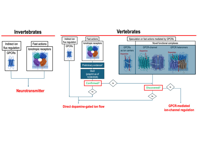
Candidate molecular routes from dopamine binding to postsynaptic ion flow. In invertebrates, GPCR-mediated modulation and ionotropic dopamine receptors establish that dopamine can support both slow modulation and fast postsynaptic transmission. In vertebrate preparations, rapid dopamine-evoked ionic responses and the recently proposed GluD-mediated ionotropic mechanism provide a rationale for a focused search for pathways coupling dopamine recognition to postsynaptic ion flow. The possibilities shown include dopamine-responsive ion channels, direct receptor–channel coupling, GPCR homomers or heteromers with emergent functions, and ion access within a dopamine-receptor core. These mechanisms are presented as experimentally distinguishable routes, with the speculative ones identified as hypotheses. a: Evidence that is discussed in the text derives from a rodent-derived hybrid cell line and chick neurons ([31, 32]); the recent preprint is available at https://doi.org/10.64898/2026.05.10.723887 (as of August 12, 2026).
Several molecular routes could couple dopamine recognition to a postsynaptic ionic response. Canonical G-protein signalling can rapidly regulate channels in the same membrane. A more direct arrangement is also plausible: ligand-induced conformational change in a dopamine GPCR could be transmitted across a stable receptor–channel complex [for a precedent in N-methyl-D-aspartate (NMDA) receptors, see below] and alter channel opening or closure. In that case, the current would pass through the associated channel, but the dopamine receptor would remain an integral part of the functional transduction unit. This hypothesis occupies an intermediate mechanistic position between a classical ligand-gated channel and a diffusible second-messenger cascade (Figure 1).
An additional observation comes from molecular-dynamics simulations of D2R. Extracellular Na+ entered the receptor spontaneously and reached the conserved allosteric Na+-binding site deep within the receptor core [39]. The trajectories therefore provide evidence for spontaneous cation access to a substantial depth of D2R; they followed entry and intra-receptor stabilization rather than complete transmembrane passage [39]. The observed entry is mechanistically important because it establishes an ion-accessible internal route whose postsynaptic relevance can now be tested by combining receptor mutagenesis, conformational-state control, sodium ion substitution, and electrophysiological recording.
Receptor–receptor interactions deserve particular attention because the functional properties of a homomer or heteromer need not equal the sum of its protomers. In a postsynaptic membrane, such complexes could alter ligand recognition, conformational propagation, G-protein coupling, trafficking, association with ion channels, or the local environment through which ions gain access to membrane proteins. Two non-exclusive hypotheses follow: dopamine binding could gate a physically associated ion channel, or a receptor assembly could acquire an emergent ion-handling function absent from an isolated protomer. These proposals remain speculative, but they are experimentally tractable and may reveal genuinely novel functions. Distinguishing them requires rapid agonist application, single-channel or whole-cell recording, disruption of defined receptor interfaces, and reconstitution of minimal complexes. The relevant endpoint in every case is a dopamine-evoked change in postsynaptic ionic conductance.
Established examples from the glutamatergic system show that dopamine receptors can physically associate with ionotropic receptors. In striatal and cortical neurons, the carboxyl tail of the D1 receptor may directly interact with the GluN1 and the GluN2A subunits of the NMDA receptor; the interaction with GluN2A contributes to the inhibition of NMDA receptor-gated currents, whereas the other attenuates NMDA receptor-mediated excitotoxicity through a phosphatidylinositol 3-kinase-dependent pathway [40]. Similarly, the D2R forms a direct complex with the GluN2B subunit within the postsynaptic density of excitatory synapses; cocaine-induced dopamine release enhances this interaction, which disrupts the association of CaMKII with GluN2B, reduces phosphorylation of this subunit at Ser1303, and inhibits NMDA receptor-mediated currents in medium-sized striatal neurons [41]. These findings show that a dopamine GPCR can be a structural and functional partner of an ion channel. In both cases, however, dopamine receptor activation modulates the response of an NMDA receptor to glutamate; no evidence indicates that dopamine itself gates the channel. They therefore illustrate the physical coupling required by the receptor–channel hypothesis, but not the ion-gating step that would have to be demonstrated.
Quite recently, delta-type glutamate receptors, known as GluDs, were reported to function as ligand-gated ion channels [42]. Notably, while this Perspective was being prepared, Wang et al. [43] (2026) reported in a preprint that GluD1 and GluD2 may act as dopamine-responsive ion channels regulated by G-protein subunits. These data provide evidence for a molecular arrangement in which dopamine responsiveness, G-protein dependence, and ion conduction converge. Mechanistically, GluD receptors differ from the established D1–D5 dopamine GPCRs in one fundamental respect. In D1–D5 receptors, dopamine binding activates G proteins that subsequently regulate downstream effectors, including ion channels, whereas in GluD1/GluD2 the receptor itself contains the ion-conducting pore and dopamine is proposed to gate that pore directly. G proteins nevertheless remain important in the GluD mechanism, but as regulators of channel gating rather than as the obligatory signalling route between dopamine binding and ion flow.
For the postsynaptic cell, the crucial implication is that dopamine could evoke a current through a receptor family not previously assigned this role. The next question is how broadly the reported mechanism operates: whether physiological dopamine concentrations evoke rapid GluD currents in native neurons, and whether those currents shape identified dopaminergic circuits in vivo.
Viewed as a working model, GluD-dependent dopamine signalling does not require a lost mammalian receptor. It suggests instead that the relevant postsynaptic channel was overlooked because it belonged to an unexpected receptor family and because its activity depended on a permissive G-protein context. If the mechanism operates in native neurons, dopamine will engage established metabotropic D1–D5 receptors and dopamine-responsive GluD channels within the same transmitter system. This dual organization would also sharpen the experimental distinction between modulation of an existing current and generation of a dopamine-evoked postsynaptic current.
The immediate priority is to extend the evidence across preparations. Independent experiments should measure dopamine binding, dopamine-evoked GluD currents, their kinetics, pharmacology, ion selectivity, concentration dependence, and occurrence in identified postsynaptic neurons. Determining the relative contributions of GluD1 and GluD2, the G-protein requirements for channel activation, and the influence of receptor–receptor interactions would define the molecular context of the response. The same strategy could test whether GluD-mediated dopaminergic signalling also operates outside the CNS.
A different outcome in any one model would narrow the mechanism without erasing the evidence obtained in another. If GluD dependence is not reproduced broadly, the rapid dopamine-evoked ionic responses in chick neurons, the rodent-derived cell line, and other preparations would still require molecular explanation. Searches should then emphasize unconventional dopamine-gated channels, receptor–channel complexes, and GPCR assemblies with emergent functions. In either case, the decisive measurements are made in the receiving cell: response latency, membrane current, conductance, reversal potential, ion selectivity, and dependence on defined molecular components.
Dopamine’s established D1–D5 receptors are GPCRs, but that fact does not establish that GPCR monomers exhaust all postsynaptic dopamine mechanisms. Importantly, quantal dopamine release can already generate temporally defined postsynaptic currents through D2R–GIRK coupling, demonstrating that rapid dopamine-mediated electrical signalling is not restricted to ionotropic mechanisms. Nevertheless, if dopamine were ultimately shown to lack any bona fide ionotropic receptor or receptor complex capable of directly converting ligand binding into ion flow, its predominant mode of action would be more appropriately viewed as neuromodulatory than as that of a classical fast neurotransmitter. The evidence reviewed here points to rapid dopamine-evoked changes in membrane conductance and ion-channel activity, while molecular studies identify several plausible architectures that could generate them. The appropriate question is therefore not whether mammals lost an ionotropic dopamine receptor, but which molecular component or assembly converts dopamine binding into postsynaptic ion flow.
Several possibilities can coexist: an unconventional dopamine-responsive channel, a dopamine receptor directly coupled to an ion channel, a homomeric or heteromeric receptor complex with an emergent function, or a GluD channel operating in a permissive G-protein context. The D2R simulations add evidence that extracellular Na+ can enter deeply into a dopamine-receptor core, although the reported trajectories concern intrareceptor access rather than complete permeation. Each observation addresses a different level of the mechanism and should therefore be tested experimentally at that level.
The broader message is that a GPCR-only account of postsynaptic dopamine signalling should be treated as a productive framework rather than as a boundary on what mechanisms can exist. Receptor–receptor and receptor–channel interactions are especially important because they can generate functions not predictable from isolated proteins. Demonstrating a genuine dopamine-gated ionotropic mechanism would therefore have consequences beyond receptor classification: it would strengthen the case for dopamine as a neurotransmitter capable of directly mediating fast postsynaptic signalling, whereas its absence would reinforce the view of dopamine primarily as a neuromodulator acting through metabotropic control of neuronal excitability. Establishing which of these mechanisms produces rapid dopamine-evoked postsynaptic currents would revise our understanding of synaptic organization, dopamine physiology, and pharmacology, while converting the present hypotheses into directly testable questions.
5-HT: 5-hydroxytryptamine
AMPA: α-amino-3-hydroxy-5-methyl-4-isoxazolepropionic acid
cAMP: cyclic adenosine monophosphate
CNS: central nervous system
EPSPs: excitatory postsynaptic potentials
GABA: gamma-aminobutyric acid
GIRKs: G protein-coupled inwardly rectifying potassium channels
GPCRs: G protein-coupled receptors
IPSPs: inhibitory postsynaptic potentials
NMDA: N-methyl-D-aspartate
VT: volume transmission
WT: wiring transmission
RF: Conceptualization, Writing—original draft, Writing—review & editing. The author read and approved the submitted version.
Rafael Franco, who is the Editor-in-Chief of Exploration of Neuroprotective Therapy, had no involvement in the decision-making or the review process of this manuscript.
Not applicable.
Not applicable.
Not applicable.
Not applicable.
The IQTC of the University of Barcelona holds a Maria de Maeztu-associated grant (CEX2021-001202-M) funded by Spanish MCIN/AEI/10.13039/501100011033. This grant does not cover Open Access publication fees. This funder had no role in study design, data collection and analysis, decision to publish, or preparation of the manuscript.
© The Author(s) 2026.
Open Exploration maintains a neutral stance on jurisdictional claims in published institutional affiliations and maps. All opinions expressed in this article are the personal views of the author(s) and do not represent the stance of the editorial team or the publisher.
Copyright: © The Author(s) 2026. This is an Open Access article licensed under a Creative Commons Attribution 4.0 International License (https://creativecommons.org/licenses/by/4.0/), which permits unrestricted use, sharing, adaptation, distribution and reproduction in any medium or format, for any purpose, even commercially, as long as you give appropriate credit to the original author(s) and the source, provide a link to the Creative Commons license, and indicate if changes were made.



View: 63
Download: 7
Times Cited: 0


