Review


Affiliation:
1Rheumatology Department, Centro de Diagnóstico de Osteoporosis y Enfermedades Reumáticas (CEDOR), Lima 15036, Perú
Email: maritzavw@gmail.com
ORCID: https://orcid.org/0000-0001-8494-5260


 ,
,
Affiliation:
2Centre for Metabolic Bone Disease, Royal National Orthopaedic Hospital, HA7 4LP London, United Kingdom
Affiliation:
2Centre for Metabolic Bone Disease, Royal National Orthopaedic Hospital, HA7 4LP London, United Kingdom
ORCID: https://orcid.org/0009-0001-8216-3828
,
Affiliation:
2Centre for Metabolic Bone Disease, Royal National Orthopaedic Hospital, HA7 4LP London, United Kingdom
ORCID: https://orcid.org/0000-0001-7576-250X
Explor Musculoskeletal Dis. 2026;4:1007135 DOI: https://doi.org/10.37349/emd.2026.1007135
Received: May 13, 2026 Accepted: July 30, 2026 Published: August 30, 2026
Academic Editor: Blanka Stiburkova, Charles University and General University Hospital, Czech Republic
Hypophosphatasia (HPP) is a rare inherited metabolic disorder caused by deficient activity of tissue non-specific alkaline phosphatase (TNSALP). The resulting accumulation of its substrates, particularly inorganic pyrophosphate (PPi) and pyridoxal-5-phosphate (PLP), impairs skeletal and dental mineralization and disrupts vitamin B6 metabolism, contributing to multisystem manifestations. The clinical spectrum is highly heterogeneous, ranging from severe, life-threatening forms in the perinatal period to milder musculoskeletal and dental phenotypes presenting in adulthood. The rarity of HPP, together with the nonspecific nature of many symptoms, contributes to frequent diagnostic delay. However, recognition of characteristic biochemical abnormalities and clinical features can facilitate earlier identification and appropriate management. This review summarizes current evidence regarding the pathophysiology, clinical manifestations, diagnosis, and management of HPP, with particular emphasis on diagnostic challenges, biomarker interpretation, and recent advances in patient care.
Hypophosphatasia (HPP) is a rare metabolic bone disease characterized by impaired mineralization of bone and teeth resulting from deficient alkaline phosphatase (ALP) activity. The clinical spectrum is broad, ranging from severe, life-threatening disease in infancy to milder forms presenting in childhood or adulthood with musculoskeletal pain, fractures, or dental abnormalities. Disease severity may vary substantially even among affected relatives, suggesting contributions from both genetic heterogeneity and additional modifying factors [1–7]. Determining the true prevalence of HPP remains challenging due to its marked phenotypic variability and substantial underdiagnosis. Severe forms are estimated to occur in approximately 1 per 100,000 to 900,000 live births in Japanese cohorts and around 1 per 300,000 in Europe and 1 per 100,000 in North America [4, 6, 8–13]. In contrast, milder forms, including adult-onset disease, are likely more common, with an estimated prevalence of approximately 1 in 6,000 in European populations [6, 10, 11, 14, 15]. Increasing evidence suggests that HPP is frequently overlooked in routine clinical practice, particularly in adults, where nonspecific symptoms and persistently low ALP levels are often unrecognized or misinterpreted. Early diagnosis is critical, as appropriate management may prevent complications, avoid inappropriate therapies such as anti-resorptive agents, and enable timely initiation of disease-specific treatment. However, significant challenges remain in identifying affected individuals and translating biochemical and genetic findings into clinical decision-making, emphasizing the need for improved diagnostic strategies and clearer guidance for clinicians.
A comprehensive, non-systematic literature search was conducted to summarize current evidence on the pathophysiology, clinical manifestations, diagnosis, and management of HPP. A structured literature search was performed in PubMed/MEDLINE, Scopus, and Google Scholar to identify relevant publications from January 2000 to March 2026. Search terms included combinations of the following keywords: “hypophosphatasia,” “ALPL,” “tissue-nonspecific alkaline phosphatase,” “low alkaline phosphatase,” “diagnosis,” “clinical manifestations,” “enzyme replacement therapy,” and “asfotase alfa.” Boolean operators (AND/OR) were applied where appropriate.
Studies were eligible for inclusion if they were peer-reviewed articles, including original research studies, clinical trials, observational studies, systematic and narrative reviews, and international consensus or guideline papers relevant to HPP. Particular emphasis was placed on recent publications, high-quality evidence, and studies addressing diagnostic strategies, disease monitoring, and therapeutic outcomes. Articles were excluded if they were non-peer-reviewed reports, conference abstracts without full text, or publications lacking sufficient clinical or methodological detail. Additional relevant studies were identified through manual screening of reference lists of selected articles. Given the narrative nature of this review, formal systematic review methodology (e.g., PRISMA) was not applied; however, efforts were made to ensure a comprehensive and balanced representation of the available literature.
HPP is caused by pathogenic variants in the ALPL gene, located on chromosome 1p36.1-p34, which encodes tissue non-specific alkaline phosphatase (TNSALP) [6, 13]. To date, more than 500 pathogenic variants have been identified, the majority of which are missense mutations, although nonsense, splice-site, and small insertion/deletion variants have also been reported. An international database of ALPL mutations is available (https://alplmutationdatabase.jku.at/) [16]. Inheritance may be autosomal recessive or autosomal dominant [3]. In general, autosomal recessive forms are associated with more severe disease due to near-complete loss of enzyme activity, whereas autosomal dominant variants, including those exerting dominant-negative effects, are more often associated with milder phenotypes [12, 13]. However, genotype-phenotype correlations remain inconsistent, and clinical severity cannot be reliably predicted from genetic findings alone. This variability suggests that additional factors may modify disease expression. Potential modifiers include residual enzyme activity associated with specific variants, compound heterozygosity, epigenetic influences, and environmental factors such as nutritional status, vitamin D levels, and mechanical loading, which may influence skeletal manifestations and timing of symptom onset [2, 4, 6, 8, 12, 13, 17].
Genetic testing helps confirm the diagnosis and determine the pattern of inheritance but is not mandatory, as some patients with clinically confirmed HPP may have negative or inconclusive results. In selected cases, evaluation of additional genes involved in phosphate metabolism and skeletal mineralization, such as runt-related transcription factor 2 (RUNX2) and ectonucleotide pyrophosphatase/phosphodiesterase 1 (ENPP1), may be considered in the context of differential diagnosis [16, 18]. Biochemical abnormalities also tend to correlate with clinical severity. Patients with severe perinatal or infantile forms typically exhibit markedly reduced serum ALP activity and significantly elevated circulating PLP levels, whereas individuals with milder childhood or adult-onset disease may show only modest biochemical alterations in PLP levels [1–6, 11, 15, 18, 19]. The clinical heterogeneity of HPP reflects the combined effects of genetic variants, residual enzyme activity, and additional modifying factors, which limits the ability to predict disease severity based on genetic findings alone. These alterations result in reduced TNSALP activity and drive the biochemical and pathophysiological changes mediating the clinical manifestations of this condition [5, 18, 19].
HPP results from deficient TNSALP activity, leading to impaired hydrolysis of extracellular phosphate-containing substrates. TNSALP is the predominant circulating isoenzyme and is widely expressed in bone, liver, kidney, lungs, and developing teeth [10–13, 20–26]. Its primary physiological substrates include inorganic pyrophosphate (PPi), phosphoethanolamine (PEA), and pyridoxal-5-phosphate (PLP), the major circulating form of vitamin B6 [5–7, 9, 11, 20–24] (Figure 1A). The accumulation of PPi, a potent inhibitor of hydroxyapatite crystal formation, is the primary driver of defective mineralization. Consequently, reduced hydrolysis of PPi impairs skeletal mineralization, manifesting as osteomalacia and rachitic changes [9–13, 24–27]. Furthermore, elevated PPi concentrations may promote calcium pyrophosphate dihydrate crystal deposition in articular and periarticular tissues, contributing to the extra-skeletal manifestations of the disease in adults [1, 2, 6, 20, 24] (Figure 1B).
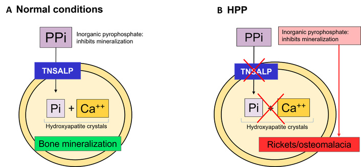
Tissue non-specific alkaline phosphatase (TNSALP)-mediated regulation of bone mineralization. (A) Under normal conditions, TNSALP hydrolyzes inorganic pyrophosphate (PPi), increasing local inorganic phosphate (Pi) availability in the extracellular matrix, which combines with calcium to form hydroxyapatite crystals that deposit on type I collagen. (B) In hypophosphatasia (HPP), reduced TNSALP activity leads to PPi accumulation, inhibition of hydroxyapatite formation, and defective bone mineralization.
TNSALP also plays a critical role in central nervous system function by regulating PLP metabolism. Under normal conditions, phosphorylated PLP cannot cross the blood–brain barrier; therefore, TNSALP-mediated dephosphorylation to pyridoxal (PL) is required to enable its transport into the brain [28–30] (Figure 2A). Once intracellular, PL is rephosphorylated to PLP, which serves as an essential cofactor for the synthesis of neurotransmitters, including gamma-aminobutyric acid (GABA), serotonin, and dopamine. In HPP, impaired dephosphorylation of PLP reduces PL availability within the central nervous system, thereby disrupting neurotransmitter synthesis. In severe infantile forms, this deficiency commonly manifests as vitamin B6-responsive seizures [3, 6, 13, 20, 30]. In contrast, adult patients may present with more subtle neuropsychiatric manifestations, such as fatigue, depression, and anxiety [10, 12, 13]. Consequently, circulating PLP levels are typically elevated and represent a sensitive biochemical marker of TNSALP deficiency (Figure 2B).
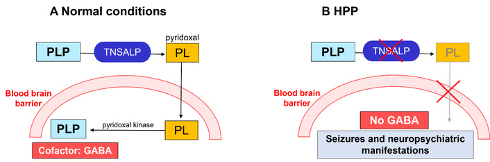
Role of non-specific alkaline phosphatase (TNSALP) in vitamin B6 metabolism and neurotransmitter synthesis. (A) TNSALP converts pyridoxal-5-phosphate (PLP) to pyridoxal (PL), allowing transport across the blood–brain barrier and re-phosphorylation to PLP within the brain. (B) In hypophosphatasia (HPP), impaired dephosphorylation of PLP limits PL availability in the brain, contributing to reduced neurotransmitter synthesis. GABA: gamma-aminobutyric acid.
HPP is classified into six clinical subtypes based on age at onset and disease presentation, reflecting a broad phenotypic spectrum that ranges from severe perinatal forms, typically inherited in an autosomal recessive manner, to milder adult presentations [2, 3, 9, 12, 30]. With the exception of odontohypophosphatasia (odonto-HPP), all forms are characterized by impaired skeletal mineralization and low ALP levels adjusted for age and sex [3, 9, 31] (Table 1).
Clinical subtypes of hypophosphatasia, inheritance patterns and key clinical manifestations.
| Subtype | Inheritance | Clinical manifestations |
|---|---|---|
| 1. Perinatal lethal | AR | Respiratory failure, seizures, hypercalcemia, early death |
| 2. Prenatal benign | AR/AD | Skeletal abnormalities present in utero with spontaneous postnatal improvement |
| 3. Infantile(< 6 months) | AR | Failure to thrive, craniosynostosis, seizures, intracranial hypertension, hypercalcemia, hypercalciuria, nephrocalcinosis, rickets, myopathy, early loss of decidual teeth |
| 4. Childhood(6 months–18 years) | AR/AD | Premature tooth loss, short stature, chronic muscle pain, myopathy, defective mineralization, osteomalacia, recurrent fractures, delayed bone healing |
| 5. Adult (> 18 years) | AR/AD | Premature tooth loss, chronic muscle pain, metatarsal fractures, recurrent fractures and pseudo-fractures, delayed bone healing, osteomalacia, enthesopathy, altered gait, pseudogout, CPPD arthritis |
| 6. odonto-HPP | AR/AD | Isolated dental disease without skeletal involvement. Painless premature exfoliation of deciduous teeth, premature tooth loss, hypoplasia of enamel and dentine, wide pulp chamber, thin and short roots, severe dental caries, periodontal disease |
The perinatal lethal form is characterized by profound skeletal hypomineralization, severe chest wall deformities, and pulmonary hypoplasia, leading to respiratory failure and early mortality [9, 22]. In contrast, the prenatal benign form presents with skeletal abnormalities detected in utero that may improve during late gestation, although residual limb deformities can persist at birth [6, 10, 20, 22]. Infantile HPP typically manifests within the first months of life with rickets, fractures, and growth impairment, and may be associated with neurological complications, including vitamin B6-dependent seizures, as well as abnormalities in cranial development [10, 17, 22].
Childhood and adult forms are generally milder but highly variable. Common manifestations include premature loss of teeth with intact roots, musculoskeletal pain, fractures, and fatigue, with symptom recurrence often observed in adulthood [10, 20, 22, 30, 32]. In adults, recurrent metatarsal fractures, femoral pseudofractures, and calcium pyrophosphate deposition disease (CPPD) are characteristic features [10, 20]. Neuropsychiatric symptoms and metabolic abnormalities, including hypercalcemia and nephrocalcinosis, may also occur [10, 33].
The odonto-HPP subtype is characterized by isolated dental manifestations without overt skeletal involvement [20, 22, 34]. Defective cementum mineralization leads to premature loss of primary teeth, particularly the mandibular incisors [10, 20, 22, 25]. Additional dental abnormalities, including defective dentin, alveolar bone loss, enlarged pulp chambers, enamel defects, and altered crown morphology, are frequently observed before the age of five and should raise clinical suspicion of HPP. Children with odonto-HPP may experience speech difficulties and impaired nutrition due to early loss of their primary teeth [10, 20, 25, 34]. Adults typically report a history of early tooth loss extending into adulthood, often accompanied by caries and periodontal disease [10, 20]. Although odonto-HPP represents the mildest end of the clinical spectrum, current genotype-phenotype correlations remain incomplete, and no specific ALPL genotype reliably predicts disease confined to the dentition. The mechanisms responsible for the apparent restriction of disease manifestations to the dentition also remain incompletely understood [20, 22, 34]. Longitudinal follow-up is recommended, as some patients may later develop skeletal manifestations [34].
The diagnosis of HPP requires integration of clinical features, biochemical findings, and imaging studies, reflecting the marked heterogeneity of the disease.
Key clinical features relevant to diagnosis reflect the broad phenotypic spectrum described above. Severe forms typically present in the perinatal or infantile period with profound hypomineralization, fractures, skeletal deformities, and respiratory compromise, and may be associated with seizures, intracranial complications, and nephrocalcinosis [6, 10, 20, 22]. In contrast, adult forms are generally milder and heterogeneous, with musculoskeletal pain, muscle weakness, enthesopathy, abnormal gait, and recurrent or poorly healing fractures, particularly involving the metatarsals and long bones. Dental and neuropsychiatric manifestations are often subtle but frequent [1, 10, 11, 13, 22, 26, 27].
Persistently low serum ALP is the biochemical hallmark of HPP and should be interpreted using age- and sex-specific reference ranges [5, 9, 34, 35]. Because reference intervals vary among laboratories and may not always be accompanied by clearly age- and sex-adjusted interpretive comments, clinicians should verify that the appropriate reference interval has been applied, particularly in children and adolescents, in whom ALP concentrations are physiologically higher during periods of skeletal growth. Additionally, proper use of blood collection devices must be ensured [5, 13, 20, 35–37]. Measurements should be confirmed on repeated testing, especially when results are borderline or discordant with the clinical presentation, and interpreted in the context of the patient’s clinical and radiographic findings [35, 36]. When uncertainty persists, consultation with the local laboratory or a metabolic bone specialist may facilitate appropriate interpretation. Alternative causes of low ALP must also be excluded (Table 2). Pre-analytical factors are also relevant, particularly the use of anticoagulants such as EDTA, citrate, and oxalate, which can artificially reduce measured ALP activity by chelating essential cofactors such as zinc and magnesium, thereby impairing enzymatic function; appropriate sample handling is therefore essential [5, 35, 36]. Notably, ALP levels may occasionally be within the laboratory reference interval or even elevated due to confounding factors such as fracture healing, medications, or other medical conditions [16, 34–36].
Conditions associated with low and elevated ALP levels in the evaluation of hypophosphatasia.
| Category | Low ALP levels | High ALP levels |
|---|---|---|
| Drugs and substances |
|
|
| Medical conditions |
|
|
| Technical factors | Blood collection tubes containing EDTA, citrate, or oxalate |
Imaging findings reflect the degree of impaired skeletal mineralization and vary across the disease spectrum. Prenatal ultrasound can detect skeletal abnormalities early in gestation, with lethality largely determined by the severity of pulmonary hypoplasia [41–43]. In postnatal life, conventional radiography remains the primary modality for identifying characteristic skeletal changes, while additional imaging techniques may be used to assess complications. Renal ultrasound may be performed when nephrocalcinosis is suspected [32, 44–47]. Dual-energy X-ray absorptiometry (DXA) has limited utility in HPP, as bone mineral density is often normal or only mildly reduced and does not reliably reflect disease severity or fracture risk [10, 13, 24, 48, 49].
International expert consensus has proposed diagnostic criteria for HPP in both children and adults [50] (Table 3). These criteria provide a practical framework for evaluating individuals with persistently low ALP levels adjusted for age and sex, in the absence of other identifiable causes [5, 50]. A diagnosis is established by the presence of either two major criteria or one major and two minor criteria. Alternative causes of low ALP, including medications and comorbid conditions, should be excluded, and potential pre-analytical factors should be considered when interpreting results [50].
Diagnostic criteria for hypophosphatasia in children and adults.
| Children | Adults | |
|---|---|---|
| Major |
|
|
| Minor |
|
|
| Diagnosis is made by meeting 2 major; or 1 major + 2 minor criteria | ||
As discussed above, measurement of TNSALP substrates, including PLP, PEA, and PPi, is incorporated into the diagnostic criteria. Among these, PLP is the most sensitive biomarker, and circulating levels generally correlate with disease severity. Vitamin B6 supplementation should be discontinued for at least one week prior to measurement. In contrast, PPi assessment is largely restricted to research settings, limiting its routine clinical use. Urinary PEA is typically elevated but has lower sensitivity and specificity, as it may be increased in other metabolic bone disorders or remain within the normal range in some patients with HPP. Additional biochemical findings may further support the diagnosis and aid in differentiation from other metabolic conditions [13, 51, 52].
Several inherited skeletal disorders should also be considered in the differential diagnosis. ENPP1-related disorders may present with rickets or osteomalacia, fractures, and impaired skeletal mineralization [52]. Likewise, RUNX2-related cleidocranial dysplasia may mimic HPP because of overlapping skeletal, dental, radiographic, and occasionally biochemical features. Characteristic findings include persistently open cranial sutures, hypoplastic or absent clavicles, delayed tooth eruption, supernumerary teeth, and delayed ossification of the pubic bones. These disorders can usually be distinguished from HPP by their characteristic clinical features, normal rather than persistently low serum ALP activity, and confirmatory genetic testing [10].
Genetic testing is increasingly available and can support diagnostic confirmation and clarify the pattern of inheritance, although it is not required for diagnosis. Next-generation sequencing (NGS), including analysis of copy number variations (CNVs) in the ALPL gene, is commonly used in patients with suspected HPP. However, negative results do not exclude the diagnosis. In such cases, additional techniques, such as multiplex ligation-dependent probe amplification (MLPA), may be required to detect gene deletions or duplications [37]. Genetic counselling and testing of first-degree relatives may facilitate early recognition of affected individuals within families [6, 9, 13, 53]. Despite these advances, early diagnosis remains difficult, and more effective strategies are needed to identify affected patients (Table 4).
Genetic, biochemical, and substrate abnormalities supporting the diagnosis of hypophosphatasia.
| Genetic | Biochemical | Substrates |
|---|---|---|
|
|
|
Systematic approaches using laboratory databases to identify persistently low ALP levels have improved case detection, particularly when combined with targeted symptom screening. Diagnostic yield appears higher when ALP levels are < 25 IU/L in the presence of musculoskeletal symptoms [15, 54, 55]. Biochemical algorithms have been developed to identify individuals with ALPL variants among patients with persistent hypophosphatasemia [55, 56]. In addition, predictive modeling and machine-learning approaches that integrate biochemical markers such as ALP and PLP with clinical variables have demonstrated promising preliminary results for identifying affected individuals within large datasets [57–61]. However, these approaches remain largely exploratory, with limited external validation and few prospective studies assessing real-world clinical performance. In current practice, careful evaluation of persistently low ALP values in the appropriate clinical context remains the most practical strategy for improving early detection of HPP. Increased clinician awareness and accurate interpretation of biochemical abnormalities therefore remain essential to reducing diagnostic delay and improving patient outcomes [54–57].
A recent expert consensus developed using a modified Delphi methodology proposed a structured framework for defining clinical severity and disease progression in adult patients with HPP. Seven consensus-based markers were identified to characterize clinical burden, together with multiple statements describing patterns of progression. Importantly, the panel distinguished between ambulatory patients and those who are non-ambulatory or functionally impaired, recognizing differences in clinical manifestations and trajectories over time [62]. Evaluation of these parameters is essential for guiding management and monitoring treatment response. The seven markers include recurrent or poorly healing fractures (particularly metatarsal fractures), fractures occurring with minimal trauma, dental manifestations such as premature loss of teeth, impaired mobility requiring assistive devices, gait abnormalities, pain, and histological evidence of osteomalacia. Progression is defined by clinically meaningful changes over time. In ambulatory adult patients, this may include low-trauma fractures, recurrent fractures, delayed or impaired fracture healing, the development of ectopic calcifications (articular, renal, or ocular), and worsening functional status with impairment in daily activities. In patients who are non-ambulatory or have significant functional impairment, progression may present with new fractures, additional calcifications, and further decline in functional capacity, including loss of independence in transfers or activities of daily living. In advanced stages, progressive loss of mobility and autonomy may be observed. Standardized functional assessments may help capture these changes longitudinally [62].
Functional assessment tools complement clinical evaluation by providing objective measures of functional status and treatment response. Validated instruments in HPP include the 6-minute walk test (6MWT) and the Lower Extremity Functional Scale (LEFS) [63, 64]. Other assessments, such as the Timed Up and Go test (TUG), Chair Stand Test (CST), and Short Physical Performance Battery (SPPB), may also be informative, although they have not been specifically validated in HPP. In addition, quality-of-life instruments, including the Short Form-36 version 2 (SF-36v2), the Health Assessment Questionnaire (HAQ), and the Brief Pain Inventory (BPI), are useful for evaluating patient-reported outcomes and monitoring changes over time [62–65].
Management of HPP requires a multidisciplinary approach involving metabolic bone specialists, dentists, orthopedic surgeons, physiotherapists, and genetic counsellors. Patients with severe infantile forms often require specialized care in an intensive care setting. In addition, supportive measures, including pain management, rehabilitation, and psychosocial support, remain essential to improve function and quality of life across the disease spectrum [6, 9, 62, 65]. Although HPP is frequently misdiagnosed as osteoporosis or osteopenia, persistently low serum ALP should prompt evaluation for HPP before initiating antiresorptive therapy, as bisphosphonates and denosumab should generally be avoided because they may exacerbate the underlying skeletal pathology and increase the risk of atypical femoral fractures [20, 62].
Enzyme replacement therapy (ERT) with AA is currently the only approved disease-specific treatment for HPP and is indicated for patients with pediatric-onset disease [6, 66, 67]. It is a recombinant human TNSALP fusion protein composed of two identical polypeptide chains, each containing 726 amino acids. Each chain includes the catalytic domain of TNSALP, a human IgG1 Fc domain, and a deca-aspartate peptide (D10) that targets the molecule to bone. AA is administered subcutaneously at a recommended dose of 6 mg/kg/week, given either as 2 mg/kg three times per week or 1 mg/kg six times per week [6, 67].
Treatment is generally well tolerated, with injection-site reactions such as erythema, rash, and pruritus being the most common adverse events. Ectopic calcifications (ophthalmic or renal), hypersensitivity reactions, and anti-drug antibodies, including neutralizing antibodies, have also been reported [6, 67–69]. Clinical studies have demonstrated substantial benefits of AA in infants and children, including improved survival, skeletal mineralization, growth, physical function, and dental manifestations [9, 67, 70–74]. Importantly, clinical benefits have also been observed when treatment is initiated later in life [9]. In adults with pediatric-onset HPP, observational data suggest improvements in pain, fracture healing, functional outcomes, and health-related quality of life, although high-quality randomized evidence remains limited [7, 9, 75, 76].
Regulatory indications for AA vary globally and can influence treatment access. In Canada and Australia, it is approved for perinatal- and infantile-onset disease [77, 78]; in the USA, UK, and Brazil, it also includes juvenile-onset patients (6 months–17 years) [79–82]; the EU uses a broader paediatric-onset designation [83–86]; and in Japan, it is approved across paediatric-onset forms with less explicitly age-defined labelling, but still focused on early-onset disease rather than unrestricted adult-onset indications [87]. Despite its efficacy, AA therapy presents challenges, including lifelong treatment burden, high cost, and limited evidence in adult populations. These considerations support the need for long-term international registries and post-marketing studies to better define treatment outcomes, long-term safety, and appropriate indications for therapy, particularly in adults with milder or variable disease manifestations [7, 76–87].
Monitoring of patients receiving AA is primarily clinical, supported by selected biochemical parameters. Interpretation of ALP levels is limited due to assay interference; however, markedly elevated circulating levels are expected during treatment [6, 67–69]. Transient increases in parathyroid hormone (PTH) may occur, particularly early after initiation, and should be interpreted in the context of calcium homeostasis. Follow-up typically includes assessment of calcium–phosphate metabolism and routine laboratory parameters, and structured monitoring schedules have been proposed [5, 23, 52] (Table 5). In children, clinical follow-up focuses on growth, motor development, respiratory status, cranial abnormalities, and dental health. Across all age groups, management aims to improve mobility, reduce pain, and preserve functional independence. Functional assessment tools and imaging studies may be used, as clinically indicated, to evaluate disease progression and response to therapy [23, 65, 88–93]. Imaging plays a key role in monitoring; in children, periodic radiological assessment evaluates rickets and mineralization defects, while across all age groups it aids in detecting fractures, pseudofractures, and skeletal response to treatment [9, 31, 37, 89–93]. In severe perinatal and infantile forms, a skeletal survey is typically performed at diagnosis and during the first year, followed by less frequent targeted imaging. In childhood-onset disease, imaging is performed at wider intervals, whereas in adults, radiological assessment is generally undertaken at baseline and thereafter according to clinical indications [37, 65, 89].
Laboratory assessment across clinical forms of hypophosphatasia treated with asfotase alfa.
| Test | Perinatal/Infantile | Childhood and adult |
|---|---|---|
| ALP | Baseline, 3, 6, 12 months and then every 6 months | Baseline, 2 weeks, 3, 6, 12 months and then annually |
| Plasma PLP, calcium, phosphate, urine PEA | Baseline, 1, 3, 6, 12 months and then annually | Baseline, 3 months and then annually |
| 25OHD | Baseline, 1, 3, 6, 12 months and then annually once normal levels are reached | Baseline, 3, 6, 12 months and then annually once normal levels are reached |
| PTH | Baseline and periodically based on calcium metabolism | Baseline and periodically based on calcium metabolism |
| Routine blood tests (CBC, liver function, electrolytes) | Baseline, 3, 6, 9, 12 months and then annually | Baseline, 6 months and then annually |
| Renal panel (creatinine, BUN) | Baseline and every 3 months | Baseline, 6 months and then annually |
| Anti-asfotase alfa antibodies (IgG) | As clinically indicated and available | As clinically indicated and available |
Novel therapeutic strategies are being explored for HPP, including next-generation enzyme replacement approaches and emerging regenerative and gene-based therapies.
Efzimfotase alfa (ALXN1850) is a second-generation recombinant ERT composed of two polypeptide chains combining the catalytic domain of human TNSALP, a human IgG2/4 Fc fragment to prolong systemic exposure, and a deca-aspartate motif for bone targeting. Compared with first-generation constructs, it incorporates an activating E108M substitution that enhances catalytic efficiency, along with removal of selected N-linked glycosylation sites (N213Q and N286Q) to improve pharmacokinetic properties [93–97]. Phase 2 and 3 clinical trials, including HICKORY, MULBERRY, and CHESTNUT, have evaluated this agent in adult patients with HPP [95, 98]. Preliminary results suggest improvements in bone mineralization and physical function, together with dose-dependent reductions in substrates such as PPi and PLP [94–97]. The therapy has generally been well tolerated, with predominantly mild injection-site reactions reported [90, 94, 95]. Although these early findings are promising, longer-term studies are needed to define durability of response, long-term safety, and its role relative to existing therapies
Ilofotase alfa, initially developed for acute kidney injury, is a recombinant ALP currently under investigation as a potential therapy for HPP [99–102]. It is a soluble chimeric enzyme derived from human intestinal ALP, in which the crown domain is replaced with the corresponding sequence from human placental ALP to enhance stability and activity [94, 101, 103, 104]. In a Phase 1b study in adults with HPP, ilofotase alfa produced dose-dependent reductions in circulating PLP and PPi levels [101], with acceptable tolerability and sustained enzymatic activity [92, 93, 101, 102]. Its pharmacokinetic profile may permit less frequent dosing compared with AA, and it is currently in early-stage clinical development, with Phase 1b studies completed [101].
Stem cell-based therapies are being explored as a potential disease-modifying approach for HPP by restoring functional osteoblast activity. MSCs are multipotent cells capable of differentiating into mesenchymal tissues, including bone and cartilage [105, 106]. Advances in cell culture, such as the development of rapidly expanding clones derived from single bone marrow cells, can facilitate large-scale production for therapeutic use [107]. Furthermore, the immunomodulatory properties of MSCs support the feasibility of allogeneic transplantation with reduced risk of immune rejection compared with other allogeneic cell therapies [108, 109]. Preclinical and limited clinical evidence has shown improvements in skeletal mineralization and growth parameters in isolated severe cases. These benefits likely arise from MSC differentiation into osteoblasts and the subsequent expression of TNSALP, as well as paracrine effects [94, 105, 106, 109, 110]. However, this approach remains experimental, with limitations including variable engraftment, uncertain long-term efficacy, and potential safety concerns, requiring further validation in controlled clinical studies [110, 111].
AAV8-mediated gene therapy for HPP delivers a functional copy of the ALPL gene to restore TNSALP activity, reducing substrate accumulation and promoting bone mineralization [94, 112, 113]. Preclinical studies suggest that a single administration may achieve sustained enzyme expression, potentially reducing the need for lifelong enzyme replacement [112–114]. While systemic intravenous delivery achieves therapeutic efficacy, intramuscular administration has been explored as a strategy to restrict vector biodistribution, thereby mitigating theoretical safety risks associated with systemic viral exposure [113, 114].
B-cell-based gene therapy represents an emerging strategy for HPP, in which B lymphocytes are genetically engineered, typically using CRISPR-Cas9, to introduce the ALPL gene into a genomic safe harbor locus (such as CCR5 or AAVS1) [115, 116]. These cells can differentiate into long-lived plasma cells capable of sustained, high-level protein secretion. Notably, TNSALP expression has been identified in several immune cell populations like B cells, supporting interest in B-cell-based delivery systems for HPP [94]. Preclinical studies have demonstrated that engineered B cells can be efficiently edited and directed toward plasma cell fates, enabling secretion of exogenous proteins both in vitro and in vivo, with evidence of long-term persistence and functional engraftment in animal models [115–118]. These advances have prompted interest in adapting this platform for HPP through ALPL gene delivery, although studies remain preclinical. This strategy may reduce the need for intensive conditioning regimens, although these aspects remain to be fully established. To date, this approach remains at the preclinical stage, with proof-of-concept studies supporting feasibility but no human trials completed [117, 118].
Pharmacological inhibition of ENPP1 has emerged as a novel therapeutic strategy for HPP by targeting the upstream regulation of PPi, a key inhibitor of mineralization. By reducing extracellular PPi generation from ATP, this approach aims to rebalance the PPi/inorganic phosphate (Pi) ratio and promote bone and dental mineralization [119, 120]. Recent preclinical studies using oral small-molecule ENPP1 inhibitors have demonstrated reductions in plasma PPi and partial improvement of skeletal mineralization in mouse models of later-onset HPP [119, 120]. While this approach remains at the preclinical stage, with no human trials completed to date, it may represent a potential oral therapeutic option, particularly for patients with later-onset disease or limited access to current therapies [119, 120].
Current research in HPP is expanding beyond conventional enzyme replacement toward regenerative, gene-based, and substrate-targeted approaches, although these strategies remain investigational and require further clinical validation.
HPP is a rare and clinically heterogeneous metabolic disorder that can present at any stage of life. The wide variability in clinical manifestations, ranging from severe skeletal and neurological disease to milder adult presentations, contributes to persistent underdiagnosis and diagnostic delay. Although recently proposed diagnostic criteria and increased clinical awareness may improve recognition, many affected individuals remain unidentified. Systematic screening strategies, including evaluation of persistently low ALP levels and the development of data-driven approaches such as artificial intelligence-assisted case finding, may help reduce diagnostic delay and improve detection in clinical practice. Biochemical and genetic testing remain central to establishing the diagnosis, particularly in individuals with suggestive clinical features. Earlier recognition is essential to facilitate timely access to appropriate care while avoiding therapies that may be unsuitable for patients with HPP. Optimal management requires a multidisciplinary approach involving metabolic bone specialists, dentists, orthopedic surgeons, and genetic counseling. ERT with AA has significantly improved outcomes in patients with pediatric-onset disease, including skeletal mineralization, physical function, and quality of life. However, important knowledge gaps remain regarding long-term outcomes, optimal treatment initiation, and disease monitoring in adults. In parallel, novel therapeutic strategies, including next-generation enzyme replacement therapies, gene-based approaches, and cell-based platforms, are under active investigation. These approaches seek to address current therapeutic limitations through complementary mechanisms, with the goal of improving long-term outcomes and expanding treatment options for patients with HPP. While early results are encouraging, their long-term efficacy, safety, and place in clinical practice remain to be established. Looking further ahead, continued advances in precision genomic medicine may ultimately enable mutation-specific therapeutic strategies, particularly for selected dominant-negative ALPL variants. Future research, including large international registries and long-term real-world studies, will be essential to better define the natural history of adult HPP, optimize diagnostic and therapeutic strategies, and determine whether emerging precision medicine approaches can be translated into effective therapies for HPP.
AA: asfotase alfa
AAV8: Adeno-associated virus serotype 8
ALP: alkaline phosphatase
CPPD: calcium pyrophosphate deposition disease
ENPP1: ectonucleotide pyrophosphatase/phosphodiesterase 1
ERT: enzyme replacement therapy
HPP: hypophosphatasia
MSC: mesenchymal stem cell
odonto-HPP: odontohypophosphatasia
PEA: phosphoethanolamine
PL: pyridoxal
PLP: pyridoxal-5-phosphate
PPi: inorganic pyrophosphate
RUNX2: runt-related transcription factor 2
TNSALP: tissue non-specific alkaline phosphatase
MV: Conceptualization, Writing—original draft, Writing—review & editing, Validation. TG: Conceptualization, Writing—original draft, Writing—review & editing, Validation. RK: Conceptualization, Writing—original draft, Writing—review & editing, Validation, Supervision. JB: Conceptualization, Writing—original draft, Writing—review & editing, Validation, Supervision. All authors read and approved the submitted version.
JB has received consultancy and speaker fees from Alexion Pharmaceuticals. RK has received consultancy and speaker fees from Alexion. There are no restrictions on the writing or publication of this article. The other authors declare no other conflicts of interest.
Not applicable.
Not applicable.
Not applicable.
The primary data for this systematic review were sourced online from databases listed in the methods. Referenced articles are accessible on PubMed/MEDLINE, Scopus, and Google Scholar. Additional supporting data are available from the corresponding author upon request.
Not applicable.
© The Author(s) 2026.
Open Exploration maintains a neutral stance on jurisdictional claims in published institutional affiliations and maps. All opinions expressed in this article are the personal views of the author(s) and do not represent the stance of the editorial team or the publisher.
Copyright: © The Author(s) 2026. This is an Open Access article licensed under a Creative Commons Attribution 4.0 International License (https://creativecommons.org/licenses/by/4.0/), which permits unrestricted use, sharing, adaptation, distribution and reproduction in any medium or format, for any purpose, even commercially, as long as you give appropriate credit to the original author(s) and the source, provide a link to the Creative Commons license, and indicate if changes were made.