 Case Report


Affiliation:
1V.A. Nasonova Research Institute of Rheumatology, Moscow 115522, Russia
2Department of Rheumatology, Russian Medical Academy of Continuous Professional Education of the Ministry of Healthcare of the Russian Federation, Moscow 125993, Russia
Email: t_reshetnyak@yahoo.com; reshetnyak.tatjana@yandex.ru
ORCID: https://orcid.org/0000-0003-3552-2522


 ,
,
Affiliation:
1V.A. Nasonova Research Institute of Rheumatology, Moscow 115522, Russia
ORCID: https://orcid.org/0000-0001-8864-7623
,
Affiliation:
1V.A. Nasonova Research Institute of Rheumatology, Moscow 115522, Russia
ORCID: https://orcid.org/0000-0001-6685-7670
,
Affiliation:
3Center for Theoretical Problems of Physico-Chemical Pharmacology, Russian Academy of Sciences, Moscow 109029, Russia
4Dmitry Rogachev National Medical Research Center of Pediatric Hematology, Oncology and Immunology, Moscow 117198, Russia
ORCID: https://orcid.org/0009-0000-4828-4063
,
Affiliation:
3Center for Theoretical Problems of Physico-Chemical Pharmacology, Russian Academy of Sciences, Moscow 109029, Russia
4Dmitry Rogachev National Medical Research Center of Pediatric Hematology, Oncology and Immunology, Moscow 117198, Russia
ORCID: https://orcid.org/0009-0006-6321-4489
,
Affiliation:
3Center for Theoretical Problems of Physico-Chemical Pharmacology, Russian Academy of Sciences, Moscow 109029, Russia
4Dmitry Rogachev National Medical Research Center of Pediatric Hematology, Oncology and Immunology, Moscow 117198, Russia
ORCID: https://orcid.org/0000-0003-4720-7319
,
Affiliation:
1V.A. Nasonova Research Institute of Rheumatology, Moscow 115522, Russia
2Department of Rheumatology, Russian Medical Academy of Continuous Professional Education of the Ministry of Healthcare of the Russian Federation, Moscow 125993, Russia
ORCID: https://orcid.org/0000-0002-6068-3080
Explor Musculoskeletal Dis. 2026;4:1007136 DOI: https://doi.org/10.37349/emd.2026.1007136
Received: May 01, 2026 Accepted: August 16, 2026 Published: September 7, 2026
Academic Editor: Fernando Pérez-Ruiz, Basque Country University, Spain
The clinical significance of neutrophil activation and NETosis in patients with antineutrophil cytoplasmic antibody (ANCA)-associated vasculitis (AAV) is the focus of ongoing research. In the presented clinical case of active granulomatosis with polyangiitis (GPA) with multi-organ damage associated with ANCA-proteinase 3 (PR3), a high proportion of neutrophils transitioning to NETosis (97.9%) was detected. This case report aimed to characterize spontaneous NETosis and neutrophil functional activity in a patient with severe active PR3-positive GPA in order to determine whether a distinct pattern of neutrophil activation could be identified. In this case, NETosis was completely represented by the suicidal variant, with no evidence of vital NETosis or extracellular DNA structures formed by other leukocytes. This observation adds to the current understanding of the key role of neutrophils and NETs in the pathogenesis of AAV. Unlike previous studies that mainly quantified circulating NET markers or stimulated NET formation in vitro, this report provides an integrated assessment of spontaneous NETosis phenotype together with neutrophil chemotaxis and thromboinflammatory behavior in an individual patient with active GPA.
Granulomatosis with polyangiitis (GPA), also known as Wegener’s granulomatosis, is a severe systemic disease belonging to the group of antineutrophil cytoplasmic antibody (ANCA)-associated vasculitis (AAV). It is characterized by the development of granulomatous inflammation and necrotizing vasculitis, predominantly affecting small vessels [1]. GPA can affect various organs and systems; however, the upper respiratory tract, lungs, and kidneys are most often affected. Neutrophils play a key role in the pathogenesis of the disease, acting as a target for ANCA on the one hand and being the main cells involved in the implementation and maintenance of the inflammatory process on the other [2–4]. The activation of neutrophils by ANCA leads to the release of proteolytic enzymes and reactive oxygen species (ROS), as well as the formation of neutrophil extracellular traps (NETs). NETs are a specific form of neutrophil death. NETs are structures composed of DNA strands and associated histone and granular proteins, including myeloperoxidase (MPO) and proteinase 3 (PR3) [5]. Although NETs play an important role in fighting pathogens, their excessive production or impaired degradation can have adverse effects in AAV. For example, they can promote thrombus formation, damage endothelial cells, activate the complement system, and serve as a source of autoantigens (PR3 and MPO) [2, 6–9]. This can further stimulate the production of ANCA and create a vicious cycle of inflammation [3, 4].
The first data indicating a possible association between NETs and AAV was obtained by Kessenbrock et al. [10], who found NETs in glomerular crescents in patients with vasculitis. They also showed that ANCA obtained from patients with small vessel vasculitis were able to induce the release of NETs from neutrophils of healthy donors pre-treated with tumour necrosis factor alpha (TNF-α). Subsequent studies detected NETs in biopsies of affected organs (e.g. kidneys, lungs and nerves) in patients with AAV [11, 12], suggesting their potential role in localised inflammatory damage.
Increased levels of NETosis markers, such as MPO-DNA complexes, citrullinated histone H3-DNA and calprotectin, are also found in the blood of patients with AAV [13–15]. In vitro studies have shown that neutrophils from patients with AAV are more likely to release NETs spontaneously than those from healthy donors [16, 17]. Assessing spontaneous NETosis could be a useful way of studying the current activity of the disease. However, the relationship between spontaneous NETosis and the activity of AAV is not yet fully understood. Moreover, previous studies have primarily focused on circulating NET biomarkers or on the ability of AAV patient serum to induce NET formation in healthy donor neutrophils, whereas detailed characterization of spontaneous NETosis phenotype together with functional neutrophil properties in individual patients remains limited [10, 13, 15, 16, 18, 19].
Therefore, the aim of this case report was to characterize the spontaneous NETosis phenotype together with neutrophil functional activity in a patient with severe active PR3-positive GPA and to explore their potential contribution to disease pathogenesis. We present a description of a clinical case of a patient with a reliable active GPA and a study of the functional activity of neutrophils and spontaneous NETosis.
Patient K., aged 56, was first admitted to the rheumatology department in January 2025 with a referral diagnosis of GPA. Complaints upon admission:
Condition after evisceration of the right eyeball;
Lack of objective vision in the left eye;
Numbness of the toes.
Figure 1 presents the chronological history of the disease as a timeline.
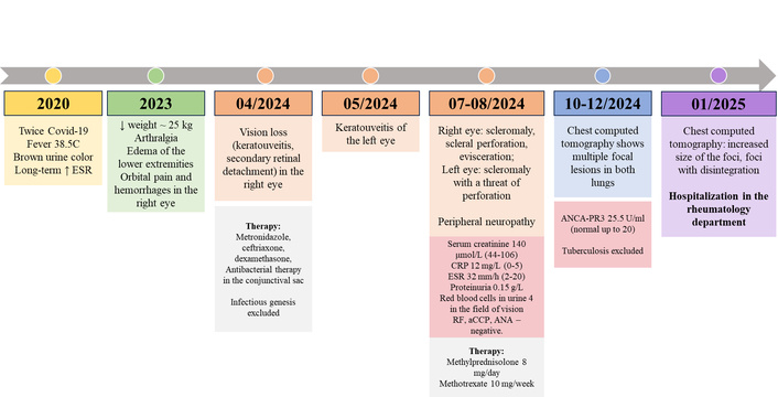
Medical history of patient K. in chronological order. aCCP: antibodies to cyclic citrullinated peptide; ANA: antinuclear antibodies; ANCA: antineutrophil cytoplasmic antibody; CRP: C-reactive protein; ESR: erythrocyte sedimentation rate; PR3: proteinase 3; RF: rheumatoid factor.
Figure 1 shows the chronological history of the disease: from the prodromal period following viral infection with SARS-CoV-2 (prolonged increase in erythrocyte sedimentation rate [ESR], weight loss, and arthralgia) to the manifestation of the disease (progressive damage to the visual organs, development of polyneuropathy, and detection of pulmonary foci with decay).
Upon admission, the condition was of moderate severity. Height: 183 cm; weight: 83 kg; body mass index: 24.78. Body temperature: 36.1°C. The skin is clean and moist with no notable features. Additionally, the right eye is anophthalmic with an ocular prosthesis, and the left eye has hyperemic conjunctiva and multiple infiltrated sclerotic areas with thinning and vascularisation of the sclera. There was no peripheral oedema. Generalized hypotrophy was noted. The lymph nodes accessible to palpation were not enlarged and were painless. The musculoskeletal system showed no pathology upon examination. No pathology was revealed upon examination of the organ systems. Admission tests are presented in Table 1.
Laboratory test results of patient K. upon admission.
| Indicators | Value | Reference range |
|---|---|---|
| Clinical blood test | ||
| Hemoglobin (g/L) | 137.00 | 120.00–140.00 |
| ESR (mm/h) | 40.00 | 2.00–20.00 |
| White blood cells (× 109/L) | 8.00 | 4.00–9.00 |
| Neutrophils (× 109/L) | 5.49 | 2.04–5.80 |
| Lymphocytes (× 109/L) | 1.96 | 1.20–3.00 |
| Platelets (× 109/L) | 251.00 | 150.00–390.00 |
| Biochemical blood test | ||
| Creatinine (μmol/L) | 106.30 | 44.00–106.00 |
| Urea (μmol/L) | 8.00 | 1.80–8.30 |
| Uric acid (μmol/L) | 358.50 | 140.00–340.00 |
| Total protein (g/L) | 58.90 | 66.00–87.00 |
| Glucose (mmol/L) | 4.4 | 3.90–6.40 |
| Immunological blood test | ||
| CRP (mg/L) | 7.60 | 0.00–5.00 |
| ANCA-PR3 (U/mL) | 36.50 | 0.00–5.00 |
| ANCA-MPO (U/mL) | 0.60 | 0.00–5.00 |
| Urine analysis | ||
| Hematuria (per hpf) | 0.00–1.00 | 0.00–1.00 |
| Leukocytes (g/L) | Negative | up to 3.00 |
| Protein (g/L) | Negative | up to 0.03 |
| Daily proteinuria (g/day) | 0.06 | up to 0.15 |
ANCA-MPO: antineutrophil cytoplasmic antibody specific for myeloperoxidase; ANCA-PR3: antineutrophil cytoplasmic antibody specific for proteinase 3; CRP: C-reactive protein; ESR: erythrocyte sedimentation rate; hpf: high power field.
Following the examinations performed, the diagnosis of ‘GPA, ANCA-PR3-positive, presenting with lung, eye and peripheral nervous system damage, as well as constitutional symptoms’ was confirmed in accordance with the classification criteria for GPA as defined by the American College of Rheumatology (ACR) and the European Alliance of Associations for Rheumatology (EULAR) [20]. The activity of GPA was assessed as high (BVASv3 = 17) due to a decrease in body weight of more than 2 kg (2 points), damage to the visual organ (6 points), lungs (3 points), and the nervous system (6 points).
The results of the instrumental studies and consultations with specialists are as follows: Computed tomography of the chest revealed multiple focal lesions of various sizes on both sides, some of which were cavitating (see Figure 2).
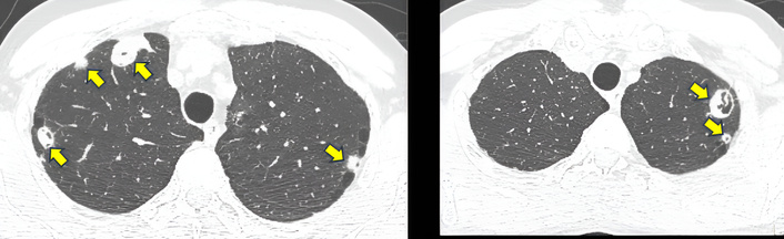
Chest CT scan of patient K. multiple heterogeneous foci are detected in both lungs (indicated by yellow arrows), some of which have signs of cavitation. Computed tomography of the paranasal sinuses showed thickening of the mucous membrane in the left frontal sinus, a polyp in the right nasal cavity, and a deviated nasal septum. Axonal sensory polyneuropathy of the lower extremities was detected on stimulation electroneuromyography. Ophthalmologist’s examination: OD—anophthalmia, OS—necrotizing scleritis. Scleral staphyloma. Marginal vascularized corneal clouding, complicated by mature cataract. Total retinal detachment. Consultation with an otorhinolaryngologist: nasal cavity polyp on the right.
The level of spontaneous NETosis was determined using the immunofluorescence method of staining leukocyte-rich plasma smears with antibodies against NETosis markers (MPO and neutrophil elastase). Detailed methods are described in previous articles [21, 22]. The functional state of neutrophils was studied in an ex vivo model of thromboinflammation described earlier [23]. The chemotaxis and adhesion of neutrophils were studied in whole blood using flat-parallel glass flow chambers coated with type I fibrillar collagen. The movement of cells was observed using fluorescence microscopy.
Data from healthy donors (n = 16) were used to interpret the spontaneous NETosis and functional activity of neutrophils. All donors underwent spontaneous NETosis and neutrophil test analysis according to the same protocol as the patient in the same laboratory. The comparison of the patient’s results with those of healthy donors was descriptive and used for contextual interpretation of the results without statistical analysis.
When spontaneous NETosis was assessed at the time of admission, the patient showed an increased level of NETs: 97.9% of the total number of analysed neutrophils were NET-positive, exceeding the range of values observed in healthy donors (Figure 3A).
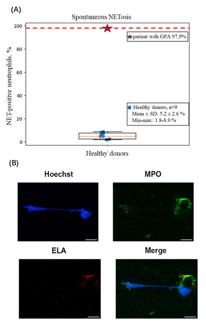
The level of spontaneous NETosis. (A) The box plot reflects the distribution of the proportion of NET-positive neutrophils in healthy donors; individual points correspond to the individual values of the donors. The point outside the distribution range (represented by a red asterisk) indicates the proportion of neutrophils that experienced NETosis in a patient with GPA. (B) Suicidal NETosis in a patient with GPA. Representative image showing a neutrophil undergoing spontaneous suicidal NETosis, the predominant form of NET formation observed in our study. The process is characterised by progressive loss of nuclear lobulation, chromatin decondensation, and rupture of the plasma membrane, leading to the extracellular release of DNA strands with granular proteins. Blue—DNA, Hoechst 33342; green—myeloperoxidase (MPO); red—neutrophil elastase. Scale bar is 10 μm. GPA: granulomatosis with polyangiitis; MPO: myeloperoxidase.
Furthermore, NETosis was exclusively of the suicidal type (sNET proportion: 97.9%) (Figure 3B), with no signs of vital NETosis (vNET) observed. Formation of extracellular traps by leukocytes other than neutrophils has not been detected. In the ex vivo thromboinflammation analysis test, the neutrophil chemotaxis rate of a patient with GPA was found to be 0.144 μm/s, which was at the upper limit of the range observed in healthy donors (see Figure 4). The number of neutrophils per experiment was 70, exceeding the median value for healthy individuals in the control group (Me: 49 [31.5; 65], min–max: 16–148 cells).
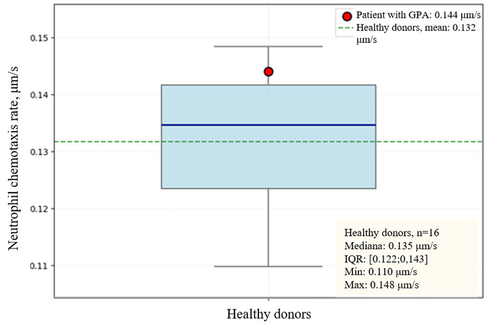
Neutrophil chemotaxis rate in patient K. with GPA and in healthy donors. GPA: granulomatosis with polyangiitis.
When observing the growth of blood clots on collagen, the patient exhibited delayed dynamics of thrombus formation. Initially, there was an absence of thrombotic structures, followed by an increase in thrombus area to 6% of the field of vision after 10 minutes, and to 20% after 25 minutes. The confidence intervals [2.5%, 97.5%] for thrombus areas in healthy donors are as follows: five minutes: 2.0%–12.5%; ten minutes: 3.7%–16.9%; and 25 minutes: 11.4%–27.0%.
In our experiments, we observed a fundamental difference in neutrophil migration patterns between healthy donors and the patient with GPA. Neutrophils from healthy donors exhibited directed chemotactic migration against the blood flow, oriented toward the chemotactic gradient generated by factors released from forming thrombi. In contrast, neutrophils from the patient with active GPA migrated at an angle approaching 90° relative to the flow direction.
Due to the progressive nature of the disease and its high activity level, as well as the involvement of vital organs and the presence of unfavourable prognostic factors such as ANCA-PR3 positivity, anti-B-cell therapy with a saturation dose of 2,000 mg of rituximab intravenously was initiated. The first infusion (1,000 mg) was administered on February 2, 2025, and the second infusion (1,000 mg) was administered on February 16, 2025; it was well tolerated. The patient received glucocorticoid therapy in the form of pulse therapy because there was an active inflammatory process present. Methylprednisolone 500 mg via an intravenous drip. The patient was prescribed a daily oral dose of 8 mg of methylprednisolone, which was continued at the same strength. The prevention of pneumocystis infection is achieved through the prescription of sulfamethoxazole/trimethoprim 480 mg twice a day.
It has been demonstrated that neutrophils from patients with AAV exhibit an increased propensity for spontaneous NETosis in vitro, even in the absence of additional stimulation, which may be indicative of their pre-activation [16, 17]. In the presented clinical case of active GPA with multi-organ damage associated with ANCA-PR3, a high proportion of neutrophils transforming into NETs was observed (97.9%). In this case, NETosis was entirely represented by the suicidal variant; no vital NETosis or extracellular DNA structures formed by other leukocytes were observed. The suicidal NETosis observed suggests that, under our experimental conditions, neutrophils were driven toward a slow, ROS-dependent death pathway with membrane permeabilisation, rather than a rapid, non-lytic expulsion of DNA. This may be due to the absence of stimuli that typically evoke vital NETosis (e.g., Staphylococcus aureus, complement factor C5a, or short-term exposure to LPS) [24–26]. In the context of GPA, this bias toward suicidal NETosis would result in a high local concentration of extracellular DNA and granule proteins, promoting thrombosis, autoantibody formation, or sterile inflammation. The lack of vital NETosis might therefore be a pathogenic switch rather than a mere observational detail [27].
van Dam et al. [18] showed in a study that the serum of patients with AAV induces a slower and more pronounced formation of NETs when incubated with healthy neutrophils and has a lytic character, compared to the serum of patients with systemic lupus erythematosus (SLE). However, these data were obtained ex vivo using neutrophils from healthy donors, so caution is required when interpreting the differences between AAV and SLE.
Due to the limited nature of our study, which is based on a single clinical case, a formal correlation between the level of spontaneous NETosis and the patient’s visual deterioration cannot be established. However, there is growing evidence that NETs contribute to the progression of various ocular surface and intraocular diseases, providing a pathophysiological rationale for a potential link between the extreme NET burden (97.9%) and the severe ocular involvement observed in this patient. Tibrewal et al. [28] demonstrated that hyperosmolar tear stress induces NET formation, which in turn aggravates ocular surface inflammation in dry eye disease. An et al. [29] showed that neutrophils, exfoliated epithelial cells, NETs, and NET-associated proteins are present in ocular surface washings and mucocellular aggregates. Eyes with higher neutrophil counts in washings exhibited more severe signs and symptoms of ocular graft-versus-host disease. These findings collectively illustrate the potential damaging effects of NETs on ocular surface tissues and are consistent with the hypothesis that near-total spontaneous NETosis could contribute to the severe ophthalmopathy and poor visual outcome in our patient with GPA. Clearly, prospective studies in larger cohorts are required to directly assess whether the quantitative level of spontaneous NETosis can serve as a predictor of visual prognosis in ANCA-associated vasculitis.
Our observations are consistent with findings suggesting that active AAV may be associated with increased NET production [10, 18, 19]. However, the relationship between NET levels and disease activity has not been confirmed by all studies. In particular, no differences were found between the active phase and remission of AAV when using circulating extracellular DNA as a marker of NETs [14].
The neutrophil chemotaxis rate in the GPA patient was at the upper limit of the healthy donor range. According to previous studies, perpendicular movement is a characteristic behavior of neutrophils during the transendothelial migration step and is mediated by integrin Mac-1 (αMβ2, CD11b/CD18) [30, 31]. Under physiological conditions, this pattern is observed during intravascular “crawling” of neutrophils as they search for endothelial junctions prior to extravasation, which is a critical step in leukocyte emigration from the bloodstream into tissues.
Under the conditions of our ex vivo assay, this behavior may be explained by several mechanisms. First, the patient’s neutrophils may have a reduced ability to sense the chemotactic gradient from thrombi, possibly due to receptor desensitization or an altered profile of chemokine receptor expression in the setting of systemic inflammation. Second, the signal for transendothelial migration (mechanotactic or integrin-mediated) in patients with active GPA may dominate over the chemotactic signal [32], shifting neutrophils into a mode of searching for endothelial junctions for tissue egress.
We hypothesize that this altered migration trajectory reflects the systemic inflammatory response characteristic of active GPA. In the presence of multi-organ involvement with pronounced inflammation, neutrophils may naturally demonstrate an enhanced propensity for transmigration across the endothelial barrier to reach sites of inflammation in target organs (lungs, eyes, peripheral nerves).
The observed delay in thrombus formation in the neutrophil assay does not contradict the well-established pro-thrombotic effect of NETs. The key physiological pathway linking NETs to thrombosis is the activation of the extrinsic coagulation cascade via tissue factor (TF). In our neutrophil assay, we use hirudin as an anticoagulant. Unlike EDTA (the anticoagulant used for NETosis smears), hirudin specifically inhibits thrombin; therefore, we are unlikely to observe the full TF-dependent NET-driven pro-thrombotic effect in our ex vivo model.
Thrombus formation delay may rather indicate platelet pre-activation in this patient with active GPA [33]. Upon blood sampling and assay setup, such pre-activated cells may display an “exhausted” phenotype, with temporarily reduced responsiveness to the collagen stimulus, which could explain the delayed onset of thrombus formation. The subsequent increase in thrombus area by 25 minutes to the mid-range of the healthy donor confidence interval suggests that the patient’s pro-thrombotic potential is nonetheless preserved and becomes engaged over time.
In our view, this delay does not represent a classical therapeutic window. Rather, it reflects an imbalance between pro- and anti-coagulant mechanisms in this patient and highlights that in our ex vivo model with hirudin, the direct contribution of NETs to thrombus initiation may be underestimated. Future studies using models that preserve the TF-dependent pathway or include physiological stimuli are needed to fully assess the role of NETs in thromboinflammation in GPA.
The presented clinical case reflects a combination of pronounced spontaneous NETosis and preserved migration capacity, alongside delayed thrombus formation, against the backdrop of high clinical activity of GPA. Our observation supplements the existing knowledge regarding the pivotal role of neutrophils and NETs in the pathogenesis of AAV. Given the central role of neutrophil activation in AAV, this area is of interest for monitoring disease activity and developing new diagnostic and therapeutic strategies. Potential therapeutic targets include inhibitors of the main stages of NET formation, drugs that enhance their degradation, and complement activation blockers.
AAV: antineutrophil cytoplasmic antibody-associated vasculitis
ANCA: antineutrophil cytoplasmic antibody
GPA: granulomatosis with polyangiitis
MPO: myeloperoxidase
NETs: neutrophil extracellular traps
PR3: proteinase 3
ROS: reactive oxygen species
SLE: systemic lupus erythematosus
TF: tissue factor
TMR: Conceptualization, Project administration, Writing—review & editing, Funding acquisition. ENV: Data curation, Writing—original draft. KSN: Visualization, Writing—review & editing. EIAA: Methodology. SVG: Methodology. ANS: Conceptualization, Project administration. AML: Supervision, Writing—review & editing. All authors read and approved the submitted version.
There are no conflicts of interest.
The study of Neutrophil activation in ANCA-associated vasculitis was approved by the local ethics committee of the V.A. Nasonova Research Institute of Rheumatology, protocol No. 20 dated October 12, 2023. The local ethics committee of the V.A. Nasonova Research Institute of Rheumatology confirmed that formal approval was not required for this case report. The study complies with the Declaration of Helsinki.
Informed consent to participate in the study was obtained from the participants.
Not applicable.
The raw data supporting the conclusions of this manuscript will be made available by the authors, without undue reservation, to any qualified.
The study was conducted with the support of the Russian Science Foundation (RSF) grant No. 25-25-00245 “Markers of NETosis in Immunoinflammatory Rheumatic Diseases.” The article was prepared within the framework of the basic scientific topic № RК 125020501434-1. Although the study of netosis in this patient was conducted as part of a fundamental study, the funders had no role in the patient’s examination, study design, data collection and analysis, decision to publish, or preparation of the manuscript.
© The Author(s) 2026.
Open Exploration maintains a neutral stance on jurisdictional claims in published institutional affiliations and maps. All opinions expressed in this article are the personal views of the author(s) and do not represent the stance of the editorial team or the publisher.
Copyright: © The Author(s) 2026. This is an Open Access article licensed under a Creative Commons Attribution 4.0 International License (https://creativecommons.org/licenses/by/4.0/), which permits unrestricted use, sharing, adaptation, distribution and reproduction in any medium or format, for any purpose, even commercially, as long as you give appropriate credit to the original author(s) and the source, provide a link to the Creative Commons license, and indicate if changes were made.



View: 380
Download: 14
Times Cited: 0


