• Open Access
    Review

    Vasopressin: a possible link between hypoxia and hypertension

    Ewa Szczepańska-Sadowska *
    Tymoteusz Żera

    Explor Med. 2022;3:414–431 DOI: https://doi.org/10.37349/emed.2022.00103

    Received: June 29, 2022 Accepted: August 03, 2022 Published: October 11, 2022

    Academic Editor: Simon W. Rabkin, Vancouver Hospital, Canada

    Abstract

    Cardiovascular and respiratory diseases are frequently associated with transient and prolonged hypoxia, whereas hypoxia exerts pro-hypertensive effects, through stimulation of the sympathetic system and release of pressor endocrine factors. This review is focused on the role of arginine vasopressin (AVP) in dysregulation of the cardiovascular system during hypoxia associated with cardiovascular disorders. AVP is synthesized mainly in the neuroendocrine neurons of the hypothalamic paraventricular nucleus (PVN) and supraoptic nucleus (SON), which send axons to the posterior pituitary and various regions of the central nervous system (CNS). Vasopressinergic neurons are innervated by multiple neuronal projections releasing several neurotransmitters and other regulatory molecules. AVP interacts with V1a, V1b and V2 receptors that are present in the brain and peripheral organs, including the heart, vessels, lungs, and kidneys. Release of vasopressin is intensified during hypernatremia, hypovolemia, inflammation, stress, pain, and hypoxia which frequently occur in cardiovascular patients, and blood AVP concentration is markedly elevated in cardiovascular diseases associated with hypoxemia. There is evidence that hypoxia stimulates AVP release through stimulation of chemoreceptors. It is suggested that acting in the carotid bodies, AVP may fine-tune respiratory and hemodynamic responses to hypoxia and that this effect is intensified in hypertension. There is also evidence that during hypoxia, augmentation of pro-hypertensive effects of vasopressin may result from inappropriate interaction of this hormone with other compounds regulating the cardiovascular system (catecholamines, angiotensins, natriuretic peptides, steroids, nitric oxide). In conclusion, current literature indicates that abnormal mutual interactions between hypoxia and vasopressin may significantly contribute to pathogenesis of hypertension.

    Keywords

    Chemoreceptors, eclampsia, hypertension, hypoxia, heart failure, syndrome of inappropriate antidiuretic hormone secretion (SIADH), vasopressin

    Introduction

    Transient hypoxia is experienced by healthy subjects whenever they are exposed to reduced oxygen content in the inspired air, however prolonged and recurrent hypoxia is one of the most serious complications of sleep apnea disorders and cardiovascular and respiratory pathologies [1, 2]. It has long been known that hypoxia induces remarkable increases of blood pressure due to stimulation of chemoreceptors [25], and more recently, it has been shown that the pressure responses tend to be greater in patients with cardiovascular diseases than in healthy subjects [1, 68]. The above findings inspired investigators to search for mechanisms responsible for hypertensive effects of hypoxia.

    It is known that hypoxia activates neurons of the autonomic nervous system (ANS) engaged in the regulation of cardiovascular functions [4, 5]. The hypoxic signals alarming the ANS are generated in specialized groups of cells of carotid and aortic chemoreceptor bodies as well as in hypoxia sensitive neurons of the brain and peripheral organs [911]. Recently, it has been shown that enhanced activity of the superior cervical ganglion causes sensitization of the carotid body in spontaneously hypertensive rats (SHR), and that this effect results in greater pressor and sympathoexcitatory responses to the carotid body stimulation [9]. In addition, deficit of oxygen may promote release of neuroendocrine factors involved in the regulation of the cardiovascular system, such as vasopressin, oxytocin, angiotensins and aldosterone [1218].

    Majority of mammals synthesize arginine vasopressin (AVP), while the pig and hippopotamus produce lysine vasopressin (lysipressin, LVP) and the non-mammal vertebrates secrete arginine vasotocin (AVT). The AVP gene consists of three exones encoding the N-terminal signal peptide, vasopressin, neurophysin II and C-terminal peptide, known as copeptin. Vasopressin and copeptin are released in equimolar quantities, but AVP shows great variability due to its binding to platelets and therefore copeptin is frequently estimated in biological samples as a surrogate of vasopressin release [1922].

    The present review draws special attention to the role of AVP in the development of hypertension during hypoxia. In particular, it discusses the mechanisms through which hypoxia modulates regulation of blood pressure and release of AVP. In addition, the consequences of enhanced release of AVP for tissues oxygenation and cardiovascular regulation are analyzed.

    Central and peripheral vasopressin systems

    Experimental and clinical studies combined with immunocytochemical and radioimmunological measurements clearly show that vasopressin is synthesized mainly in the hypothalamus, specifically in the magnocellular (MNC) and parvocellular (PNC) neuroendocrine cells of the paraventricular nucleus (PVN), supraoptic nucleus (SON), and suprachiasmatic nucleus. The prevailing number of the MNC neurons send axons to the posterior pituitary, however some of the axons end in the median eminence (ME) of the hypothalamus. Axons of the MCN neurons form close contacts with capillaries either in the posterior pituitary or in the ME, where AVP is released into the blood [2325]. The PNC innervate the neighboring hypothalamic neurons. They send also multiple projections to several regions of the central nervous system (CNS), namely to the bed nucleus of the stria terminalis (BNST), the medial amygdaloid nucleus, the dorsomedial hypothalamic nucleus, the locus coeruleus (LC), the vertical diagonal band of Broca and the olfactory bulb, as well as to the nucleus ambiguous, nucleus of the solitary tract (NTS), the lateral habenular nucleus, the basal nucleus of Meynert, the substantia nigra, the ventral hippocampus, the central gray, the rostral ventrolateral medulla (RVLM) and the spinal cord [23, 24, 2633].

    Experimental and clinical studies provide evidence that both peripheral and central release of vasopressin are influenced by a variety of regulatory factors [3436]. AVP release is considerably elevated during hypernatremia and hyperosmolality, hypovolemia, heamorrhage, hypoxia, hyperthermia, stress and pain and it is significantly decreased during hypoosmolality, hypervolemia and elevation of blood pressure. Mechanisms involved in the regulation of vasopressinergic system by these challenges have been discussed extensively in several previous studies [21, 22, 26, 35, 3743]. Among factors affecting electrophysiological properties of vasopressinergic neurons and release of vasopressin are noradrenaline, gamma-aminobutyric acid (GABA) and glutamate. It has been shown that neurosecretory cells of SON and PVN nuclei receive inputs from A1 noradrenergic, GABAergic and glutamatergic neurons of the brain stem and forebrain [4451]. Effectiveness of these neurotransmitters is modulated by several locally acting neuropeptides and by gasotransmitters, specifically by angiotensin II, cytokines, and nitric oxide, and by some other local factors [5257].

    Acting in the brain and peripheral organs, vasopressin exerts several regulatory effects, which are of primary importance for regulation of water-electrolyte balance, blood pressure, breathing, metabolism, behavior, learning and memory, sensitivity to stress and pain [21, 22, 26, 27, 32, 38, 42, 5865]. As discussed below, hypoxia belongs to potent stimulators of the vasopressinergic systems, and accumulating evidence indicates that vasopressin also plays a significant role in the regulation of breathing [3739, 66].

    Regulatory functions of vasopressin are mediated by V1a receptors (V1aRs), V1bRs and V2Rs, which are present in multiple regions of the CNS and in several peripheral organs (Figure 1) [22, 24, 6775]. Vasopressin can also interact with oxytocin receptors [21, 76]. Besides, specificity of neuroregulatory effects of vasopressin in the CNS depends on interaction with several neurotransmitters and neuromodulators [26, 27, 33, 42, 60, 62, 63, 7779]. In peripheral organs, V1Rs have been detected in testis, superior cervical ganglion, carotid bodies, liver, kidney, thymus, heart, blood vessels, lung, spleen, uterus, and breast [58, 70, 8083].

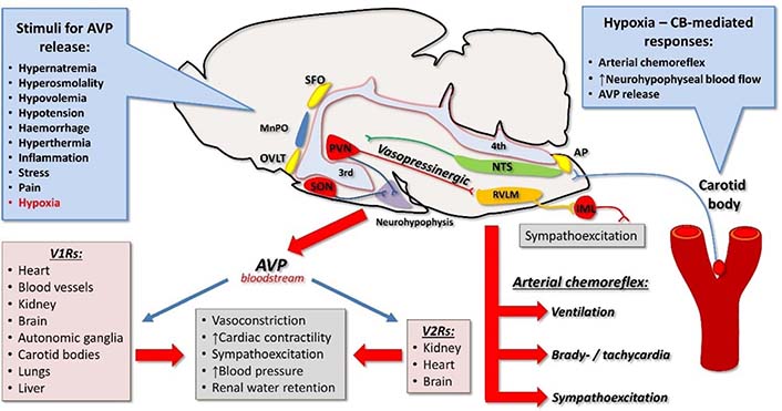
    Vasopressin, hypoxia, and cardiovascular system. Vasopressin is released from the neurohypophysis into the bloodstream in response to numerous stimuli, including hypoxia. Carotid body-mediated responses to hypoxia depend on its connections to the CNS via NTS. These responses include activation of arterial chemoreflex, increase in neurohypophysial blood flow, and release of AVP. Vasopressin exerts its cardiovascular actions by binding to V1Rs and V2Rs. Vasopressinergic pathways from the PVN to the brainstem are activated by hypoxia and involve activation of the RVLM and sympathoexcitation. AP: area postrema; CB: carotid body; IML: intermediolateral nucleus; MnPO: median pre-optic nucleus; OVLT: organum vasculosum of the lamina terminalis; SFO: subfornical organ; 3rd: third cerebral ventricle; 4th: fourth cerebral ventricle; ↑: increase

    V2Rs were found mainly in the kidney, but there is also evidence for their presence in other organs (Figure 1) [8486]. Specifically, V2R binding sites were detected in endothelial cells, where they can participate in the regulation of von Willebrand factor secretion [87, 88]. V2R messenger RNA (mRNA) and binding sites have also been identified in the heart and the brain of the rat in early postnatal life [59, 89].

    Several studies indicate that secretion of vasopressin, expression of its receptors and its action are significantly affected by multiple homeostatic and pathological challenges such as hypernatremia, hyponatremia, dehydration, hypovolemia, hypervolemia, stress, hypertension, cardiac failure, neurological and psychiatric diseases [35, 69, 90106].

    Role of chemoreceptors in blood pressure regulation in relevance with vasopressin

    Arterial chemoreceptors include the carotid bodies strategically located at the bifurcation of the common carotid artery and the aortic bodies situated in the aortic arch. Hypoxia is the primary stimulus for arterial chemoreceptors, especially the carotid bodies (Figure 1). Activation of the arterial chemoreceptors triggers the arterial chemoreflex, which leads to sympathoexcitation, changes in cardiovagal balance, increase in phrenic nerve activity, and pulmonary ventilation and arousal [10, 107, 108]. The involvement of carotid bodies in driving sympathetic activity in hypertension, obstructive sleep apnea, and heart failure is well documented in preclinical and clinical studies. However, the role of aortic bodies is less defined and still requires further investigations.

    Increased activity of the arterial chemoreceptors, especially the carotid bodies, has been postulated and found to be causative for experimental arterial hypertension [8, 109113] and confirmed by clinical studies [114116]. Increased activity of the arterial chemoreflex is seen before the onset of hypertension in SHR [117], which is in line with findings in normotensive young adults with family history of hypertension, who often manifest increased sensitivity of arterial chemoreflex [8]. There seems to be a selective enhancement of the sympathetic response, with tonically increased activity of the arterial chemoreflex evoked from the carotid bodies as well as increased sensitivity of the chemoreceptors to hypoxic stimuli in hypertension [10, 118, 119]. There is also accumulating evidence that purinergic P2X receptors in the carotid body are involved in this selective drive to the sympathetic system [120122], thus suggesting that distinct mediators may be involved in specific components of the arterial chemoreflex [10]. The causative role of arterial chemoreceptors in hypertension is further demonstrated by blood pressure lowering effects of removal, denervation, or suppression with hyperoxia of the carotid bodies in animal models of hypertension and in hypertensive patients [110, 112, 114116].

    In this light, it is essential to note that hypoxia increases neurohypophyseal blood flow [123125] and elevates plasma levels of AVP both in animals and humans [39, 126128] (Figure 1). The effect of hypoxia on AVP release at least in part depends on the sensory input from the carotid bodies [124, 126]. It has been postulated that the increase in plasma AVP concentration helps in preventing the hypoxia-induced vasodilation, supporting in this way peripheral vascular resistance [129, 130].

    Furthermore, AVP released into the circulation may directly target chemosensitive glomus cells of the carotid bodies, which express V1aRs [83] as well as G protein q/11 and phosphokinase C that are key intracellular components of the V1aR signaling [131]. These findings indicate that circulating AVP may affect the glomus cells expressing V1aRs and presumably modulate the activity of the carotid body, thus, providing a potential feedback loop between the hypoxia-mediated release of AVP and activity of the chemoreceptors (Figure 2). In addition, decreased carotid body blood flow activates arterial chemoreflex and has been suggested as a putative mechanism involved in elevated activity of the reflex in hypertension [109, 132]. Thus, it is probable that AVP may also enhance arterial chemoreceptors’ activity by increasing vascular resistance of the carotid body artery thereby decreasing the carotid body blood flow with resultant sensitization of the chemoreceptors.

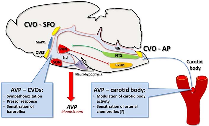
    Vasopressin targets circumventricular organs and carotid body. Vasopressin present in the bloodstream contributes to the neural control of cardiovascular system by binding to its receptors in the circumventricular organs in the brain. Vasopressin may also act on V1aRs expressed in the carotid body, however, its sensitizing effect on the carotid body activity and arterial chemoreflex (?) awaits further elucidation, especially under conditions of hypertension. CVO: circumventricular organ

    Vasopressin released into the bloodstream also binds to the circumventricular organs that lack the blood-brain barrier. In particular, V1aRs are expressed in the area postrema [133, 134] and binding of AVP to these receptors alters neuronal activity in the surrounding cardiovascular centers [135, 136] and inhibits phrenic nerve activity [133]. In contrast, it has been suggested that AVP acting locally in the carotid bodies may increase its sensitivity [83] and thus counterbalance the inhibitory effects of the neurohormone binding to V1aRs in the area postrema. This action could fine-tune respiratory and hemodynamic responses to hypoxia [38, 83]. In addition, AVP binding to its receptors in the area postrema sensitizes the arterial baroreflex thus, it helps to maintain blood pressure buffering responses [22, 137]. Despite the sensitizing effect on the baroreflex, intracerebroventricular infusions of AVP that allow for binding of AVP with its receptors in the circumventricular organs trigger pressor response under most experimental paradigms [22, 137] (Figure 2). The question arises whether systemically released AVP modulates engagement of chemoreceptors in regulation of blood pressure in normotensive subjects. In this regard, it has been shown that systemic inhibition of V1aRs do not affect pressor response to acutely evoked arterial chemoreflex with cyanide [138]. Thus, it appears that in normotensive rats AVP plays an insignificant role in hemodynamic response to arterial chemoreflex activation. Undoubtedly, the role of AVP and its V1aR in the carotid body under conditions of arterial hypertension awaits further elucidation.

    Altered regulation of vasopressin release in cardiovascular and respiratory diseases

    Function of vasopressin system in cardiovascular diseases

    Hypertension and other cardiovascular diseases are frequently complicated by cardiac failure, which is associated with hypoxia. It has been shown that hypoxia strongly promotes activation of the hypothalamic vasopressinergic neurons and increases release of vasopressin or its surrogate marker copeptin [12, 14, 16, 139, 140].

    There is also evidence that heart failure results in significant reorganization of central and peripheral components of vasopressinergic system. It has been found that acute cardiac failure induced by myocardial infarction causes significant elevation of c-Fos protein expression in magnocellular neurosecretory vasopressinergic neurons of the PVN and SON [141, 142] and that the activation results from inappropriate stimulation of these neurons by glutamatergic N-methyl-D-aspartate (NMDA) receptors [143, 144]. It should be noted that hypertension and heart failure are associated with reduced inhibition of the PVN neurons by GABAergic and nitrergic transmission [145, 146]. The study of Kc et al. [16] provided evidence that local hypoxia of PVN neurons stimulates subpopulation of vasopressinergic neurons innervating the RVLM. In addition, the authors found that expression of V1aRs in the RVLM is elevated in rats exposed to chronic intermittent hypoxia and that blockade of V1aRs in the RVLM significantly attenuates blood pressure and heart rate responses to intermittent hypoxia [16].

    Experimental studies revealed that hypertension has significant impact on the brain vasopressin receptors in specific brain regions. Hence, AVP mRNA expression and V1aR expression were found to be higher in the PVN of the SHR than in the PVN of Wistar-Kyoto (WKY) rats [147]. In hypertensive renin transgenic TGR(mRen2)27 rats, expression of V1bR mRNA was elevated in the mesencephalon-pontine region [148]. Enhanced engagement of V1aR in regulation of blood pressure was found in SHR, deoxycorticosterone acetate (DOCA)-salt hypertension and transgenic TGR(mRen2)27 hypertension [147152].

    Multiple clinical studies revealed increased plasma AVP concentration in patients with heart failure and reduced ejection fraction [153155]. Evidence of elevated concentration of AVP was also found in patients with left ventricle hypertrophy and reduced level of atrial natriuretic peptide [156]. It should be noted that in patients with heart failure, plasma AVP level was elevated in spite of normal or even reduced sodium concentration and osmolality—the symptoms which are illustrative for the syndrome of inappropriate vasopressin secretion (SIADH). Presence of water retention, hypoosmolality and hyponatremia in heart failure is usually interpreted as a result of arterial underfilling, which causes unloading of baroreceptors [157] and elevation of non-osmotic release of AVP [158160].

    In majority of vascular beds, AVP elicits vasoconstriction and a decrease of blood flow due to stimulation of V1Rs [42, 161], however, in the pulmonary circulation, it can promote vasodilation [162164]. Pulmonary vessels are resistant to vasoconstrictory action of vasopressin [164166] and in vitro experiments on the pulmonary artery provide evidence for its relaxing effect [167]. In the cerebral circulation, vasopressin can cause either increase of the blood flow through an action mediated by V2Rs [85] or restriction of the flow, presumably through stimulation of V1Rs. Interestingly, it appears that the vasoconstrictory action of vasopressin in the cerebral circulation may predominate in hyponatremia, which is an unavoidable attribute of SIADH [168, 169]. The traumatic brain injury which is associated with hypoxia causes significant increase of V1aR in the frontoparietal cortex [104]. Vasopressin interacts synergistically with proinflammatory cytokines and it is likely that its vasoconstrictory action on cerebral vessel may worsen cerebral blood flow in the traumatic shock [104, 170].

    Hypoxia and enhanced involvement of vasopressin in regulation of blood pressure in cardiovascular diseases

    Several studies provide evidence that hypertension and heart failure are associated with inappropriate regulation of AVP release and its function [42, 90, 94, 171174]. It has been postulated that elevated concentration of blood AVP in heart failure and hypertension worsens health state due to excessive stimulation of V2Rs which promotes water retention, and because of enhanced stimulation of V1R which causes vasoconstriction and elevation of total peripheral resistance [173176]. Moreover, chronic intravenous infusion of V1 agonist in normotensive Sprague-Dawley (SD) rats resulted in the development of hypertension [92] and other experimental studies employing V1aR knockout (V1aRKO) mice and mice with myocyte-selective transgenic overexpression of cardiac V1aR suggested that permanent stimulation of these receptors may play an essential role in the development of cardiac hypertrophy [175].

    It has been shown that administration of V2R antagonists (tolvaptan, lixivaptan) exerts positive effects in animal and human heart failure, manifested by normalization of electrolytes status, diminution of myocardial infarction-induced fibrosis and macrophage infiltration, as well as by improvement of the left ventricle function [177184]. Moreover, some long-term trials revealed that administration of tolvaptan improves long-term prognosis in patients with congestive heart failure [185, 186]. However, other authors were not able to prove effectiveness of tolvaptan on the long-term survival of patients with heart failure [160].

    It has been reported that administration of conivaptan, which is a dual V1a and V2 vasopressin antagonist, significantly improves hemodynamic parameters and renal functions of patients with heart failure [173, 174, 187189]. In canine model of tachypacing-induced heart failure, chronic application of pecavaptan (BAY1753011), which is another dual V1aR/V2R antagonist, exerted diuretic effects and significantly improved vital hemodynamic parameters, as shown by elevation of cardiac output and diminution of total peripheral resistance [190]. The above effects were significantly relative to placebo and tolvaptan alone. Affinity of pecavaptan to human and canine V1aR/V2R is similar [190], and therefore it is likely that similar beneficial effects of pecavaptan would occur in human patients with heart failure.

    Hypertension is one of the serious complications of eclampsia. Excessive stimulation of vasopressinergic system, associated with hypoxia and inappropriate tissue oxygenation has been found in the pre-eclampsia state [191193]. Moreover, infusion of vasopressin into pregnant mice resulted in pathological changes, which are typical for pre-eclampsia (hypertension and placental oxidative stress) and which could be prevented by blockade of AVP receptors. Accordingly, it has been suggested that elevated secretion of vasopressin contributes to the development of hypertension in pre-eclampsia [194]. However, exposure of maternal rats to hypoxia increases also expression of V2R in newborn rats [195, 196], whereas stimulation of V2Rs causes dilation of pulmonary vessels [88, 162, 163]. Thus, it is also likely that upregulation of V2R in newborns may belong to mechanisms protecting from negative consequences of maternal hypoxia.

    Septic shock is another pathological state which causes inflammatory symptoms, tissue hypoxia and activation of the vasopressinergic system [197, 198]. During experimental model of septic shock, blood AVP concentration initially increases up to 500 pg/mL and subsequently decreases to levels that are disproportionately low in relation to hypotensive effects of shock [199]. Because survival of patients with septic shock is significantly improved by combined infusions of vasopressin and corticosteroids in comparison to vasopressin alone or noradrenaline [198, 200], it is likely that prolonged sepsis may somehow disorganize regulation of vasopressin release and consequently result in defective cardiovascular regulation.

    Finally, it is worthy to note that some experimental studies suggest that in emergency states AVP may exert some protective effects in the brain. For instance, in a rat model of asphyxial cardiac arrest, which imitates hemodynamic disturbances occurring during cardiopulmonary resuscitation, intraperitoneal administration of vasopressin was found to reduce neuronal apoptosis in the hippocampus [201].

    Conclusions

    Hypoxia and elevated blood AVP concentration are frequent attributes of cardiovascular diseases and both exert pro-hypertensive effects. There is strong evidence that hypoxia potently stimulates central and systemic release of AVP and that numerous actions of this hormone determine responsiveness of the cardiovascular system to hypoxia (Figures 1 and 2). Survey of literature carried out in the present review draws attention to mechanisms underlying mutual interactions between hypoxia and activation of systemic and central vasopressin systems in health and cardiovascular diseases. It is concluded that during hypoxia, vasopressin exerts pro-hypertensive effects via intensification of stimulation of chemoreceptors and through interactions with other cardiovascular regulatory factors, mainly catecholamines, angiotensins, natriuretic peptides, steroid hormones and gasotransmitters These effects are potentiated in hypertension and cardiac failure. However, vasopressin may also exert some positive effects through actions mediated by receptors located in specific cardiovascular regions.

    Abbreviations

    AVP:

    arginine vasopressin

    CNS:

    central nervous system

    GABA:

    gamma-aminobutyric acid

    mRNA:

    messenger RNA

    NTS:

    nucleus of the solitary tract

    PVN:

    paraventricular nucleus

    RVLM:

    rostral ventrolateral medulla

    SHR:

    spontaneously hypertensive rats

    SON:

    supraoptic nucleus

    V1aRs:

    V1a receptors

    Declarations

    Acknowledgments

    The authors are grateful to Professor Agnieszka-Cudnoch-Jędrzejewska—head of the Department of Experimental and Clinical Physiology and other colleagues of the Department for friendly support during preparation of the manuscript.

    Author contributions

    ESS conceived conception and design of the paper and wrote the first draft of the manuscript; TŻ contributed to conception of the paper and wrote sections of the manuscript. Both authors contributed to manuscript revision, read and approved the submitted version.

    Conflicts of interest

    The authors declare that they have no conflicts of interest.

    Ethical approval

    Not applicable.

    Consent to participate

    Not applicable.

    Consent to publication

    Not applicable.

    Availability of data and materials

    Not applicable.

    Funding

    This work was supported by the Medical University of Warsaw Scientific Projects, Poland. The funder had no role in study design, data collection and analysis, decision to publish, or preparation of the manuscript.

    Copyright

    © The Author(s) 2022.

    References

    Javaheri S, Redline S. Insomnia and risk of cardiovascular disease. Chest. 2017;152:43544. [DOI] [PubMed] [PMC]
    Karnati S, Seimetz M, Kleefeldt F, Sonawane A, Madhusudhan T, Bachhuka A, et al. Chronic obstructive pulmonary disease and the cardiovascular system: vascular repair and regeneration as a therapeutic target. Front Cardiovasc Med. 2021;8:649512. [DOI] [PubMed] [PMC]
    Hirooka Y, Polson JW, Potts PD, Dampney RA. Hypoxia-induced Fos expression in neurons projecting to the pressor region in the rostral ventrolateral medulla. Neuroscience. 1997;80:120924. [DOI] [PubMed]
    Sun MK, Jeske IT, Reis DJ. Cyanide excites medullary sympathoexcitatory neurons in rats. Am J Physiol. 1992;262:R1829. [DOI] [PubMed]
    Horn EM, Waldrop TG. Modulation of the respiratory responses to hypoxia and hypercapnia by synaptic input onto caudal hypothalamic neurons. Brain Res. 1994;664:2533. [DOI] [PubMed]
    Javaheri S, Barbe F, Campos-Rodriguez F, Dempsey JA, Khayat R, Javaheri S, et al. Sleep apnea: types, mechanisms, and clinical cardiovascular consequences. J Am Coll Cardiol. 2017;69:84158. [DOI] [PubMed] [PMC]
    Li T, Chen Y, Gua C, Wu B. Elevated oxidative stress and inflammation in hypothalamic paraventricular nucleus are associated with sympathetic excitation and hypertension in rats exposed to chronic intermittent hypoxia. Front Physiol. 2018;9:840. [DOI] [PubMed] [PMC]
    Trzebski A, Tafil M, Zoltowski M, Przybylski J. Increased sensitivity of the arterial chemoreceptor drive in young men with mild hypertension. Cardiovasc Res. 1982;16:16372. [DOI] [PubMed]
    Felippe ISA, Zera T, da Silva MP, Moraes DJA, McBryde F, Paton JFR. The sympathetic nervous system exacerbates carotid body sensitivity in hypertension. Cardiovasc Res. 2022;[Epub ahead of print]. [DOI] [PubMed]
    Zera T, Moraes DJA, da Silva MP, Fisher JP, Paton JFR. The logic of carotid body connectivity to the brain. Physiology (Bethesda). 2019;34:26482. [DOI] [PubMed]
    Iturriaga R, Andrade DC, Del Rio R. Enhanced carotid body chemosensory activity and the cardiovascular alterations induced by intermittent hypoxia. Front Physiol. 2014;5:468. [DOI] [PubMed] [PMC]
    Anderson RJ, Pluss RG, Berns AS, Jackson JT, Arnold PE, Schrier RW, et al. Mechanism of effect of hypoxia on renal water excretion. J Clin Invest. 1978;62:76977. [DOI] [PubMed] [PMC]
    Carmichael CY, Wainford RD. Hypothalamic signaling mechanisms in hypertension. Curr Hypertens Rep. 2015;17:39. [DOI] [PubMed] [PMC]
    Coldren KM, Li DP, Kline DD, Hasser EM, Heesch CM. Acute hypoxia activates neuroendocrine, but not presympathetic, neurons in the paraventricular nucleus of the hypothalamus: differential role of nitric oxide. Am J Physiol Regul Integr Comp Physiol. 2017;312:R98295. [DOI] [PubMed] [PMC]
    Cooke M, Cruttenden R, Mellor A, Lumb A, Pattman S, Burnett A, et al. A pilot investigation into the effects of acute normobaric hypoxia, high altitude exposure and exercise on serum angiotensin-converting enzyme, aldosterone and cortisol. J Renin Angiotensin Aldosterone Syst. 2018;19:1470320318782782. [DOI] [PubMed] [PMC]
    Kc P, Balan KV, Tjoe SS, Martin RJ, Lamanna JC, Haxhiu MA, et al. Increased vasopressin transmission from the paraventricular nucleus to the rostral medulla augments cardiorespiratory outflow in chronic intermittent hypoxia-conditioned rats. J Physiol. 2010;588:72540. [DOI] [PubMed] [PMC]
    Lam SY, Liu Y, Ng KM, Liong EC, Tipoe GL, Leung PS, et al. Upregulation of a local renin-angiotensin system in the rat carotid body during chronic intermittent hypoxia. Exp Physiol. 2014;99:22031. [DOI] [PubMed]
    Yamashita K, Ito K, Endo J, Matsuhashi T, Katsumata Y, Yamamoto T, et al. Adrenal cortex hypoxia modulates aldosterone production in heart failure. Biochem Biophys Res Commun. 2020;524:1849. [DOI] [PubMed]
    Acher R, Chauvet J. Structure, processing and evolution of the neurohypophysial hormone-neurophysin precursors. Biochimie. 1988;70:1197207. [DOI] [PubMed]
    Morgenthaler NG, Struck J, Alonso C, Bergmann A. Assay for the measurement of copeptin, a stable peptide derived from the precursor of vasopressin. Clin Chem. 2006;52:1129. [DOI] [PubMed]
    Szczepanska-Sadowska E, Wsol A, Cudnoch-Jedrzejewska A, Żera T. Complementary role of oxytocin and vasopressin in cardiovascular regulation. Int J Mol Sci. 2021;22:11465. [DOI] [PubMed] [PMC]
    Szczepanska-Sadowska E, Żera T, Sosnowski P, Cudnoch-Jedrzejewska A, Puszko A, Misicka A. Vasopressin and related peptides; potential value in diagnosis, prognosis and treatment of clinical disorders. Curr Drug Metab. 2017;18:30645. [DOI] [PubMed]
    Buijs RM, De Vries GJ, Van Leeuwen FW, Swaab DF. Vasopressin and oxytocin: distribution and putative functions in the brain. Prog Brain Res. 1983;60:11522. [DOI] [PubMed]
    Ginsburg M, Jayasena K. The distribution of proteins that bind neurohypophysial hormones. J Physiol. 1968;197:6576. [DOI] [PubMed] [PMC]
    Bichet DG. Regulation of thirst and vasopressin release. Annu Rev Physiol. 2019;81:35973. [DOI] [PubMed]
    Buijs RM, Hermes MH, Kalsbeek A. The suprachiasmatic nucleus-paraventricular nucleus interactions: a bridge to the neuroendocrine and autonomic nervous system. Prog Brain Res. 1998;119:36582. [DOI] [PubMed]
    Buijs RM, Hurtado-Alvarado G, Soto-Tinoco E. Vasopressin: an output signal from the suprachiasmatic nucleus to prepare physiology and behaviour for the resting phase. J Neuroendocrinol. 2021;33:e12998. [DOI] [PubMed]
    Ginsberg SD, Hof PR, Young WG, Morrison JH. Noradrenergic innervation of vasopressin- and oxytocin-containing neurons in the hypothalamic paraventricular nucleus of the macaque monkey: quantitative analysis using double-label immunohistochemistry and confocal laser microscopy. J Comp Neurol. 1994;341:47691. [DOI] [PubMed]
    Romijn HJ, van Uum JF, Emmering J, Goncharuk V, Buijs RM. Colocalization of VIP with AVP in neurons of the human paraventricular, supraoptic and suprachiasmatic nucleus. Brain Res. 1999;832:4753. [DOI] [PubMed]
    Rogers CN, Ross AP, Sahu SP, Siegel ER, Dooyema JM, Cree MA, et al. Oxytocin- and arginine vasopressin-containing fibers in the cortex of humans, chimpanzees, and rhesus macaques. Am J Primatol. 2018;80:e22875. [DOI] [PubMed] [PMC]
    Sawchenko PE, Swanson LW. Immunohistochemical identification of neurons in the paraventricular nucleus of the hypothalamus that project to the medulla or to the spinal cord in the rat. J Comp Neurol. 1982;205:26072. [DOI] [PubMed]
    Swaab DF, Fliers E, van Gool WA. Immunocytochemical localization of vasopressin in the human brain; its possible consequences for therapeutic strategies in aging and dementia. Prog Brain Res. 1986;65:10513. [DOI] [PubMed]
    Wood CE, Chen HG, Bell ME. Role of vagosympathetic fibers in the control of adrenocorticotropic hormone, vasopressin, and renin responses to hemorrhage in fetal sheep. Circ Res. 1989;64:51523. [DOI] [PubMed]
    Simon-Oppermann C, Gray D, Szczepańska-Sadowska E, Simon E. Vasopressin in blood and third ventricle CSF of dogs in chronic experiments. Am J Physiol. 1983;245:R5418. [DOI] [PubMed]
    Szczepańska-Sadowska E, Gray D, Simon-Oppermann C. Vasopressin in blood and third ventricle CSF during dehydration, thirst, and hemorrhage. Am J Physiol. 1983;245:R54955. [DOI] [PubMed]
    Szczepańska-Sadowska E, Simon-Oppermann C, Gray D, Simon E. Control of central release of vasopressin. J Physiol (Paris). 1984;79:4329. [PubMed]
    King TL, Kline DD, Ruyle BC, Heesch CM, Hasser EM. Acute systemic hypoxia activates hypothalamic paraventricular nucleus-projecting catecholaminergic neurons in the caudal ventrolateral medulla. Am J Physiol Regul Integr Comp Physiol. 2013;305:R111223. [DOI] [PubMed] [PMC]
    Proczka M, Przybylski J, Cudnoch-Jędrzejewska A, Szczepańska-Sadowska E, Żera T. Vasopressin and breathing: review of evidence for respiratory effects of the antidiuretic hormone. Front Physiol. 2021;12:744177. [DOI] [PubMed] [PMC]
    Raff H, Shinsako J, Keil LC, Dallman MF. Vasopressin, ACTH, and corticosteroids during hypercapnia and graded hypoxia in dogs. Am J Physiol. 1983;244:E4538. [DOI] [PubMed]
    Szczepanska-Sadowska E. Plasma ADH increase and thirst suppression elicited by preoptic heating in the dog. Am J Physiol. 1974;226:15561. [DOI] [PubMed]
    Szczepańska-Sadowska E, Simon-Oppermann C, Gray DA, Simon E. Plasma and cerebrospinal fluid vasopressin and osmolality in relation to thirst. Pflugers Arch. 1984;400:2949. [DOI] [PubMed]
    Szczepanska-Sadowska E, Czarzasta K, Cudnoch-Jedrzejewska A. Dysregulation of the renin-angiotensin system and the vasopressinergic system interactions in cardiovascular disorders. Curr Hypertens Rep. 2018;20:19. [DOI] [PubMed] [PMC]
    Lévy F, Kendrick KM, Goode JA, Guevara-Guzman R, Keverne EB. Oxytocin and vasopressin release in the olfactory bulb of parturient ewes: changes with maternal experience and effects on acetylcholine, gamma-aminobutyric acid, glutamate and noradrenaline release. Brain Res. 1995;669:197206. [DOI] [PubMed]
    Brefel C, Lazartigues E, Tran MA, Gauquelin G, Geelen G, Gharib C, et al. Central cardiovascular effects of acetylcholine in the conscious dog. Br J Pharmacol. 1995;116:217582. [DOI] [PubMed] [PMC]
    Hillhouse EW, Milton NG. Effect of noradrenaline and gamma-aminobutyric acid on the secretion of corticotrophin-releasing factor-41 and arginine vasopressin from the rat hypothalamus in vitro. J Endocrinol. 1989;122:71923. [DOI] [PubMed]
    Savci V, Goktalay G, Ulus IH. Intracerebroventricular choline increases plasma vasopressin and augments plasma vasopressin response to osmotic stimulation and hemorrhage. Brain Res. 2002;942:5870. [DOI] [PubMed]
    Wang L, Ennis M, Szabó G, Armstrong WE. Characteristics of GABAergic and cholinergic neurons in perinuclear zone of mouse supraoptic nucleus. J Neurophysiol. 2015;113:75467. [DOI] [PubMed] [PMC]
    Zaninetti M, Blanchet C, Tribollet E, Bertrand D, Raggenbass M. Magnocellular neurons of the rat supraoptic nucleus are endowed with functional nicotinic acetylcholine receptors. Neuroscience. 1999;95:31923. [DOI] [PubMed]
    Zaninetti M, Tribollet E, Bertrand D, Raggenbass M. Nicotinic cholinergic activation of magnocellular neurons of the hypothalamic paraventricular nucleus. Neuroscience. 2002;110:28799. [DOI] [PubMed]
    Smith DW, Buller KM, Day TA. Role of ventrolateral medulla catecholamine cells in hypothalamic neuroendocrine cell responses to systemic hypoxia. J Neurosci. 1995;15:797988. [DOI] [PubMed] [PMC]
    Hermes ML, Ruijter JM, Klop A, Buijs RM, Renaud LP. Vasopressin increases GABAergic inhibition of rat hypothalamic paraventricular nucleus neurons in vitro. J Neurophysiol. 2000;83:70511. [DOI] [PubMed]
    Dawson CA, Jhamandas JH, Krukoff TL. Activation by systemic angiotensin II of neurochemically identified neurons in rat hypothalamic paraventricular nucleus. J Neuroendocrinol. 1998;10:4539. [DOI] [PubMed]
    Loń S, Szczepańska-Sadowska E, Szczypaczewska M. Evidence that centrally released arginine vasopressin is involved in central pressor action of angiotensin II. Am J Physiol. 1996;270:H16773. [DOI] [PubMed]
    Raber J, Bloom FE. Arginine vasopressin release by acetylcholine or norepinephrine: region-specific and cytokine-specific regulation. Neuroscience. 1996;71:74759. [DOI] [PubMed]
    Raber J, Bloom FE. IL-2 induces vasopressin release from the hypothalamus and the amygdala: role of nitric oxide-mediated signaling. J Neurosci. 1994;14:618795. [DOI] [PubMed] [PMC]
    Sladek CD, Kapoor JR. Neurotransmitter/neuropeptide interactions in the regulation of neurohypophyseal hormone release. Exp Neurol. 2001;171:2009. [DOI] [PubMed]
    Tobin VA, Bull PM, Arunachalam S, O’Carroll AM, Ueta Y, Ludwig M. The effects of apelin on the electrical activity of hypothalamic magnocellular vasopressin and oxytocin neurons and somatodendritic peptide release. Endocrinology. 2008;149:613645. [DOI] [PubMed] [PMC]
    Evora PR, Pearson PJ, Schaff HV. Arginine vasopressin induces endothelium-dependent vasodilatation of the pulmonary artery. V1-receptor-mediated production of nitric oxide. Chest. 1993;103:12415. [DOI] [PubMed]
    Gutkowska J, Miszkurka M, Danalache B, Gassanov N, Wang D, Jankowski M. Functional arginine vasopressin system in early heart maturation. Am J Physiol Heart Circ Physiol. 2007;293:H226270. [DOI] [PubMed]
    Pizzirusso A, Oliva P, Maione S, D’Amico M, Rossi F, Berrino L. Role of vasopressin on excitatory amino acids mediated pressor responses in the periaqueductal gray area. Naunyn Schmiedebergs Arch Pharmacol. 1998;357:5148. [DOI] [PubMed]
    Szczepańska-Sadowska E. Hemodynamic effects of a moderate increase of the plasma vasopressin level in conscious dogs. Pflugers Arch. 1973;338:31322. [DOI] [PubMed]
    Wacker D, Ludwig M. The role of vasopressin in olfactory and visual processing. Cell Tissue Res. 2019;375:20115. [DOI] [PubMed] [PMC]
    Yang J, Yang Y, Xu HT, Chen JM, Liu WY, Lin BC. Arginine vasopressin induces periaqueductal gray release of enkephalin and endorphin relating to pain modulation in the rat. Regul Pept. 2007;142:2936. [DOI] [PubMed]
    Burrell LM, Phillips PA, Rolls KA, Buxton BF, Johnston CI, Liu JJ. Vascular responses to vasopressin antagonists in man and rat. Clin Sci (Lond). 1994;87:38995. [DOI] [PubMed]
    Landgraf R, Neumann I, Holsboer F, Pittman QJ. Interleukin-1 beta stimulates both central and peripheral release of vasopressin and oxytocin in the rat. Eur J Neurosci. 1995;7:5928. [DOI] [PubMed]
    Iovino M, Guastamacchia E, Giagulli VA, Fiore G, Licchelli B, Iovino E, et al. Role of central and peripheral chemoreceptors in vasopressin secretion control. Endocr Metab Immune Disord Drug Targets. 2013;13:2505. [DOI] [PubMed]
    Corbani M, Marir R, Trueba M, Chafai M, Vincent A, Borie AM, et al. Neuroanatomical distribution and function of the vasopressin V1B receptor in the rat brain deciphered using specific fluorescent ligands. Gen Comp Endocrinol. 2018;258:1532. [DOI] [PubMed]
    Chodobski A, Wojcik BE, Loh YP, Dodd KA, Szmydynger-Chodobska J, Johanson CE, et al. Vasopressin gene expression in rat choroid plexus. Adv Exp Med Biol. 1998;449:5965. [DOI] [PubMed]
    Góźdź A, Szczepańska-Sadowska E, Maśliński W, Kumosa M, Szczepańska K, Dobruch J. Differential expression of vasopressin V1a and V1b receptors mRNA in the brain of renin transgenic TGR(mRen2)27 and Sprague-Dawley rats. Brain Res Bull. 2003;59:399403. [DOI] [PubMed]
    Góźdź A, Szczepańska-Sadowska E, Szczepańska K, Maśliński W, Luszczyk B. Vasopressin V1a, V1b and V2 receptors mRNA in the kidney and heart of the renin transgenic TGR(mRen2)27 and Sprague Dawley rats. J Physiol Pharmacol. 2002;53:34957. [PubMed]
    Ostrowski NL, Lolait SJ, Young WS 3rd. Cellular localization of vasopressin V1a receptor messenger ribonucleic acid in adult male rat brain, pineal, and brain vasculature. Endocrinology. 1994;135:151128. [DOI] [PubMed]
    Noszczyk B, Lon S, Szczepańska-Sadowska E. Central cardiovascular effects of AVP and AVP analogs with V1, V2 and ‘V3’ agonistic or antagonistic properties in conscious dog. Brain Res. 1993;610:11526. [DOI] [PubMed]
    Schorscher-Petcu A, Dupré A, Tribollet E. Distribution of vasopressin and oxytocin binding sites in the brain and upper spinal cord of the common marmoset. Neurosci Lett. 2009;461:21722. [DOI] [PubMed]
    Young LJ, Toloczko D, Insel TR. Localization of vasopressin (V1a) receptor binding and mRNA in the rhesus monkey brain. J Neuroendocrinol. 1999;11:2917. [DOI] [PubMed]
    Manning M, Misicka A, Olma A, Bankowski K, Stoev S, Chini B, et al. Oxytocin and vasopressin agonists and antagonists as research tools and potential therapeutics. J Neuroendocrinol. 2012;24:60928. [DOI] [PubMed] [PMC]
    Song Z, Albers HE. Cross-talk among oxytocin and arginine-vasopressin receptors: relevance for basic and clinical studies of the brain and periphery. Front Neuroendocrinol. 2018;51:1424. [DOI] [PubMed] [PMC]
    Currás-Collazo MC, Gillard ER, Jin J, Pandika J. Vasopressin and oxytocin decrease excitatory amino acid release in adult rat supraoptic nucleus. J Neuroendocrinol. 2003;15:18290. [DOI] [PubMed]
    Szczepanska-Sadowska E, Wsol A, Cudnoch-Jedrzejewska A, Czarzasta K, Żera T. Multiple aspects of inappropriate action of renin-angiotensin, vasopressin, and oxytocin systems in neuropsychiatric and neurodegenerative diseases. J Clin Med. 2022;11:908. [DOI] [PubMed] [PMC]
    Szmydynger-Chodobska J, Zink BJ, Chodobski A. Multiple sites of vasopressin synthesis in the injured brain. J Cereb Blood Flow Metab. 2011;31:4751. [DOI] [PubMed] [PMC]
    Lolait SJ, O’Carroll AM, Mahan LC, Felder CC, Button DC, Young WS 3rd, et al. Extrapituitary expression of the rat V1b vasopressin receptor gene. Proc Natl Acad Sci U S A. 1995;92:67837. [DOI] [PubMed] [PMC]
    Phillips PA, Abrahams JM, Kelly JM, Mooser V, Trinder D, Johnston CI. Localization of vasopressin binding sites in rat tissues using specific V1 and V2 selective ligands. Endocrinology. 1990;126:147884. [DOI] [PubMed]
    Simon JS, Brody MJ, Kasson BG. Characterization of a vasopressin-like peptide in rat and bovine blood vessels. Am J Physiol. 1992;262:H799805. [DOI] [PubMed]
    Żera T, Przybylski J, Grygorowicz T, Kasarełło K, Podobińska M, Mirowska-Guzel D, et al. Vasopressin V1a receptors are present in the carotid body and contribute to the control of breathing in male Sprague-Dawley rats. Peptides. 2018;102:6874. [DOI] [PubMed]
    Hirasawa A, Hashimoto K, Tsujimoto G. Distribution and developmental change of vasopressin V1A and V2 receptor mRNA in rats. Eur J Pharmacol. 1994;267:715. [DOI] [PubMed]
    Koźniewska E, Szczepańska-Sadowska E. V2-like receptors mediate cerebral blood flow increase following vasopressin administration in rats. J Cardiovasc Pharmacol. 1990;15:57985. [DOI] [PubMed]
    Juul KV, Bichet DG, Nielsen S, Nørgaard JP. The physiological and pathophysiological functions of renal and extrarenal vasopressin V2 receptors. Am J Physiol Renal Physiol. 2014;306:F93140. [DOI] [PubMed]
    Kaufmann JE, Oksche A, Wollheim CB, Günther G, Rosenthal W, Vischer UM. Vasopressin-induced von Willebrand factor secretion from endothelial cells involves V2 receptors and cAMP. J Clin Invest. 2000;106:10716. [DOI] [PubMed] [PMC]
    Kaufmann JE, Iezzi M, Vischer UM. Desmopressin (DDAVP) induces NO production in human endothelial cells via V2 receptor- and cAMP-mediated signaling. J Thromb Haemost. 2003;1:8218. [DOI] [PubMed]
    Kato Y, Igarashi N, Hirasawa A, Tsujimoto G, Kobayashi M. Distribution and developmental changes in vasopressin V2 receptor mRNA in rat brain. Differentiation. 1995;59:1639. [DOI] [PubMed]
    Cudnoch-Jedrzejewska A, Szczepanska-Sadowska E, Dobruch J, Gomolka R, Puchalska L. Brain vasopressin V(1) receptors contribute to enhanced cardiovascular responses to acute stress in chronically stressed rats and rats with myocardial infarcton. Am J Physiol Regul Integr Comp Physiol. 2010;298:R67280. [DOI] [PubMed]
    Cudnoch-Jedrzejewska A, Puchalska L, Szczepanska-Sadowska E, Wsol A, Kowalewski S, Czarzasta K. The effect of blockade of the central V1 vasopressin receptors on anhedonia in chronically stressed infarcted and non-infarcted rats. Physiol Behav. 2014;135:20814. [DOI] [PubMed]
    Cowley AW Jr, Szczepanska-Sadowska E, Stepniakowski K, Mattson D. Chronic intravenous administration of V1 arginine vasopressin agonist results in sustained hypertension. Am J Physiol. 1994;267:H7516. [DOI] [PubMed]
    Faraco G, Wijasa TS, Park L, Moore J, Anrather J, Iadecola C. Water deprivation induces neurovascular and cognitive dysfunction through vasopressin-induced oxidative stress. J Cereb Blood Flow Metab. 2014;34:85260. [DOI] [PubMed] [PMC]
    Dobruch J, Cudnoch-Jedrzejewska A, Szczepanska-Sadowska E. Enhanced involvement of brain vasopressin V1 receptors in cardiovascular responses to stress in rats with myocardial infarction. Stress. 2005;8:27384. [DOI] [PubMed]
    Goncharuk VD, van Heerikhuize J, Dai JP, Swaab DF, Buijs RM. Neuropeptide changes in the suprachiasmatic nucleus in primary hypertension indicate functional impairment of the biological clock. J Comp Neurol. 2001;431:32030. [DOI] [PubMed]
    Manaenko A, Fathali N, Khatibi NH, Lekic T, Hasegawa Y, Martin R, et al. Arginine-vasopressin V1a receptor inhibition improves neurologic outcomes following an intracerebral hemorrhagic brain injury. Neurochem Int. 2011;58:5428. [DOI] [PubMed] [PMC]
    Paczwa P, Budzikowski AS, Szczepańska-Sadowska E. Enhancement of central pressor effect of AVP in SHR and WKY rats by intracranial N(G)-nitro-L-arginine. Brain Res. 1997;748:5161. [DOI] [PubMed]
    Paczwa P, Szczepańska-Sadowska E Loń S, Ganten SL, Ganten D. Role of central AT1 and V1 receptors in cardiovascular adaptation to hemorrhage in SD and renin TGR rats. Am J Physiol. 1999;276:H191826. [DOI] [PubMed]
    Szczepańska-Sadowska E. Neuropeptides in neurogenic disorders of the cardiovascular control. J Physiol Pharmacol. 2006;57 Suppl 11:3153. [PubMed]
    Szczepańska-Sadowska E. The activity of the hypothalamo-hypophysial antidiuretic system in conscious dogs. I. The influence of isoosmotic blood volume changes. Pflugers Arch. 1972;335:13946. [DOI] [PubMed]
    Szczepanska-Sadowska E, Stepniakowski K, Skelton MM, Cowley AW Jr. Prolonged stimulation of intrarenal V1 vasopressin receptors results in sustained hypertension. Am J Physiol. 1994;267:R121725. [DOI] [PubMed]
    Szczepanska-Sadowska E, Cudnoch-Jedrzejewska A, Ufnal M, Zera T. Brain and cardiovascular diseases: common neurogenic background of cardiovascular, metabolic and inflammatory diseases. J Physiol Pharmacol. 2010;61:50921. [PubMed]
    Szmydynger-Chodobska J, Szczepańska-Sadowska E, Chodobski A. Effect of arginine vasopressin on CSF composition and bulk flow in hyperosmolar state. Am J Physiol. 1990;259:R12508. [DOI] [PubMed]
    Szmydynger-Chodobska J, Chung I, Koźniewska E, Tran B, Harrington FJ, Duncan JA, et al. Increased expression of vasopressin v1a receptors after traumatic brain injury. J Neurotrauma. 2004;21:1090102. [DOI] [PubMed]
    Szmydynger-Chodobska J, Fox LM, Lynch KM, Zink BJ, Chodobski A. Vasopressin amplifies the production of proinflammatory mediators in traumatic brain injury. J Neurotrauma. 2010;27:144961. [DOI] [PubMed] [PMC]
    Yilmaz A, Buijs FN, Kalsbeek A, Buijs RM. Neuropeptide changes in the suprachiasmatic nucleus are associated with the development of hypertension. Chronobiol Int. 2019;36:107287. [DOI] [PubMed]
    Marshall JM. Peripheral chemoreceptors and cardiovascular regulation. Physiol Rev. 1994;74:54394. [DOI] [PubMed]
    Iturriaga R, Alcayaga J, Chapleau MW, Somers VK. Carotid body chemoreceptors: physiology, pathology, and implications for health and disease. Physiol Rev. 2021;101:1177235. [DOI] [PubMed] [PMC]
    Przybylski J. Do arterial chemoreceptors play a role in the pathogenesis of hypertension? Med Hypotheses. 1981;7:12731. [DOI] [PubMed]
    Abdala AP, McBryde FD, Marina N, Hendy EB, Engelman ZJ, Fudim M, et al. Hypertension is critically dependent on the carotid body input in the spontaneously hypertensive rat. J Physiol. 2012;590:426977. [DOI] [PubMed] [PMC]
    Paton JF, Sobotka PA, Fudim M, Engelman ZJ, Hart EC, McBryde FD, et al. The carotid body as a therapeutic target for the treatment of sympathetically mediated diseases. Hypertension. 2013;61:513. [DOI] [PubMed]
    McBryde FD, Abdala AP, Hendy EB, Pijacka W, Marvar P, Moraes DJ, et al. The carotid body as a putative therapeutic target for the treatment of neurogenic hypertension. Nat Commun. 2013;4:2395. [DOI] [PubMed]
    Segiet A, Smykiewicz P, Kwiatkowski P, Żera T. Tumour necrosis factor and interleukin 10 in blood pressure regulation in spontaneously hypertensive and normotensive rats. Cytokine. 2019;113:18594. [DOI] [PubMed]
    Narkiewicz K, Ratcliffe LE, Hart EC, Briant LJ, Chrostowska M, Wolf J, et al. Unilateral carotid body resection in resistant hypertension: a safety and feasibility trial. JACC Basic Transl Sci. 2016;1:31324. [DOI] [PubMed] [PMC]
    Sinski M, Lewandowski J, Przybylski J, Zalewski P, Symonides B, Abramczyk P, et al. Deactivation of carotid body chemoreceptors by hyperoxia decreases blood pressure in hypertensive patients. Hypertens Res. 2014;37:85862. [DOI] [PubMed]
    Siński M, Lewandowski J, Przybylski J, Bidiuk J, Abramczyk P, Ciarka A, et al. Tonic activity of carotid body chemoreceptors contributes to the increased sympathetic drive in essential hypertension. Hypertens Res. 2012;35:48791. [DOI] [PubMed]
    Tan ZY, Lu Y, Whiteis CA, Simms AE, Paton JF, Chapleau MW, et al. Chemoreceptor hypersensitivity, sympathetic excitation, and overexpression of ASIC and TASK channels before the onset of hypertension in SHR. Circ Res. 2010;106:53645. [DOI] [PubMed] [PMC]
    McBryde FD, Hart EC, Ramchandra R, Paton JF. Evaluating the carotid bodies and renal nerves as therapeutic targets for hypertension. Auton Neurosci. 2017;204:12630. [DOI] [PubMed]
    Paton JF, Ratcliffe L, Hering D, Wolf J, Sobotka PA, Narkiewicz K. Revelations about carotid body function through its pathological role in resistant hypertension. Curr Hypertens Rep. 2013;15:27380. [DOI] [PubMed] [PMC]
    Pijacka W, Moraes DJ, Ratcliffe LE, Nightingale AK, Hart EC, da Silva MP, et al. Purinergic receptors in the carotid body as a new drug target for controlling hypertension. Nat Med. 2016;22:11519. [DOI] [PubMed] [PMC]
    Moraes DJA, da Silva MP, Spiller PF, Machado BH, Paton JFR. Purinergic plasticity within petrosal neurons in hypertension. Am J Physiol Regul Integr Comp Physiol. 2018;315:R96371. [DOI] [PubMed]
    Bardsley EN, Pen DK, McBryde FD, Ford AP, Paton JFR. The inevitability of ATP as a transmitter in the carotid body. Auton Neurosci. 2021;234:102815. [DOI] [PubMed]
    Wilson DA, Hanley DF, Feldman MA, Traystman RJ. Influence of chemoreceptors on neurohypophyseal blood flow during hypoxic hypoxia. Circ Res. 1987;61:II94101. [DOI] [PubMed]
    Hanley DF, Wilson DA, Feldman MA, Traystman RJ. Peripheral chemoreceptor control of neurohypophysial blood flow. Am J Physiol. 1988;254:H74250. [DOI] [PubMed]
    Raff H. Endocrine adaptation to hypoxia. In: Terjung R, editor. Comprehensive physiology. New Jersey: Wiley; 2011. pp. 125975. [DOI]
    Share L, Levy MN. Effect of carotid chemoreceptor stimulation on plasma antidiuretic hormone titer. Am J Physiol Content. 1966;210:15761. [DOI]
    Koller EA, Bührer A, Felder L, Schopen M, Vallotton MB. Altitude diuresis: endocrine and renal responses to acute hypoxia of acclimatized and non-acclimatized subjects. Eur J Appl Physiol Occup Physiol. 1991;62:22834. [DOI] [PubMed]
    Wang BC, Sundet WD, Goetz KL. Vasopressin in plasma and cerebrospinal fluid of dogs during hypoxia or acidosis. Am J Physiol. 1984;247:E44955. [DOI] [PubMed]
    Walker BR. Role of vasopressin in the cardiovascular response to hypoxia in the conscious rat. Am J Physiol. 1986;251:H131623. [DOI] [PubMed]
    Louwerse AM, Marshall JM. The role of vasopressin in the regional vascular responses evoked in the spontaneously breathing rat by systemic hypoxia. J Physiol. 1993;470:46372. [DOI] [PubMed] [PMC]
    Zhou T, Chien MS, Kaleem S, Matsunami H. Single cell transcriptome analysis of mouse carotid body glomus cells. J Physiol. 2016;594:422551. [DOI] [PubMed] [PMC]
    Brognara F, Felippe ISA, Salgado HC, Paton JFR. Autonomic innervation of the carotid body as a determinant of its sensitivity: implications for cardiovascular physiology and pathology. Cardiovasc Res. 2021;117:101532. [DOI] [PubMed]
    Yang SJ, Lee KZ, Wu CH, Lu KT, Hwang JC. Vasopressin produces inhibition on phrenic nerve activity and apnea through V(1A) receptors in the area postrema in rats. Chin J Physiol. 2006;49:31325. [PubMed]
    Hindmarch CC, Fry M, Smith PM, Yao ST, Hazell GG, Lolait SJ, et al. The transcriptome of the medullary area postrema: the thirsty rat, the hungry rat and the hypertensive rat. Exp Physiol. 2011;96:495504. [DOI] [PubMed]
    Smith PM, Lowes VL, Ferguson AV. Circulating vasopressin influences area postrema neurons. Neuroscience. 1994;59:18594. [DOI] [PubMed]
    Cai Y, Hay M, Bishop VS. Stimulation of area postrema by vasopressin and angiotensin II modulates neuronal activity in the nucleus tractus solitarius. Brain Res. 1994;647:2428. [DOI] [PubMed]
    Japundžić-Žigon N, Lozić M, Šarenac O, Murphy D. Vasopressin & oxytocin in control of the cardiovascular system: an updated review. Curr Neuropharmacol. 2020;18:1433. [DOI] [PubMed] [PMC]
    Fernandes LG, Antunes VR, Bonagamba LG, Machado BH. Pressor response to chemoreflex activation in awake rats: role of vasopressin and adrenal medulla. Physiol Behav. 2005;84:3944. [DOI] [PubMed]
    Ostergaard L, Rudiger A, Wellmann S, Gammella E, Beck-Schimmer B, Struck J, et al. Arginine-vasopressin marker copeptin is a sensitive plasma surrogate of hypoxic exposure. Hypoxia (Auckl). 2014;2:14351. [DOI] [PubMed] [PMC]
    Maruyama NO, Mitchell NC, Truong TT, Toney GM. Activation of the hypothalamic paraventricular nucleus by acute intermittent hypoxia: implications for sympathetic long-term facilitation neuroplasticity. Exp Neurol. 2019;314:18. [DOI] [PubMed] [PMC]
    Kc P, Dick TE. Modulation of cardiorespiratory function mediated by the paraventricular nucleus. Respir Physiol Neurobiol. 2010;174:5564. [DOI] [PubMed] [PMC]
    Roy RK, Augustine RA, Brown CH, Schwenke DO. Acute myocardial infarction activates magnocellular vasopressin and oxytocin neurones. J Neuroendocrinol. 2019;31:e12808. [DOI] [PubMed]
    Ferreira-Neto HC, Biancardi VC, Stern JE. A reduction in SK channels contributes to increased activity of hypothalamic magnocellular neurons during heart failure. J Physiol. 2017;595:642942. [DOI] [PubMed] [PMC]
    Ferreira-Neto HC, Stern JE. Functional coupling between NMDA receptors and SK channels in rat hypothalamic magnocellular neurons: altered mechanisms during heart failure. J Physiol. 2021;599:50720. [DOI] [PubMed] [PMC]
    Haywood JR, Mifflin SW, Craig T, Calderon A, Hensler JG, Hinojosa-Laborde C. gamma-aminobutyric acid (GABA)—a function and binding in the paraventricular nucleus of the hypothalamus in chronic renal-wrap hypertension. Hypertension. 2001;37:6148. [DOI] [PubMed]
    Zhang K, Zucker IH, Patel KP. Altered number of diaphorase (NOS) positive neurons in the hypothalamus of rats with heart failure. Brain Res. 1998;786:21925. [DOI] [PubMed]
    Pietranera L, Saravia F, Roig P, Lima A, De Nicola AF. Mineralocorticoid treatment upregulates the hypothalamic vasopressinergic system of spontaneously hypertensive rats. Neuroendocrinology. 2004;80:10010. [DOI] [PubMed]
    Moriguchi A, Ferrario CM, Brosnihan KB, Ganten D, Morris M. Differential regulation of central vasopressin in transgenic rats harboring the mouse Ren-2 gene. Am J Physiol. 1994;267:R78691. [DOI] [PubMed]
    Jackiewicz E, Szczepanska-Sadowska E, Dobruch J. Altered expression of angiotensin AT1a and vasopressin V1a receptors and nitric oxide synthase mRNA in the brain of rats with renovascular hypertension. J Physiol Pharmacol. 2004;55:72537. [PubMed]
    Milik E, Szczepanska-Sadowska E, Cudnoch-Jedrzejewska A, Dobruch J. Down-regulation of V1a vasopressin receptors in the cerebellum after myocardial infarction. Neurosci Lett. 2011;499:11923. [DOI] [PubMed]
    Pittman QJ. Vasopressin and central control of the cardiovascular system: a 40-year retrospective. J Neuroendocrinol. 2021;33:e13011. [DOI] [PubMed]
    Szczepańska-Sadowska E, Paczwa P, Loń S, Ganten D. Increased pressor function of central vasopressinergic system in hypertensive renin transgenic rats. J Hypertens. 1998;16:150514. [DOI] [PubMed]
    Goldsmith SR, Francis GS, Cowley AW Jr, Levine TB, Cohn JN. Increased plasma arginine vasopressin levels in patients with congestive heart failure. J Am Coll Cardiol. 1983;1:138590. [DOI] [PubMed]
    Riegger GA, Liebau G, Bauer E, Kochsiek K. Vasopressin and renin in high output heart failure of rats: hemodynamic effects of elevated plasma hormone levels. J Cardiovasc Pharmacol. 1985;7:15. [DOI] [PubMed]
    Francis GS. Neuroendocrine activity in congestive heart failure. Am J Cardiol. 1990;66:D339. [DOI] [PubMed]
    Chirinos JA, Sardana M, Oldland G, Ansari B, Lee J, Hussain A, et al. Association of arginine vasopressin with low atrial natriuretic peptide levels, left ventricular remodelling, and outcomes in adults with and without heart failure. ESC Heart Fail. 2018;5:9119. [DOI] [PubMed] [PMC]
    Dunlap ME, Kinugawa T, Sica DA, Thames MD. Cardiopulmonary baroreflex control of renal sympathetic nerve activity is impaired in dogs with left ventricular dysfunction. J Card Fail. 2019;25:81927. [DOI] [PubMed]
    Ishikawa SE, Schrier RW. Pathophysiological roles of arginine vasopressin and aquaporin-2 in impaired water excretion. Clin Endocrinol (Oxf). 2003;58:117. [DOI] [PubMed]
    Kitada S, Kikuchi S, Sonoda H, Yoshida A, Ohte N. Elevation of arginine vasopressin levels following loop diuretic therapy as a prognostic indicator in heart failure. J Int Med Res. 2016;44:143042. [DOI] [PubMed] [PMC]
    Lanfear DE, Sabbah HN, Goldsmith SR, Greene SJ, Ambrosy AP, Fought AJ et al.; EVEREST trial investigators. Association of arginine vasopressin levels with outcomes and the effect of V2 blockade in patients hospitalized for heart failure with reduced ejection fraction: insights from the EVEREST trial. Circ Heart Fail. 2013;6:4752. [DOI] [PubMed] [PMC]
    Cowley AW Jr. Vasopressin and blood pressure regulation. Clin Physiol Biochem. 1988;6:15062. [PubMed]
    Johns RA. Desmopressin is a potent vasorelaxant of aorta and pulmonary artery isolated from rabbit and rat. Anesthesiology. 1990;72:85864. [DOI] [PubMed]
    Russ RD, Resta TC, Walker BR. Pulmonary vasodilatory response to neurohypophyseal peptides in the rat. J Appl Physiol (1985). 1992;73:4738. [DOI] [PubMed]
    Sarkar J, Golden PJ, Kajiura LN, Murata LA, Uyehara CF. Vasopressin decreases pulmonary-to-systemic vascular resistance ratio in a porcine model of severe hemorrhagic shock. Shock. 2015;43:47582. [DOI] [PubMed]
    Sugawara Y, Mizuno Y, Oku S, Goto T. Effects of vasopressin during a pulmonary hypertensive crisis induced by acute hypoxia in a rat model of pulmonary hypertension. Br J Anaesth. 2019;122:43747. [DOI] [PubMed] [PMC]
    Wallace AW, Tunin CM, Shoukas AA. Effects of vasopressin on pulmonary and systemic vascular mechanics. Am J Physiol. 1989;257:H122834. [DOI] [PubMed]
    Liao LM, Zhou L, Wang CR, Hu JY, Lu YJ, Huang S. Opposing responses of the rat pulmonary artery and vein to phenylephrine and other agents in vitro. BMC Pulm Med. 2021;21:189. [DOI] [PubMed] [PMC]
    Aleksandrowicz M, Kozniewska E. Effect of vasopressin-induced chronic hyponatremia on the regulation of the middle cerebral artery of the rat. Pflugers Arch. 2018;470:104754. [DOI] [PubMed] [PMC]
    Aleksandrowicz M, Klapczynska K, Kozniewska E. Dysfunction of the endothelium and constriction of the isolated rat’s middle cerebral artery in low sodium environment in the presence of vasopressin. Clin Exp Pharmacol Physiol. 2020;47:75964. [DOI] [PubMed]
    Szmydynger-Chodobska J, Gandy JR, Varone A, Shan R, Chodobski A. Synergistic interactions between cytokines and AVP at the blood-CSF barrier result in increased chemokine production and augmented influx of leukocytes after brain injury. PLoS One. 2013;8:e79328. [DOI] [PubMed] [PMC]
    Cudnoch-Jedrzejewska A, Dobruch J, Puchalska L, Szczepańska-Sadowska E. Interaction of AT1 receptors and V1a receptors-mediated effects in the central cardiovascular control during the post-infarct state. Regul Pept. 2007;142:8694. [DOI] [PubMed]
    Milik E, Szczepanska-Sadowska E, Dobruch J, Cudnoch-Jedrzejewska A, Maslinski W. Altered expression of V1a receptors mRNA in the brain and kidney after myocardial infarction and chronic stress. Neuropeptides. 2014;48:25766. [DOI] [PubMed]
    Goldsmith SR, Elkayam U, Haught WH, Barve A, He W. Efficacy and safety of the vasopressin V1A/V2-receptor antagonist conivaptan in acute decompensated heart failure: a dose-ranging pilot study. J Card Fail. 2008;14:6417. [DOI] [PubMed]
    Urbach J, Goldsmith SR. Vasopressin antagonism in heart failure: a review of the hemodynamic studies and major clinical trials. Ther Adv Cardiovasc Dis. 2021;15:1753944720977741. [DOI] [PubMed] [PMC]
    Wasilewski MA, Grisanti LA, Song J, Carter RL, Repas AA, Myers VD, et al. Vasopressin type 1A receptor deletion enhances cardiac contractility, β-adrenergic receptor sensitivity and acute cardiac injury-induced dysfunction. Clin Sci (Lond). 2016;130:201727. [DOI] [PubMed] [PMC]
    Wasilewski MA, Myers VD, Recchia FA, Feldman AM, Tilley DG. Arginine vasopressin receptor signaling and functional outcomes in heart failure. Cell Signal. 2016;28:22433. [DOI] [PubMed] [PMC]
    Abraham WT, Shamshirsaz AA, McFann K, Oren RM, Schrier RW. Aquaretic effect of lixivaptan, an oral, non-peptide, selective V2 receptor vasopressin antagonist, in New York Heart Association functional class II and III chronic heart failure patients. J Am Coll Cardiol. 2006;47:161521. [DOI] [PubMed]
    Finley JJ 4th, Konstam MA, Udelson JE. Arginine vasopressin antagonists for the treatment of heart failure and hyponatremia. Circulation. 2008;118:41021. [DOI] [PubMed]
    Gheorghiade M, Konstam MA, Burnett JC Jr, Grinfeld L, Maggioni AP, Swedberg K et al.; Efficacy of Vasopressin Antagonism in Heart Failure Outcome Study With Tolvaptan (EVEREST) Investigators. Short-term clinical effects of tolvaptan, an oral vasopressin antagonist, in patients hospitalized for heart failure: the EVEREST Clinical Status Trials. JAMA. 2007;297:133243. [DOI] [PubMed]
    Lei L, Mao Y. Hormone treatments in congestive heart failure. J Int Med Res. 2018;46:206381. [DOI] [PubMed] [PMC]
    Rehsia NS, Dhalla NS. Potential of endothelin-1 and vasopressin antagonists for the treatment of congestive heart failure. Heart Fail Rev. 2010;15:85101. [DOI] [PubMed]
    Udelson JE, Orlandi C, Ouyang J, Krasa H, Zimmer CA, Frivold G, et al. Acute hemodynamic effects of tolvaptan, a vasopressin V2 receptor blocker, in patients with symptomatic heart failure and systolic dysfunction: an international, multicenter, randomized, placebo-controlled trial. J Am Coll Cardiol. 2008;52:15405. [DOI] [PubMed]
    Udelson JE, Bilsker M, Hauptman PJ, Sequeira R, Thomas I, O’Brien T, et al. A multicenter, randomized, double-blind, placebo-controlled study of tolvaptan monotherapy compared to furosemide and the combination of tolvaptan and furosemide in patients with heart failure and systolic dysfunction. J Card Fail. 2011;17:97381. [DOI] [PubMed]
    Yamazaki T, Nakamura Y, Shiota M, Osada-Oka M, Fujiki H, Hanatani A, et al. Tolvaptan attenuates left ventricular fibrosis after acute myocardial infarction in rats. J Pharmacol Sci. 2013;123:5866. [DOI] [PubMed]
    Imamura T, Kinugawa K. Tolvaptan improves the long-term prognosis in patients with congestive heart failure with preserved ejection fraction as well as in those with reduced ejection fraction. Int Heart J. 2016;57:6006. [DOI] [PubMed]
    Nakamura M, Sunagawa O, Kinugawa K. Tolvaptan improves prognosis in responders with acute decompensated heart failure by reducing the dose of loop diuretics. Int Heart J. 2018;59:8793. [DOI] [PubMed]
    Goldsmith SR, Gilbertson DT, Mackedanz SA, Swan SK. Renal effects of conivaptan, furosemide, and the combination in patients with chronic heart failure. J Card Fail. 2011;17:9829. [DOI] [PubMed]
    Goldsmith SR, Udelson JE, Gheorghiade M. Dual vasopressin V1a/V2 antagonism: the next step in neurohormonal modulation in patients with heart failure? J Card Fail. 2018;24:1124. [DOI] [PubMed]
    Kolkhof P, Pook E, Pavkovic M, Kretschmer A, Buchmüller A, Tinel H, et al. Vascular protection and decongestion without renin-angiotensin-aldosterone system stimulation mediated by a novel dual-acting vasopressin V1a/V2 receptor antagonist. J Cardiovasc Pharmacol. 2019;74:4452. [DOI] [PubMed]
    Mondritzki T, Mai TA, Vogel J, Pook E, Wasnaire P, Schmeck C, et al. Cardiac output improvement by pecavaptan: a novel dual-acting vasopressin V1a/V2 receptor antagonist in experimental heart failure. Eur J Heart Fail. 2021;23:74350. [DOI] [PubMed] [PMC]
    Opichka MA, Rappelt MW, Gutterman DD, Grobe JL, McIntosh JJ. Vascular dysfunction in preeclampsia. Cells. 2021;10:3055. [DOI] [PubMed] [PMC]
    Tuten A, Oncul M, Kucur M, Imamoglu M, Ekmekci OB, Acıkgoz AS, et al. Maternal serum copeptin concentrations in early- and late-onset pre-eclampsia. Taiwan J Obstet Gynecol. 2015;54:3504. [DOI] [PubMed]
    Zulfikaroglu E, Islimye M, Tonguc EA, Payasli A, Isman F, Var T, et al. Circulating levels of copeptin, a novel biomarker in pre-eclampsia. J Obstet Gynaecol Res. 2011;37:1198202. [DOI] [PubMed]
    Sandgren JA, Deng G, Linggonegoro DW, Scroggins SM, Perschbacher KJ, Nair AR, et al. Arginine vasopressin infusion is sufficient to model clinical features of preeclampsia in mice. JCI Insight. 2018;3:e99403. [DOI] [PubMed] [PMC]
    Ding H, Luo Y, Hu K, Huang H, Liu P, Xiong M, et al. Hypoxia in utero increases the risk of pulmonary hypertension in rat offspring and is associated with vasopressin type-2 receptor upregulation. Mol Med Rep. 2020;22:417382. [DOI] [PubMed] [PMC]
    Gardner DS, Fletcher AJ, Bloomfield MR, Fowden AL, Giussani DA. Effects of prevailing hypoxaemia, acidaemia or hypoglycaemia upon the cardiovascular, endocrine and metabolic responses to acute hypoxaemia in the ovine fetus. J Physiol. 2002;540:35166. [DOI] [PubMed] [PMC]
    Yang H, Du L, Zhang Z. Potential biomarkers in septic shock besides lactate. Exp Biol Med (Maywood). 2020;245:106672. [DOI] [PubMed] [PMC]
    Russell JA, Walley KR, Gordon AC, Cooper DJ, Hébert PC, Singer J et al.; Dieter Ayers for the Vasopressin and Septic Shock Trial Investigators. Interaction of vasopressin infusion, corticosteroid treatment, and mortality of septic shock. Crit Care Med. 2009;37:8118. [DOI] [PubMed]
    Oliveira-Pelegrin GR, Ravanelli MI, Branco LG, Rocha MJ. Thermoregulation and vasopressin secretion during polymicrobial sepsis. Neuroimmunomodulation. 2009;16:4553. [DOI] [PubMed]
    Bauer SR, Lam SW, Cha SS, Oyen LJ. Effect of corticosteroids on arginine vasopressin-containing vasopressor therapy for septic shock: a case control study. J Crit Care. 2008;23:5006. [DOI] [PubMed]
    Ma C, Zhu Z, Wang X, Zhao G, Liu X, Li R. Vasopressin decreases neuronal apoptosis during cardiopulmonary resuscitation. Neural Regen Res. 2014;9:6229. [DOI] [PubMed] [PMC]