• Open Access
    Review

    The impact of tumour pH on cancer progression: strategies for clinical intervention

    Carol Ward 1
    James Meehan 1
    Mark E Gray 2
    Alan F Murray 3
    David J Argyle 2
    Ian H Kunkler 1
    Simon P Langdon 1*

    Explor Target Antitumor Ther. 2020;1:71–100 DOI: https://doi.org/10.37349/etat.2020.00005

    Received: November 30, 2019 Accepted: February 05, 2020 Published: April 28, 2020

    Academic Editor: Nicola Normanno, Istituto Nazionale Tumori “Fondazione Pascale” Via Mariano Semmola, Italy

    Abstract

    Dysregulation of cellular pH is frequent in solid tumours and provides potential opportunities for therapeutic intervention. The acidic microenvironment within a tumour can promote migration, invasion and metastasis of cancer cells through a variety of mechanisms. Pathways associated with the control of intracellular pH that are under consideration for intervention include carbonic anhydrase IX, the monocarboxylate transporters (MCT, MCT1 and MCT4), the vacuolar-type H+-ATPase proton pump, and the sodium-hydrogen exchanger 1. This review will describe progress in the development of inhibitors to these targets.

    Keywords

    Tumour pH, acidosis, inhibitor, hypoxia, carbonic anhydrase IX, sodium-hydrogen exchanger 1, monocarboxylate transporter 1, monocarboxylate transporter 4, vacuolar-type H+-ATPase proton pump

    Introduction

    The manifestation of acidic pH in solid tumours is closely connected to the development of areas of low O2 concentration (hypoxia). Although angiogenesis occurs in tumours, the resulting vasculature is often malformed and chaotic, causing minimal O2 perfusion within cancers and a decreased capacity to deliver nutrients or remove metabolic waste from rapidly proliferating cells. Hypoxic regions develop in tumour regions that are more than 100 μm away from blood vessels (Figure 1). At such locations, there is often a focal necrotic zone, surrounded by a peri-necrotic area, featuring hypoxic and acidic conditions. Cells survive in this environment via adaptive modifications which are largely under the control of the transcription factor hypoxia-inducible factor-1 (HIF-1), a heterodimer formed from α and β subunits. The α subunit is extremely unstable in normoxic O2 concentrations, with a short half-life of several minutes because of the O2-dependent activation of prolyl hydroxylases; these proteins hydroxylate HIF-1α on two proline residues (402 and 564), directing the interaction of HIF-1α with the Von Hippel-Lindau factor (an E3 ligase), which ubiquitinates the protein and targets it for proteasomal destruction [1]. Conversely, in hypoxia, hydroxylation is inhibited and HIF-1α is stabilised, combines with HIF-1β to form HIF-1, translocates to the nucleus, and activates transcription of target genes. HIF-1α can be stabilised independently of hypoxia by the increased expression of growth factor receptors and the dysregulation of oncogenes, such as the amplification of c-myc in cancer cells, and by intracellular lactate accumulation [2, 3].

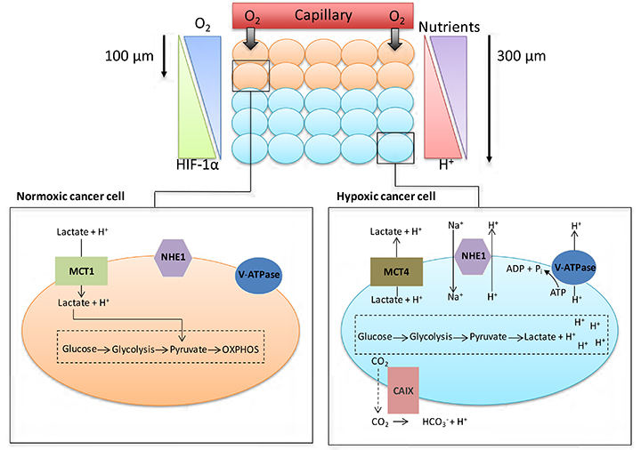
    Cancer cell metabolism, HIF and pH. As cancer cells proliferate, some cells are pushed further away from blood vessels, decreasing the levels of O2 and nutrients available to these cells. Normoxic cells can produce energy through oxidative phosphorylation (OXPHOS); hypoxic cancer cells are unable to acquire energy through OXPHOS due to low O2 levels. Activation of the HIF family of transcription factors is one of the principle oxygen-responsive signalling pathways that allows the adaptation of hypoxic cancer cells to this hostile microenvironment. HIF signalling shifts energy production from OXPHOS in the mitochondria towards glycolysis, allowing hypoxic cancer cells to continue to produce energy despite the low O2 levels. The increased dependency on glycolysis in hypoxic cells leads to the production of increased amounts of H+ ions, which can lead to changes in the pHi of cancer cells if not dealt with. To help cope with the excess H+ ions being produced, cancer cells upregulate/activate a number of pH regulating proteins; these proteins include CAIX, NHE1, MCT4 and V-ATPase

    To maintain intracellular energy levels, cells in hypoxic conditions favour glycolysis, leading to an increased utilisation of glucose and the production of lactate [3]. Many cancer cells use glycolysis for energy production even in O2 concentrations that permit oxidative respiration (aerobic glycolysis or the Warburg effect) [4]. HIF-1 activation in hypoxic tumours enhances the expression of various glycolytic enzymes and glucose transporters such as lactate dehydrogenase-α and Glut1, facilitating increased glycolysis [5, 6]. This generates lactate and hydrogen ions that should decrease intracellular pH (pHi), but because modulation of pHi affects crucial cell processes such as adhesion, proliferation, metabolism and apoptosis, it is finely controlled [7]. Cancer cells maintain pHi partially through increased activation and expression of HIF1-dependent genes [6], such as SLC16A3 [monocarboxylate transporter 4 (MCT4)] which removes lactate along with hydrogen ions from tumour cells, or CA9 [carbonic anhydrase IX (CAIX)] which facilitates the formation of bicarbonate and hydrogen ions from CO2 and H2O [8, 9]. However, HIF-2α also plays a role in adaptation of tumour cells to acidosis [10].

    Low pH and hypoxia can also activate the sodium-hydrogen exchanger 1 (NHE1), which exchanges intracellular H+ ions for extracellular sodium ions [6, 7]. Mechanisms such as these ensure tumour cell pHi remains neutral to slightly alkaline, ranging between 7.1-7.7, as transporters, enzymes and ion pumps maintain the internal cellular environment, while concurrently inducing acidification (acidosis) of the tumour microenvironment (TME) [11, 12]. Normal cellular pHi is generally about 7.2 and frequently lower than cancer cell pHi [13]. pHi has been measured by techniques that include 31P-magnetic resonance spectroscopy [14], 14C-benzoic acid distribution [15] and use of pH-sensitive fluorescent probes, e.g., BCECFAM probe [14, 15]. Normal cells have an extracellular pH (pHe) value around 7.4 which contrasts with the pHe of cancer cells which can typically vary between 6.7-7.1 [13]. However, this can decrease to as low as 6.0 for cancer cells 300 mm from tumour blood vessels, as lactic acid and protons accumulate extracellularly because the malformed tumour vasculature inhibits their removal, thus reducing the buffering capacity of the extracellular environment [11, 13]. This acidic microenvironment can be toxic to normal cells, but cancer cells adapt and survive [11, 13, 14]. Indeed, studies indicate that while the proliferation rate of normal cells is maximal at pHe 7.3, tumour cell proliferation is highest at pHe 6.8 [14].

    Consequences of acidosis for cancer progression and treatment

    Effect on radiosensitivity and chemosensitivity

    The survival strategies adopted by cancer cells in the hypoxic and acidic TME promote radiotherapy and chemotherapy resistance and are clearly connected to the evolution of in situ to invasive cancer [11]. For radiotherapy to work effectively, O2 must be present at the time of radiation to generate free radicals producing DNA breaks leading to cell death. Under hypoxic conditions, there may be insufficient O2 for this radiosensitisation. Hypoxia hinders the homologous recombination (HR), non-homologous end-joining and mismatch repair DNA pathways, as well as inhibiting the G1/S cell cycle checkpoint, allowing DNA errors to accrue and chromosomal instability to increase, while the alkaline pHi of tumour cells hinders mitotic arrest initiated by activated DNA damage checkpoints [13, 16].

    Treatment resistance is also linked to acidic pHe, through changes in drug structure and charge (for example, doxorubicin becomes highly charged, inhibiting uptake across cell membranes in low pH) [13, 17], with acidosis also leading to resistance to apoptosis in irradiated cancer cells [18].

    Effect on cancer cell function

    Alkaline pHi inhibits apoptosis since activation of caspases requires acidic pHi [19], with the basic pHi also promoting DNA synthesis and cell proliferation [20], thus facilitating increases in cell number and tumour volume. These processes lead to higher rates of mutation in cancer cells [5, 13] and augment cellular survival, causing progression of the disease. A pHi threshold around 7.1–7.2 exists below which growth factors fail to stimulate G1 progression and cell cycle entry [21].

    Acidic pHe promotes migration, invasion and metastasis of cancer cells through varied mechanisms; in tumours, the zones exhibiting the most extensive invasion correlate with those displaying the lowest pH, while increased acidity in tumours correlates with poorer prognosis [11, 2124]. Xenograft studies illustrate a two-fold increase in lung metastases when melanoma cells are exposed to pH 6.8 for 48 h before implantation [22]. Acidic pHe increases the expression of proteinases and the activity of metalloproteases (MMPs) such as MMP-1, 2 and 9, which degrade extracelular matrix (ECM) components, thus aiding the invasion and migration of cancer cells [2426].

    Effect on the tumor microenvironment

    Tumours contain stromal elements including cells of the immune system that should recognise and remove tumour cells and maintain tumour latency [27]. Tumour acidity has been described as a “global protection shield” by which cancer cells neutralise the activity of antitumor immune cells and convert regulatory immune cells to become allies [27].

    Cytotoxic T (CytT) cells and natural killer (NK) cells are involved in anti-tumour defences, and large numbers of these cells in a tumour correlate with positive prognosis [28]. Hypoxia and acidosis interfere with immune cell function, hampering this natural defence; acidosis in particular obstructs the activation of both CytT and NK cells, facilitating tumour progression [27, 2932]. These cells lose their functionality undergoing anergy followed by apoptosis at low pHe [27].

    Lactate or acidic pHe inhibits maturation of dendritic cells (DCs) from myeloid precursors, thus hindering antigen presentation [29]. Additionally, acidic pHe can increase expression of cytokines and growth factors [such as IL-8, IL-6 and vascular endothelial growth factor (VEGF)] that support the growth and progression of tumours and angiogenesis, while lactate can activate TGF-β to suppress immune cell function [3234]. Lactate may also inhibit cytokine release from DCs and CytT cells and interfere with signal transduction [35, 36]. A recent study illustrates that the counteraction of acidic pHe opposes anergic unresponsivenesss in human and murine tumour-infiltrating T lymphocytes [37]. Myeloid-derived suppressor cells (MDSCs) strongly suppress innate and adaptive immunity by inhibiting the anti-tumour functions of T and NK cells, facilitating the maturation of regulatory T cells and hindering the development of DCs. Increased production of lactate by tumour cells promotes MDSC development, suggesting an important role for lactate in the evolution of an immunosuppressive TME [38].

    Increased lactate concentrations also stimulate the development of a cancer stem cell phenotype [39]. In glioma, acidic pHe enhanced cell malignancy, promoting expression of stem cell markers such as Oct4 and Nanog, and increasing VEGF levels, via an acidic pH-driven increase in HIF-2α mRNA and protein [40]. Additionally, lactate has been shown to attract human mesenchymal stem cells to tumour cells and heighten stem cell migration [41].

    Activated T cells rely on glycolysis for energy production, but high concentrations of extracellular lactate in the microenvironment prevent lactate export from these cells, since movement is dependent on a lactate gradient between the cytosol and the extracellular space [36]. High extracellular lactate concentrations therefore affect activated T cell function. Consequently, acidic pHe, in concert with increased lactate, may have synergistic effects on the inhibition of immune function in cancer [38]. Lactate also increases cancer cell migration [42], and causes the release of VEGF from endothelial cells, linking lactate with angiogenesis [43]. This is supported by in vivo work demonstrating that lactate can cause IL-8-dependent angiogenesis and tumour growth in xenograft models [44]. Lactate accumulation in several tumour types correlates with patient survival, metastatic potential and radioresistance [19, 20, 45]; radioresistance may be caused by the antioxidant effects of lactate [46]. The effects of acidic pHe on tumour development and treatment are summarised in Figure 2. When considered in combination, the consequences of acidosis on tumour growth and progression are substantial.

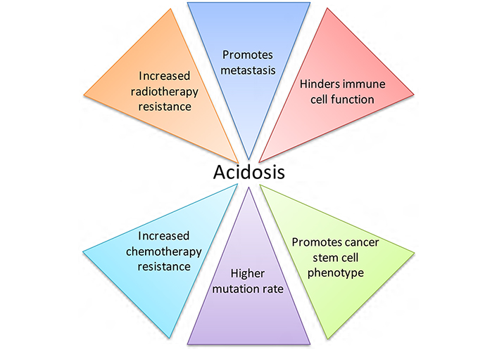
    The effects of acidic pHe on tumour development and treatment. Acidosis is a microenvironmental factor that impacts tumour progression greatly, contributing to tumour growth and negatively affecting both radiotherapy and chemotherapy treatment

    Potential therapeutic strategies

    Given the significant roles that acidosis and pH regulation play in the proliferation, survival, invasion and metastasis of cancer cells, this is an area ripe for therapeutic exploitation in the treatment of solid tumours. Here we expand on several possible targets for intervention, in particular CAIX, monocarboxylate transporter 1 (MCT1) and 4, the vacuolar-type H+-ATPase proton pump (V-ATPase), and NHE1, all of which have significant functions in the control of cellular pHi [9, 13, 47], as illustrated in Figure 3. Other ion exchangers and transporters are involved in pHi regulation, but their roles in cancer progression are still unclear. A family of proton sensing G protein-coupled receptors [48] that can be activated by acidic extracellular pH may be further targets for tumour therapy.

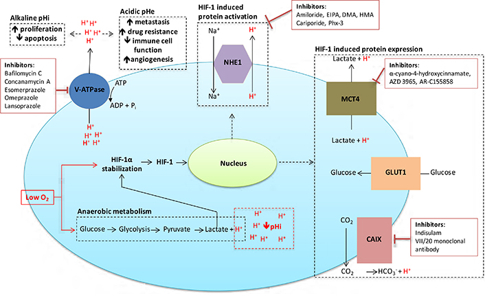
    Inhibitors targeted against pH regulatory molecules with the cell. Various drugs targeting the main pH regulating proteins of cancer cells are outlined, along with the effects that alkaline pHi/acidic pHe have on tumour development and treatment

    NHE1

    While there are nine NHE isoforms, this review concentrates on NHE1. Even though this protein is ubiquitously expressed, it has been firmly associated with cell transformation and malignant progression [49], suggesting it has strong potential as a therapeutic target for cancer [50]. This isoform localises to the plasma membrane and is involved in the homeostasis of pHi and cell volume, as well as migration, cell cycle progression, proliferation and cell death [7, 47, 51, 52]. It operates in concert with many signalling pathways and may function as a framework for the interaction of signalling complexes [53]. NHE1 activity is stimulated by hypoxia, decreased pHi, cell transformation, and downstream effectors of epidermal growth factor receptors (EGFR) such as ERK and p90Rsk, B-Raf, G protein-coupled receptors, PKC, RhoA, and integrin receptors [6, 5459]. The NHE1 protein plays a significant role in maintaining pHi by importing extracellular Na+ for intracellular H+ in a 1:1 ratio [7]. In solid cancers, this leads to acidification of the TME [60], increasing the metastatic potential of cancer cells [61]. For example, higher activity and expression of NHE1 is proposed to increase the potential for invasion and survival of breast and melanoma cancer cells [58, 6062].

    The leading cause of treatment failure in cancer is from metastatic spread of the primary tumour, in which NHE1 is strongly implicated through increased metalloproteinase activity leading to digestion of extracellular matrix components [63]. NHE1 interacts with CD44, a transmembrane glycoprotein that functions in cell adhesion, migration, invasion and survival [64]. This interaction activates hyaluronidase-2 and cathepsin B allowing invasion of breast tumour cells [24]. A positive correlation between CD44 and NHE1 has been described, where knockdown of CD44 caused a decrease in NHE1 mRNA and protein expression which was associated with the suppression of migration and invasion of breast cancer cells; this was reversed by increasing NHE1 expression in cells where CD44 expression was impaired [65]. NHE-1 is expressed in lamellipodia and invadopodia, where it functions in cell motility and directional migration [66, 67]. CD44 is also located in invadopodia, where it stimulates NHE1 activity and invasion via the RhoA effector ROCK1 [23, 58].

    NHE1 secures the cytoskeleton to the cell membrane by attaching to ezrin/radixin/moesin proteins and talin, which connect to actin, thus in part, explaining the involvement of NHE1 in cell motility [62, 68]. It partially controls the pH-dependent activation/integration of cytoskeletal components involved in the regulation of actin polymerisation, such as ADF/cofilin, talin and gelsolin, allowing refashioning of cell-substrate adhesion complexes required for migration [62, 69, 70]. As part of these focal contacts, NHE1 may co-operate with integrins and other cell signalling complexes [7]. Increased expression of the ErbB2 tyrosine kinase is associated with many types of cancer. In its N-terminally truncated isoform (ErbB2t), it becomes constitutively active leading to poorer patient prognosis [71]. This is associated with an NHE1-dependent mechanism, since ErbB2t expression is related to acidification of the pericellular space which is sensitive to NHE1 inhibition [72]. NHE1 regulates motility of breast cancer cells expressing ErbB2t and stimulates EMT development [73].

    Because acidic pHi is optimal for activation of apoptotic proteins such as caspases and endonucleases, proteins that maintain alkaline pHi can reduce activation of apoptotic pathways and protect against programmed cell death [17]. Increased expression of NHE1 leads to decreased responsiveness of several known inducers of apoptosis in tumour cells, while reduced expression sensitises cells to apoptosis [74, 75]. NHE1 is a caspase-3 substrate, therefore cleavage of NHE1 may be part of the apoptotic process [76]. The PKB/Akt survival pathway is stimulated by association of NHE1 and ezrin, and in turn, PKB/Akt can stimulate NHE1 activity [77, 78], suggesting that NHE1 may increase cancer cell survival per se, regardless of its role in pHi maintenance. However, NHE1 inhibition can block Akt phosphorylation, suggesting that Akt activity may be dependent on alkaline pHi [79]. Inhibition of NHE1 sensitises cancer cells to etoposide, paclitaxel and daunorubicin [74, 80, 81]. However, paclitaxel itself can inhibit NHE1, and this may be causal in the induction of apoptosis induced by this and other cancer therapeutics, or the inhibition may be due to apoptotic cleavage of NHE1 as previously mentioned [75, 7981]. Additionally, NHE1 inhibition or depletion reversed cisplatin resistance in breast cancer cells, doxorubicin resistance in human colon cancer cells [69, 80, 82], and caused growth arrest, lowering of pHi and increased responsiveness to cell death inducers in cholangiocarcinoma cells [74, 83]. In leukaemia cells, NHE1 inhibition reduced proliferation and VEGF synthesis [84], suggesting that NHE1 may play a role in angiogenesis. In support of this, a recent study demonstrated that NHE1 suppression using siRNA could inhibit HIF-1α-induced angiogenesis in a human umbilical vein endothelial cells (HUVEC) model [85]. Peroxisome proliferator-activator receptor gamma ligands reduce cancer cell division by repressing NHE1 expression [80], while xenograft models demonstrate that deficient NHE1 activity or NHE1 deletion inhibits tumour growth [86, 87]. However, earlier work using the NHE1 inhibitor amiloride demonstrated a delay or inhibition of apoptosis induced by radiation in HL60 cells [88], although this is not a solid tumour model. Taken together, these studies suggest targeting NHE1 activity may induce growth arrest in cancer cells and increase sensitivity to anticancer treatment.

    Some specific inhibitors of NHE1 (see Table 1), such as cariporide (HOE642) and eniporide have concluded phase II/phase III clinical trials [89, 90] in a cardiological or ischaemic-reperfusion injury setting. An early study using cariporide suggested that NHE1 inhibitors could have therapeutic benefit in reperfusion injury, leading to further clinical trials [9193]. Although cariporide reduced myocardial infarcts, it was linked to increased cerebrovascular events, causing the trials to be stopped [90, 92]. This may have been due to the use of higher cumulating doses of the drug [94], but these trials allowed a tolerable dosing schedule to be developed. Cariporide has never been used clinically in cancer patients, but if the undesirable side effects could be dissassociated from therapeutic gain by minimising the dose, it could form part of a treatment schedule. Its use in cancer patients is supported by early preclinical studies which demonstrated activity in leukemia and cholangiocarcinoma cells [12, 9597]. Recently, preclinical activity of cariporide has been demonstrated across a broad range of cancer types [98103]. The drug has been shown to inhibit invasion of head and neck squamous cancer [98] and EGFR driven invasion of pancreatric cancer [99]. Cariporide can sensitise breast cancer cells to doxorubicin both in vitro and in vivo [100] and can inhibit accelerated net acid extrusion in human colon cancer biopsies [101]. The agent has also been shown to modulate pHi of human glioblastoma xenografts implanted into mice brains [102]. Combined with the MCT1 inhibitor, AR-C155858, it has an additive inhibitory effect on leukemia cells [103].

    Therapeutic targets and inhibitors

    Target/InhibitorExamples of preclinical sensitivityReferences
    NHE1
      AmilorideMammary, prostate, gastric, hepatoma[104]
      Ethylisopropylamiloride (EIPA)Bladder, breast cancer[109]
      Dimethylamiloride (DMA)Bladder, breast cancer[109]
      Hexamethyleneamiloride (HMA)Leukemia[110]
      CariporideLeukemia, cholangiocarcinoma[12, 96103]
      Phx-3Gastric cancer[111, 12]
    MCT1/MCT4
      α-cyano-4-hydroxycinnamateLung cancer[116]
      AZD 3965Small cell lung cancer[131]
      AR-C155858Rat transformed fibroblasts[121]
    V-ATPases
      Balifomycin CPancreatic cancer, colon cancer[157, 159]
      Concanamycin ASubmandibular cancer[162]
      EsomerprazoleBreast cancer, esophageal cancer[171, 172]
      OmeprazoleMelanoma[168, 169]
      LansoprazolePancreatic cancer, breast cancer[166, 170]
    CAIX
      IndisulamLung cancer[208]
      VII/20 Monoclonal antibodyColon cancer[198]
    Display full size

    Amiloride, another NHE1 inhibitor, is well tolerated as a diuretic [104] and direct antitumoural, antimetastatic and antiangiogenic effects have been reported [105107]. Analogues of amiloride that are more specific NHE1 inhibitors such as ethylisopropylamiloride (EIPA), dimethylamiloride (DMA), and hexamethyleneamiloride (HMA) also have anticancer potential [108110]. A phenoxazine structure, Phx-3, has also shown interesting preclinical activity [111]. Improved specificity and/or delivery of NHE1 inhibitors may allow further exploitation of this anti-cancer target.

    Monocarboxylate transporters (MCTs) 1, 4 and associated molecules

    MCTs 1 and 4 enable movement of lactate and protons across cell membranes and therefore also function in pHi regulation [112]. These isoforms are ubiquitously expressed, with MCT4 expression strongly enhanced in glycolytic tissues [112]. MCTs levels are increased in many cancers such as breast, colorectal, ovarian, and prostate, and are associated with disease progression and poor prognosis [113115].

    The activation of HIF-1 suppresses the expression of MCT1 while increasing that of MCT4; this maintains an economical use of glucose in the tumour [8] since the lactate produced by hypoxic cells is exported along with hydrogen ions from the cell by MCT4, and can be transported into cells in more oxygenated areas by MCT1 [8, 116]. Where O2 concentrations are sufficient for oxidative phosphorylation, cells can use lactate in the tricarboxylic acid cycle. This allows for the development of a symbiotic association between cells in solid tumours [116]. Stromal cells, such as tumour-associated fibroblasts and endothelial cells, demonstrate increased MCT1 expression and can therefore ulitise lactate [44, 117]. Blocking this symbiotic mechanism should prevent lactate use by normoxic tumour cells, increasing dependency on glucose, and decreasing glucose availability for hypoxic tumour cells. Although inhibition of MCT4 alone could be sufficient to block this symbiotic relationship one study using a mouse model has shown that MCT1 inhibition can also effectively target cells in hypoxic areas of lung and colorectal tumours and increased the efficacy of radiation treatment [116]. Therefore, obstructing the activity of MCT1 or MCT4 may be a useful therapeutic target, both in terms of disrupting the efficient usage of glucose and lactate, and also for intervention in pH regulation.

    Both MCT1 and 4 are associated with cancer cell invasion and drug resistance [118, 119]. By obstructing intracellular acidification, MCTs also inhibit the induction of apoptosis [13], while lactate import into vascular endothelial cells via MCT1 stimulates angiogenesis [120]. Therefore, MCT inhibitors could decrease tumour growth by reducing neovascularisation and cell survival, while limiting invasion and counteracting drug resistance. Preclinical studies indicate that reducing MCT1 activity diminishes the growth of a diverse range of tumours such as lymphoma, glioma, and colon cancer [116, 121123]. The MCT1 inhibitor α-cyano-4-hydroxycinnamate sensitised colon cancer cells to cisplatin by a mechanism involving the down regulation of MDRI and MRP1 genes, which are involved in multidrug resistance (MDR) [122]. This may be via an effect on pHi since prior reports have illustrated that pHi has a pivotal involvement in MDR development [124] through regulating MDR gene expression [125]. Disturbing the function of MCTs causes intracellular lactic acid to accrue and this in turn reduces glucose transport, ATP production and pHi, hindering growth of human cancer cells [126]. In vivo preclinical studies established that MCT1 inhibitors delayed the onset of tumour growth; growth was inhibited in mice co-treated with both MCT1 inhibitors and metformin, with many mice not developing tumours at all [126]. In another report, inhibition of MCTs increased cell death in human colon cancer cells; this was associated with decreased lactate secretion and a fall in pHi [127]. Reduction in lactate secretion has been linked to increased radiosensitivity in small cell lung cancer xenografts treated with an MCT1 inhibitor [128]. Therefore, MCT inhibitors are appealing therapeutic agents (see Table 1). Preclinical studies have shown that MCT inhibitors exhibit efficacy with some of these drugs now in clinical trials [129, 130]. For example, AZD3965 is undergoing phase I clinical trials in prostate and gastric cancer and diffuse large B cell lymphoma (http://www.clinicaltrials.gov/show/NCT01791595) [131].

    Plasma membrane expression of MCT1 and MCT4 requires co-expression of the ancillary molecule CD147, which appears to be HIF-1α-dependent in lung, breast and liver cancer [132, 133]. CD147 is an obligatory assembly factor for MCTs [133]. In breast and pancreatic cancer cells, CD147 silencing strongly reduces glycolysis and lactate secretion [134, 135], and may prevent transformation and tumour development via MCT loss of function [121]. Glucose deficiency can induce ROS-dependent stabilisation of both MCT1 and CD147 [135]. These MCT1-CD147 complexes facilitate tumour cell migration towards glucose, and thus migration away from the low glucose TME [135]. Such complexes are often found colocalised with β1 integrin in the lamellipodia of migrating cells [8] and can act as a framework for other proteins functioning in migration [136]. Consequently, expression of CD147 is increased in metastatic breast cancer cells and other aggressive tumours [112115, 137], while studies show that decreasing CD147 expression inhibits migration of invasive cancer cells [138]. CD147 also stimulates production of MMPs, which facilitate migration [139]. In A549 cells, increased CD147 expression was found to reduce apoptosis, while also increasing the invasive potential of these cells in hypoxic conditions [130]. Several reports further suggest a role for CD147 in radioresistance. For example, high expression levels of CD147 in cervical squamous cell carcinoma patients were found to be indicative of radiation resistance and poor prognosis [140]. In vitro and in vivo studies showed that CD147 knockdown significantly enhanced the radiosensitivity of hepatocellular carcinoma cells and decreased radiation induced migration and invasion [141] through a mechanism that inhibited β1 integrin signalling. Therefore, CD147 is another focus for cancer treatment that could be used to disrupt tumour pH homeostasis and glucose/lactate symbiosis.

    H+-associated ATPases

    V-ATPases are ATP-dependent multi-subunit enzymes that translocate protons across membranes, playing a significant role in the maintenance of pHi homeostasis, and are involved in cell survival [142]. They comprise of a V0 region, composed of five different subunits, that is involved in proton transport and a V1 sector, involving eight different subunits, which is concerned with ATP hydrolysis [142]. They are primarily situated in the membranes of endosomes and lysosomes and the plasma membrane of some tumour cells [142144]. The role of V-ATPases in the control of vacuolar pH influences the pharmacokinetics of chemotherapeutic drugs and may be involved in multidrug resistance mechanisms and the metastatic potential of tumour cells [143146]. Irradiated cancer cells can develop autophagic vesicles which are acidified by V-ATPase; this acidification is restricted by V-ATPase inhibitors and can sensitise these cells to radiation [147]. Therefore, the inhibition of this enzyme is a plausible intervention strategy in cancer treatment (see Table 1).

    Increased expression of V-ATPases occurs in various tumours such as hepatocellular carcinoma, oral squamous cell cancer, pancreatic cancer and melanoma [148, 149]. Studies illustrate that V-ATPase inhibitors or siRNA knockdown of V-ATPases can restrain tumour growth and metastatic potential, increase cell death and resensitise tumour cells to chemotherapeutics [146, 150153]. Inhibition of V-ATPase induces loss of cell adhesion and cell death in colon carcinoma cells and antiproliferative activity in other human carcinoma cell lines (such as ovarian, breast, lung, prostate, hepatocellular, melanoma and renal), and has been shown to reverse trastuzumab resistance in breast cancer cells by interference with HER2 signalling pathway and cellular location [150, 153155]. In vitro models demonstrate that V-ATPase inhibitors or siRNA knockdown hinder the invasion of breast cancer cells through matrigel [143, 144]. Rab27B-dependent invasive growth and metastasis of breast cancer is reliant on V-ATPase activity [156], with the V-ATPase V1E protein expressed in the majority of breast cancer patients with poor prognosis [156]. Classic V-ATPase inhibitors such as bafilomycin A1 and concanamycin A restrain tumour growth in vitro and in vivo through decreasing the pHi of cancer cells, and have also been shown to induce apoptosis [157159]. They inhibit the interaction of HIF-1α with VHL, increasing HIF-1α expression in cancer cells, but without enhancing transcription of HIF-1 target genes. Rather, this HIF-1α interferes with binding of C-MYC to the CIP1 promoter, causing increased expression of p21CIP1 and inhibiting cell cycle progression [160].

    Bafilomycin A1 and concanamycin A are unsuitable for clinical use because of toxicity. However, recent studies have determined thata subunit of the V0 domain determines the intracellular location of V-ATPase. There are 4 different isoforms of this subunit, and of these the a3 and a4 isoforms are involved in targetting V-ATPase to the plasma membrane [143, 161]. Higher plasma membrane expression of V-ATPase is associated with metastatic cancer cells and knockdown of these subunits inhibits the invasion of breast cancer cells and bone metastasis of mouse melanoma cells [143, 148, 161]. Concanamycin A has been shown to induce apoptosis in human submandibular gland ductal cancer cells [162]. Therefore, future strategies may involve specific targeting of the a3 or a4 subunits, thus reducing overall toxicity by limiting the inhibition of V-ATPase expressed intracellularly.

    Another approach involves the use of proton pump inhibitors (PPIs), such as esomerprozole and omeprazole that target H+/K+ ATPases, since they also inhibit V-ATPase activity; these drugs have long been used efficaciously as therapy for acid-related diseases such as peptic ulcers with minimal side effects [163, 164]. These compounds have anti-inflammatory, immunomodulatory and anti-metastatic properties; they can act as anti-oxidants, interact with neutrophils, monocytes, epithelial and endothelial cells, and inhibit the binding of cancer cell integrins [165]. For example, the PPI lansoprazole prevented cancer cell binding to ECM components [166] and the formation of lung metastases by murine colon cancer cells [165].

    PPIs sensitise cancer cells to cytotoxics such as cisplatin, 5-fluorouracil and vinblastine [145], and because PPIs are weak bases, the active protonated form of the drug accrues in the acidic TME [167]. These compounds induced cell death in human B-cell xenografts via a ROS-dependent, caspase-independent mechanism [152], and have shown antitumour activity against melanoma in vitro and in vivo via a caspase and pH-dependent mechanism [168]. Pretreatment with omeprazole increased cisplatin effectiveness in a murine melanoma model [145] and inhibited invasion and metastasis of breast cancer cells by downregulating the expression of MMP-9 and the chemokine receptor 4 (CXCR4) [169]. Another compound, lansoprazole, induced apoptosis in breast cancer cells and xenografts, inhibiting tumour growth in vivo, with no evidence of systemic toxicity [170], while an additional pre-clinical study has shown that the PPI esomeprazole can increase the effectivess of doxorubicin in triple negative breast cancer cells [171]. The compound has also demonstrated acivity against esophageal cancer cell lines [172]. Furthermore, esomeprazole enhanced the activity of tumour-infiltrating T lymphocytes in a murine melanoma model, delaying cancer progression and increasing the efficacy of anticancer vaccines [37], suggesting that PPIs may be useful in reversing the immune evasion mechanisms dependent on acidic pHe and also increasing the effectiveness of anti-cancer immunotherapy treatments.

    PPI use may decrease the development of oesophageal adenocarcinoma and hepatoblastoma [173, 174]. Over a 5 year period, one report confirmed that PPIs such as omeprazole, esomeprozole, rabeprazole, pantoprazole or lansoprazole significantly decreased both the growth and progression of Barrett’s oesophagus [174]. A multi-centre phase II clinical trial examining the ability of esomeprazole to sensitise osteosarcoma to neo-adjuvant chemotherapy found an increased response, particularly in chondroblastic osteosarcoma, with no serious side effects reported by patients [175]. In a phase I/II clinical study in veterinary canine and feline patients with spontaneously occurring tumours showing chemotherapy resistance, 67.6% of treated animals demonstrated partial or complete clinical response with a high dosage PPI/chemotherapy treatment [176]. Combination treatments of high dose esomeprazole with cisplatin or docetaxel in metastatic breast cancer in a phase II clinical study (ClinicalTrials.gov Identifier: NCT01069081) reported enhanced responses to chemotherapy with no increase in toxicity [177]. This study demonstrated an increased objective response rate and time to progression with combination treatment and was particularly effective in triple negative breast cancer patients.

    Carbonic anhydrase IX

    Carbonic anhydrases (CAs) catalyse the reversible conversion of CO2 and H2O to HCO3 and H+. These proteins are ubiquitously expressed and are mainly involved in pHi regulation; this review considers the role of CAIX, since increased expression of this protein is associated with poor prognosis in several cancer types [9]. The extracellular HCO3 generated by CAIX is transferred into the cytosol where it limits acidification, while protons remain at the cell surface lowering the pHe [178]. CAIX is a dimeric protein with an N-terminal proteoglycan domain, a CA catalytic domain, and a transmembrane region with a small cytoplasmic tail at the C-terminus [179].

    Activation of HIF-1 increases the expression of CAIX, particularly in solid, hypoxic tumours [47]. Expression is limited in normal tissues [9], and knockout of the mouse CAIX homolog causes little abnormality other than gastric hyperplasia [180], suggesting that toxicity in normal tissue by strategies that target this molecule should be limited. CAIX enhances tumour cell survival and growth by regulating pHi homeostasis [14, 47], but it may also influence survival signalling pathways, since limiting CAIX expression significantly affected cell growth and survival independently of O2 concentration in breast tumour models [181]. Further, EGF can induce phosphorylation of the intracellular domain of CAIX, causing an interaction with PI3K and Akt activation in some cancer cells such as clear cell renal cancer [182]. Disregulated PI3K activation in tumours can enhance CAIX expression via a mechanism dependent on HIF-1α translation by mTOR [183]. Recent in vivo research demonstrated that CAIX knockdown reduced xenograft tumour volume in breast and colon cancer models, and when used in concert with antiangiogenic therapy, reduced growth additively [14, 184, 185]. Conversely, CAIX overexpression in a colon cancer spheroid and xenograft model showed enhanced rates of growth and increased expression of the proliferation marker Ki-67 [185].

    CAIX is strongly associated with the cell membrane and cell adhesion, via an interaction with β-catenin that may inhibit E-cadherin-dependent cell-cell adhesion [179, 186]; it functions in both cell migration and invasion. At the C-terminal intracellular tail, Thr-443 can be phosphorylated by PKA in hypoxic conditions, allowing migration to occur [187] via modification of transcriptional activity and proteins involved with EMT and cytoskeletal composition [188], and also in alterations in Rho/ROCK signalling that can stimulate paxillin, and focal adhesion turnover [188, 189]. Recently, CAIX was found to be involved in cancer cell migration and MMP14-mediated invasion by supplying the protons needed for the catalytic activity of MMP14 [190].

    The invasive potential of human ductal breast cancer spheroids, along with renal and ovarian cancer cells, was decreased when CAIX expression was reduced or activity was inhibited using novel sulfamate inhibitors of CAIX [191193]. These inhibitors reduced proliferation, enhanced the rates of apoptosis and inhibited metastasis in breast cancer xenografts models, while also exhibiting the potential to reverse the invasion of human breast tumour biopsy tissue [185, 192, 194, 195]. Basal like breast cancers frequently express CAIX and have been associated with high levels of brain, lung and bone metastases [194], leading to suggestions that targeting of CAIX may be a possible approach to combat metastatic disease [194, 195]. Of note, in vivo studies conducted with these inhibitors did not appear to induce toxic responses.

    Several novel inhibitors and antibodies targeting CAIX have been assessed as potential anti-cancer treatments [195198] (see Table 1). SLC-0111/WBI5111 inhibits both tumour growth, metastasis and stem cell numbers either alone or in combination with other anti-cancer therapeutics and is undergoing clinical trials (NTC02215850) [199, 200]. One monoclonal antibody has proved efficacious in colorectal cancer xenografts [198]. Several clinical trials have been undertaken to assess the effectiveness of the antibody girentuximab (cG250), the most clinically advanced monoclonal, in renal cancer (NCT00087022, NCT02002312). Phase I and II trials established safety, tolerability and a positive effect on the disease either alone or in combination with IL-2 treatment [201203]. In a phase III trial NCT00087022, cG250 showed no effects on disease free survival on non-metastatic renal cell carcinoma patients; however, this study did not stratify patients by CAIX expression. Subsequent analysis demonstrated significant improvement in patients with high CAIX expression, suggesting patient selection is necessary for CAIX-based therapies [204]. CAIX mAbs have been developed that allow delivery of radioisotopes and cytotoxic drugs to cancer cells [205, 206]. A phase II trial of 177Lu-girentuximab in metastatic clear cell renal carcinoma attained stable disease in 9 of 14 patients [207]. Another inhibitor indisulam, which demonstrated promising preclinical activity [208], has undergone phase II clinical trials for stage IV melanoma, renal clear cell carcinoma and metastatic breast cancer [209], although in advanced NSCLC patients there was little response when it was used as second-line therapy [210].

    Novel CAIX inhibitors have been found to increase sensitivity to radiation [192, 211], suggesting possible combination treatment strategies. Knockdown of CAIX alone or both CAIX and CAXII enhanced the response of tumour cells to radiation, implying that CAIX may protect cells by maintaining alkaline pHi and preventing cell death [212]. Another study demonstrated the sensitisation of renal cell carcinoma to radiation using CAIX inhibitors, where increased levels of apoptosis were observed [213]. Similar results were found in a breast cancer model, where proteomics suggested that CAIX inhibitors in concert with radiation reduced the expression of anti-apoptotic proteins, while increasing that of pro-apoptotic proteins [192]. These results are supported by other studies in xenograft models of colon cancer [211, 214]. CAIX may also affect cancer cell responses to radiation via mechanism that are not pH-dependent, such as interactions with EGFR, PI3K/AKT and NF-kB signalling pathways [215]. Further new inhibitors are in pre-clinical assessment [192, 195, 196], and a phase I clinical trial using a novel dual CAIX inhibitor/radiosensitiser DTP348 has been recently announced (NCT02216669).

    Novel pH targets

    Proton sensing G protein-coupled receptors

    A family of five G-protein coupled receptors (GPCRs) involved in proton sensing influence many tumour cell activities such as proliferation, apoptosis, metastasis and angiogenesis; these proteins may therefore offer further targets for anticancer treatments [216226]. These GPCRs [GPR4, GPR65 (TDAG8), GPR68 (OGR1), GPR81 (HCAR1) and GPR132 (G2A)] are activated by acidic pHe via protonation of histidine residues [216, 217, 225230]. They are not directly involved in proton translocation, but link to various signalling pathways, such as cAMP, phospholipase C/Ca2+, and Rho [216, 217, 225, 226, 231].

    Enhanced activation or expression of proton transporters is linked to increased tumour progression, but the stimulation or overexpression of some GPCRs is associated with anti-tumour effects. For example, activation of GPR4 in acidic conditions reduces migration, invasion and the metastatic potential of tumours, while repressing vascular growth [217, 219]; this GPCR activates the Gs, G12/13 and Gq G-protein pathways [216, 220, 221, 232]. In melanoma and prostate cancer cells, ectopic expression of GPR4 activated by acidic pH decreases migration and melanoma lung metastases, but it does not significantly decrease growth of the primary tumour [220, 232]. However, GPR4 deficiency in murine models reduces tumour allograft growth by interfering with angiogenesis since GPR4 is expressed on endothelial cells [217, 223]; however, overexpression of GPR4 can transform fibroblasts [233]. This suggests that GPR4 may have both anti and pro-tumour effects dependant on cell type, which may limit the clinical application of compounds that increase or limit GPR4 activation, until further research clarifies the possible mechanisms involved.

    GPR68 overexpression impeded migration of both breast and prostate tumour cells and metastasis of prostate cancer cells; although this effect may be independent of pH [219, 234]. It also inhibited ovarian cancer cell migration and augmented adhesion to several ECM proteins [222]. However, GPR68 expression appears to be lower in metastic tumours when compared with the primary growth [218]. GPR68 deficiency significantly reduced tumourigenesis of melanoma cells in a mouse model [235]. Recent research suggests that this GPCR can partially control the interactions between cancer-associated fibroblasts (CAFs) and pancreatic ductal adenocarcinoma (PDAC) cells. TNF-α, secreted by PDAC cells, enhances expression of GPR68 on CAFs via a mechanism involving cAMP/PKA/CREB signaling; activated GPR68 causes secretion of IL-6 from CAFs and this in turn increases the proliferation of PDAC cells [236]. Another study has shown that the transformation of mesenchymal stem cells to CAFs in acidic pHe is dependent on GPR68 [237]. Hypoxia may also control the expression of GPR68 in some cells such as macrophages since HIF-1α binds to the promoter of this gene and its expression is further enhanced by acidic pHe [238].

    GPR65 is usually confined to lymphoid cells, but it behaves as an oncogene in lymphoid and other cancers [239]. GPR65 is activated at pH levels lower than 7.4, but activation is maximal between pH 6.2 to 6.8, which is within levels measured in the TME [226, 228]; the ability of this molecule to act in a tumourigenic manner is dependent on its pH sensor activities [240]. High expression of GPR65 mRNA has been detected in kidney, ovary, colon and breast tumours amongst others [233]. Increased levels of GPR65 were associated with the growth of lung cancer cells and the transformation of mammary epithelial cells [233, 234]. In a murine model, overexpression of GPR65 increased tumour growth via a mechanism that may involve adaptation to acidic conditions through activation of PKA and ERK; knockdown of this receptor inhibited survival of lung cancer cells in an acidic environment [240]. Enhanced cAMP (which activates PKA) production in response to acidic pHe by this sensor has also been reported [228, 229]. Acidic activation of GPR65 upregulates expression of Bcl-2 via MEK/ERK signaling and can inhibit apoptosis of glutamine starved cells [241]. Lymphoma cells and lymphocytes express high levels of GPR65 [242]. In a mouse model, GPR65 knock-out does not produce abnormalities, suggesting this receptor might be a potential therapeutic target [243].

    GPR81, a G protein-coupled lactate receptor, is expressed in some tumour cells; stimulation of this receptor helps modulate the effects of lactate by modifying MCT and CD147 expression, aiding tumour growth and metastasis [227]. The GPR81/lactate pathway also suppresses immune surveillance; GPR81 activation causes inhibition of PKA via a decrease in intracellular cAMP levels, aiding activation of the PDL1/PD-1 immune checkpoint and diminished T cell function [244].

    GPR132 was initially discovered to inhibit tumour development by hindering cell cycle progression and mitosis; but high levels of expression in fibroblasts caused transformation in one study [245, 246]. Recently, GPR132 has been shown to be involved as a macrophage lactate sensor in the acidic TME during metastasis of breast cancer. Lactate stimulates GPR132 on tumour macrophages, promoting the development of an M2 phenotype that enables migration and invasion of tumour cells [247]. Clinical data show a positive correlation in breast cancer between GPR132, M2 macrophages, metastasis and poor patient prognosis, while GPR132 deletion in a mouse model of breast cancer inhibits metastasis to the lung [248]. Interestingly, expression of GPR132 is almost non-existent in breast cancer cells, suggesting that anti-metastatic effects occur via stromal elements in the tumour [248]. GPR132 expression appears to be suppressed by PPARg activators such as the thiazolidinediones, which may suggest these as possible anti-metastic treatments in breast cancer [248].

    Further research is required to fully understand the role of pH-sensing GPCRs in particular cancer types, but small molecule modulators are under development and evaluation; this may help to define their functions. GPR4, GPR68 and GPR65 antagonists are now available [249, 250].

    Histone acetylation

    Histone acetylation causes chromatin to adopt an open state, increasing the accessibility of transcription factors. Histone acetylation may also be involved in regulation of pHi [251]. A fall in pHi was associated with lower levels of histone acetylation, which was dependent on the actions of histone deacetylases (HDACs), which are overexpressed in many cancers. Activation of HDACs caused an increase in acetate anions which were transported extracellularly along with protons via MCTs. Increased pHi caused a reversal of transportation. This suggests that histone acetylation can function to regulate pHi and that inhibition of HDACs may form another strategy to compromise tumour cell pH regulation [251]. Interestingly, decreased histone acetylation has been associated with more aggressive cancers and poorer prognosis in breast, prostate and pancreatic cancers [252255], with overexpression of particular HDACs correlating with disease-free survival, overall survival and poor patient prognosis in several solid tumour types [256], while HDAC inhibitors (HDACi) are known to lower pHi in colon carcinoma cells and xenografts [255]. Numerous preclinical studies have shown increased radiation sensitivity in reponse to co-treatment with HDACi. For example, studies in prostate cancer and NSCLC cell lines showed that HDACi could increase radiosensitivity in a time dependent manner in both hypoxic and normoxic conditions, increasing apoptosis and DNA damage [257, 258]. HR-related gene expression can be reduced by these inhibitors, which may partially explain their ability to radiosensitise cells [259].

    There are currently 14 HDACi involved in over 130 clinical trials as monotherapy or in combination with radiation/chemotherapy in a variety of tumours [256]; three are now FDA-approved for use in cancer treatment [260, 261]. Clinical trials in prostate, kidney and bladder cancer were recently reviewed [262]. However, little or no response has been seen in most solid tumours to HDACi alone; most trials now use HDACis in combination with other therapies [261, 263]. HDACi such as vorinostat and valproic acid have been assessed in combination with radiotherapy [259]; a phase I trial (NCT00670553) on prostate cancer has concluded. Vorinostat sensitises glioblastoma cells to radiation, and clinical trials (NCT00731731) of this HDACi in combination with temozolomide and radiotherapy have been undertaken [264]. Phase II studies have also been conducted in combination treatment with paclitaxel (in advanced gastric cancer) [265], and tamoxifen (in hormone therapy resistant breast cancer), where a clinical benefit rate of 40% was achieved [266].

    Innovative technology exploiting the tumour microenvironment

    Although the lowered pH in the TME can be linked to cancer progression and poor prognosis, it can also be exploited for cancer detection and treatment. By designing lytic peptides that are activated in the acidic TME, several groups have reported success in treating xenograft models of human cancer. Makovitzki et al., demonstrated the reduction of prostate tumour volumes and angiogenesis using these peptides [267]. Recent studies showed that a technology using the pH low insertion peptide (pHLIP), a peptide that forms α-helix at acidic pH and inserts across cell membranes, could be applied to target tumour cells and image acidic tissue by positron emission tomography in solid tumours in breast and prostate xenografts and in spontaneous murine breast cancer models [268270]. A recent study suggests that differential uptake of pHLIPs which depend on acidic pHe, may be useful as a predictive biomarker of tumour prognosis [271]. pHLIP-coated liposomes can also be used to insert nanopores caused by the insertion of gramicidin A into the membranes of cancer cells. This hydrophobic peptide forms a β-helix channel that allows protons to enter the cell and decreases intracellular pH, thus inducing apoptosis [272]. Further, because of the lytic action of these peptides, resistance mechanisms are unlikely to develop [267].

    Similarly, conditions in the tumour can be utilised to activate pro-drugs that are triggered or become more potent at acidic pH to differentially kill cancer cells. Low pH can increase resistance to some cancer chemotherapeutics by altering charge and structure [13, 25], but pro-drugs have been designed to preferentially target cancer cells and release drugs in the lysosomal or endosomal compartment (pH 5.0). For example, a pro-drug that targets αvβ3 integrin which is overexpressed in certain cancers, and also contains both a fluorescent reporter group and a detachable form of doxorubicin, was recently reported to induce apoptosis in vitro in αvβ3 positive glioma cells [273]. Similarly, cytotoxic responses were achieved in cancer cells using pH-responsive silica prodrug nanoparticles to enable the controlled release of doxorubicin in endosomes [274]. This strategy can help overcome intrinsic and acquired multidrug resistance, allowing high intracellular drug concentrations to accumulate. Polyethelyne glycol (PEG) can increase the circulation time of nanoparticles and can be combined with pH-sensitive polymers to allow shedding of PEG in the TME, allowing access of nanoparticles to tumour cells [275]. Cell penetrating peptides such as trans-activator of transcription (TAT) can allow drugs direct access into a cell, but they act in a non-specific manner. This can be overcome using TAT in a smart micelle that shields TAT at normal body pH, but exposes it in the TME if pH falls below 7.0 [276]. Other strategies use the activation of MMPs by the acidic TME to cleave linkers that free cell penetrating peptides only in tumour tissue; these can also be utilised to detect and monitor primary and metastatic tumour growth [277, 278]. The use of pH-sensitive nano-systems including polymers, peptides, as well as inorganic materials, for drug delivery in cancer therapy has been recently reviewed [277, 279]. Phase I clinical trials using cell penetrating peptides have been reported in high grade gliomas and advanced carcinomas [267, 268]. The glioma study used an inhibitor of p53 ubiquitination and reported convincing data showing antitumour activity, with few adverse effects [280]; cell penetrating peptides linked with SN38, the active metabolite of irinotecan, was found to stabilise some advanced carcinomas [281].

    The inconsistent nature of the vasculature in tumours means that conditions in the microenvironment can be extremely variable in terms of pH and oxygenation. The ability to measure such changes in real-time would allow treatment to be optimised for maximum efficacy. Radiation treatment is most effective when O2 concentrations are high, whereas pro-drugs could be preferentially used when pH is low. At present, O2 and metabolic markers can be observed in real-time experiments in vitro using microphysiometry methodologies [47]. However, the development of wireless biosensors that can be implanted into solid tumours, to monitor both pH and hypoxia in vivo and in real-time is under way; see http://www.see.ed.ac.uk/drupal/impact. This would allow monitoring of tumour conditions to optimise radation and chemotherapy. Such systems have already been used to deliver drugs in an ocular system, and this technology could be further combined with O2 and pH sensing [282]. Recently an implantable device was developed that can test up to 16 different drugs/concentration/combinations in tumours in vivo [283]. The device was delivered using a biopsy needle, with tumour tissue removed for analysis using a coring needle. A combination of these implantable technologies would greatly improve drug development and would allow the treatment and monitoring of cancer in a truly personalised manner.

    Conclusions

    pH is commonly dysregulated in solid tumours, therefore interference with pH regulation is a promising strategy for anti-cancer drug development [13]. Taken together, the acid-base transporters and proton-sensing receptors described above are important for cancer cells to sense and adapt to the acidic tumour microenvironment. Further research is warranted to validate these pH regulators as potential targets for cancer therapy and chemoprevention. As further research into the exploitation of the TME is undertaken, more targets are likely to emerge in this exciting field. Technological advances in drug design and delivery that exploit the acidic nature of solid tumours, in concert with the ability to monitor environmental conditions and treatment responses in real-time, mean that the adaptation of tumour cells to low pH conditions may be exploited in the clinic in the near future.

    Abbreviations

    CAIX:

    carbonic anhydrase IX/9

    CyT:

    cytotoxic T cells

    DC:

    dendritic cells

    ECM:

    extracelular matrix

    EGFR:

    epidermal growth factor receptor

    EIPA:

    ethylisopropylamiloride

    GPCR:

    G-protein coupled receptor

    HDAC:

    histone deacetylases

    HIF-1:

    hypoxia-inducible factor-1

    HR:

    homologous recombination

    HUVEC:

    human umbilical vein endothelial cells

    MCT1:

    monocarboxylate transporter 1

    MCT4:

    monocarboxylate transporter 4

    MCTs:

    monocarboxylate transporters

    MDSC:

    myeloid-derived suppressor cells

    MMP:

    metalloproteases

    MDR:

    multidrug resistant

    NHE1:

    sodium-hydrogen exchanger 1

    NK cells:

    natural killer cells

    OXPHOS:

    oxidative phosphorylation

    PEG:

    polyethelyne glycol

    pHe:

    extracellular pH

    pHi:

    intracellular pH

    pHlip:

    pH low insertion peptide

    PPI:

    protein pump inhibitor

    TME:

    tumour microenvironment

    V-ATPase:

    vacuolar-type H+-ATPase proton pump

    VEGF:

    vascular endothelial growth factor

    Declarations

    Author contributions

    CW prepared the majority of the first draft while all other authors contributed further sections. All authors contributed to manuscript revision, read and approved the submitted version.

    Conflicts of interest

    The authors declare that they have no conflicts of interest.

    Ethical approval

    Not applicable.

    Consent to participate

    Not applicable.

    Consent to publication

    Not applicable.

    Availability of data and materials

    Not applicable.

    Funding

    We gratefully acknowledges support from the 7th Framework Program of the European Union (METOXIA project; HEALTH-F2-2009-222741), from the UK Engineering and Physical Sciences Research Council [IMPACT programme Grant (EP/K-34510/1)] and from the Edinburgh Cancer Centre research endowment/Edinburgh and Lothians Health Foundation fund.

    Copyright

    © The Author(s) 2020.

    References

    Wang GL, Jiang BH, Rue EA, Semenza GL. Hypoxia-inducible factor 1 is a basic-helix-loop-helix-PAS heterodimer regulated by cellular O2 tension. Proc Natl Acad Sci U S A. 1995;92:55104. [DOI] [PubMed] [PMC]
    Doe MR, Ascano JM, Kaur M, Cole MD. Myc posttranslationally induces HIF1 protein and target gene expression in normal and cancer cells. Cancer Res. 2011;72:94957. [DOI] [PubMed] [PMC]
    Ippolito L, Morandi A, Giannoni E, Chiarugi P. Lactate: a metabolic driver in the tumour landscape. Trends Biochem Sci. 2019;44:15366. [DOI] [PubMed]
    Warburg O. On respiratory impairment in cancer cells. Science. 1956;124:26970. [PubMed]
    Ward C, Langdon SP, Mullen P, Harris AL, Harrison DJ, Supuran CT, et al. New strategies for targeting the hypoxic tumour microenvironment in breast cancer. Cancer Treat Rev. 2013;39:1719. [DOI] [PubMed]
    Semenza GL. Regulation of cancer cell metabolism by hypoxia-inducible factor 1. Semin Cancer Biol. 2009;19:126. [DOI] [PubMed]
    Putney LK, Denker SP, Barber DL. The changing face of the Na+/H+ exchanger, NHE1: structure, regulation, and cellular actions. Ann Rev Pharmacol Toxicol. 2002;42:52752. [DOI] [PubMed]
    Ullah MS, Davies AJ, Halestrap P. The plasma membrane lactate transporter MCT4, but not MCT1, is up-regulated by hypoxia through a HIF-1α-dependent mechanism. J Biol Chem. 2006;281:90307. [DOI] [PubMed]
    Swietach P, Hulikova A, Vaughan-Jones RD, Harris AL. New insights into the physiological role of carbonic anhydrase IX in tumour pH regulation. Oncogene. 2010;29:650921. [DOI] [PubMed]
    Corbet C, Draoui N, Polet F, Pinto A, Drozak X, Riant O, et al. The SIRT1/HIF2α axis drives reductive glutamine metabolism under chronic acidosis and alters tumor response to therapy. Cancer Res. 2014;74:550719. [DOI] [PubMed]
    Gatenby RA, Smallbone K, Maini PK, Rose F, Averill J, Nagle RB, et al. Cellular adaptations to hypoxia and acidosis during somatic evolution of breast cancer. Br J Cancer. 2007;97:64653. [DOI] [PubMed] [PMC]
    Harguindey S, Arranz JL, Orozco JDP, Rauch C, Fais S, Cardone RA, et al. Cariporide and other new and powerful NHE1 inhibitors as potentially selective anticancer drugs--an integral molecular/biochemical/metabolic/clinical approach after one hundred years of cancer research. J Trans Med. 2013;11:282. [DOI]
    Webb BA, Chimenti M, Jacobson MP, Barber DL. Dysregulated pH: a perfect storm for cancer progression. Nat Rev Cancer. 2011;11:6717. [DOI] [PubMed]
    Zhang X, Lin Y, Gillies RJ. Tumor pH and its measurement. J Nucl Med. 2010;51:116770. [DOI] [PubMed] [PMC]
    Chiche J, IIc K, Laferrière J, Trottier E, Dayan F, Mazure NM, et al. Hypoxia-inducible carbonic anhydrase IX and XII promote tumor cell growth by counteracting acidosis through the regulation of the intracellular pH. Cancer Res. 2009;69:35868. [DOI] [PubMed]
    Bristow RG, Hill RP. Hypoxia and metabolism. Hypoxia, DNA repair and genetic instability. Nat Rev Cancer. 2008;8:18092. [DOI] [PubMed]
    Gerweck LE, Vijayappa S, Kozin S. Tumor pH controls the in vivo efficacy of weak acid and base chemotherapeutics. Mol Cancer Ther. 2006;5:12759. [DOI] [PubMed]
    Hunter A, Hendrikse A, Renan M, Abratt R. Does the tumor microenvironment influence radiation-induced apoptosis? Apoptosis. 2006;11:172735. [DOI] [PubMed]
    Matsuyama S, Llopis J, Deveraux QL, Tsien RY, Reed JC. Changes in intramitochondrial and cytosolic pH: early events that modulate caspase activation during apoptosis. Nat Cell Biol. 2000;2:31825. [DOI] [PubMed]
    Schreiber R. Ca2+ signalling, intracellular pH and cell volume in cell proliferation. J Membr Biol. 2005;205:12937. [DOI] [PubMed]
    Parks SK, Chiche J, Pouysségur J. Disrupting proton dynamics and energy metabolism for cancer therapy. Nat Rev Cancer. 2013;13:61123. [DOI] [PubMed]
    Walenta S, Wetterling M, Lehrke M, Schwickert G, Sundfør K, Rofstad EK, et al. High lactate levels predict likelihood of metastases, tumor recurrence, and restricted patient survival in human cervical cancers. Cancer Res. 2000;60:91621. [PubMed]
    Estrella V, Chen T, Lloyd M, Wojtkowiak J, Cornnell HH, Ibrahim-Hashim A, et al. Acidity generated by the tumor microenvironment drives local invasion. Cancer Res. 2013;73:152435. [DOI] [PubMed] [PMC]
    Rofstad EK, Matiesen B, Kindem K, Galappathi K. Acidic extracellular pH promotes experimental metastasis of human melanoma cells in athymic nude mice. Cancer Res. 2006;66:6699707. [DOI] [PubMed]
    Bourguignon LY, Singleton PA, Diedrich F, Stern R, Gilad E. CD44 interaction with Na+-H+ exchanger (NHE1) creates acidic microenvironments leading to hyaluronidase-2 and cathepsin B activation and breast tumor cell invasion. J Biol Chem. 2004;279:269917007. [DOI] [PubMed]
    Gatenby RA, Gawlinski ET, Gmitro AF, Kaylor B, Gillies RJ. Acid-mediated tumor invasion: a multidisciplinary study. Cancer Res. 2006;66:521623. [DOI] [PubMed]
    Kim R, Emi M, Tanabe K. Cancer immunoediting from immune surveillance to immune escape. Immunology. 2007;121:114. [DOI] [PubMed] [PMC]
    Fridman WH, Galon J, Pagès F, Tartour E, Sautès-Fridman C, Kroemer G. Prognostic and predictive impact of intra- and peritumoral immune infiltrates. Cancer Res. 2011;71:56015. [DOI] [PubMed]
    Kareva I, Hahnfeldt P. The emerging “Hallmarks” of metabolic reprogramming and immune evasion: distinct or linked? Cancer Res. 2013;73:273742. [DOI] [PubMed]
    Lardner A. The effects of extracellular pH on immune function. J Leukoc Biol. 2001;69:52230. [DOI] [PubMed]
    Mendler AN, Hu B, Prinz PU, Kreutz M, Gottfried E, Noessner E. Tumor lactic acidosis suppresses CTL function by inhibition of p38 and JNK/c-Jun activation. Int J Cancer. 2012;131:63340. [DOI] [PubMed]
    Xu L, Fukumura D, Jain RK. Acidic extracellular pH induces vascular endothelial growth factor (VEGF) in human glioblastoma cells via ERK1/2 MAPK signalling pathway: mechanism of low pH-induced VEGF. J Biol Chem. 2002;277:1136874. [DOI] [PubMed]
    Seliger C, Leukel P, Moeckel S, Jachnik B, Lottaz C, Kreutz M, et al. Lactate-modulated induction of THBS-1 activates transforming growth factor (TGF)-beta2 and migration of glioma cells in vitro. PloS One. 2013;8:e78935. [DOI] [PubMed] [PMC]
    Rafiee P, Nelson VM, Manley S, Wellner M, Floer M, Binion DG, et al. Effect of curcumin on acidic pH-induced expression of IL-6 and IL-8 in human esophageal epithelial cells (HET-1A): role of PKC, MAPKs, and NF-kB. Am J Physiol Gastrointest Liver Physiol. 2009;296:G38898. [DOI] [PubMed]
    Gottfried E, Kunz-Schughart LA, Ebner S, Mueller-Klieser W, Hoves S, Andreesen R, et al. Tumor-derived lactic acid modulates dendritic cell activation and antigen expression. Blood. 2006;107:201321. [DOI] [PubMed]
    Fischer K, Hoffmann P, Voelkl S, Meidenbauer N, Ammer J, Edinger M, et al. Inhibitory effect of tumor cell-derived lactic acid on human T cells. Blood. 2007;109:38129. [DOI] [PubMed]
    Calcinotto A, Filipazzi P, Grioni M, Iero M, De Milito A, Ricupito A, et al. Modulation of microenvironment acidity reverses anergy in human and murine tumor-infiltrating T lymphocytes. Cancer Res. 2012;72:274656. [DOI] [PubMed]
    Husain Z, Huang Y, Seth P, Sukhatme VP. Tumor-derived lactate modifies antitumor immune response: effect on myeloid-derived suppressor cells and NK cells. J Immunol. 2013;191:148695. [DOI] [PubMed]
    Martinez-Outschoorn UE, Prisco M, Ertel A, Tsirigos A, Lin Z, Pavlides S, et al. Ketones and lactate increase cancer cell “stemness,” driving recurrence, metastasis and poor clinical outcome in breast cancer: achieving personalized medicine via Metabolo-Genomics. Cell Cycle. 2011;10:127186. [DOI] [PubMed] [PMC]
    Hjelmeland AB, Wu Q, Heddleston JM, Choudhary GS, MacSwords J, Lathia JD, et al. Acidic stress promotes a glioma stem cell phenotype. Cell Death Differ. 2011;18:82940. [DOI] [PubMed] [PMC]
    Rattigan YI, Patel BB, Ackerstaff E, Sukenick G, Koutcher JA, Glod JW, et al. Lactate is a mediator of metabolic cooperation between stromal carcinoma associated fibroblasts and glycolytic tumor cells in the tumor microenvironment. Exp Cell Res. 2012;318:32635. [DOI] [PubMed] [PMC]
    Goetze K, Walenta S, Ksiazkiewicz M, Kunz-Schughart LA, Mueller-Klieser W. Lactate enhances motility of tumor cells and inhibits monocyte migration and cytokine release. Int J Oncol. 2011;39:45363. [DOI] [PubMed]
    Beckert S, Farrahi F, Aslam RS, Scheuenstuhl H, Königsrainer A, Hussain MZ, et al. Lactate stimulates endothelial cell migration. Wound Repair Regen. 2006;14:3214. [DOI] [PubMed]
    Végran F, Boidot R, Michiels C, Sonveaux P, Feron O. Lactate influx through the endothelial cell monocarboxylae transporter MCT1 supports an NF-κB/IL-8 pathway that drives tumor angiogenesis. Cancer Res. 2011;71:255060. [DOI] [PubMed]
    Sattler UG, Meyer SS, Quennet V, Hoerner C, Knoerzer H, Fabian C, et al. Glycolytic metabolism and tumour response to fractionated irradiation. Radiother Oncol. 2010;94:1029. [DOI] [PubMed]
    Groussard C, Morel I, Chevanne M, Monnier M, Cillard J, Delamarche A. Free radical scavenging and antioxidant effects of lactate ion: an in vitro study. J Appl Physiol. 2000;89:16975. [DOI] [PubMed]
    Pettersen EO, Ebbesen P, Gieling RG, Williams KJ, Dubois L, Lambin P, et al. Targeting tumour hypoxia to prevent cancer metastasis. From biology, biosensing and technology to drug development: the METOXIA consortium. J Enzyme Inhib Med Chem. 2015;30:689721. [DOI] [PubMed]
    Damaghi M, Wojtkowiak JW, Gillies RJ. pH sensing and regulation in cancer. Front Physiol. 2013;4:370. [DOI] [PubMed] [PMC]
    Reshkin SJ, Bellizzi A, Caldiera S, Albarani V, Malanchi I, Poignee M, et al. Na+/H+ exchanger-dependent intracellular alkalinization is an early event in malignant transformation and plays a central role in the development of subsequent transformation-associated phenotypes. FASEB J. 2000;14:218597. [DOI] [PubMed]
    Provost JJ, Wallert MA. Inside out: targeting NHE1 as an intracellular and extracellular regulator of cancer progression. Chem Biol Drug Des. 2013:81:85101. [DOI] [PubMed]
    Pedersen SF. The Na+/H+ exchanger NHE1 in stress-induced signal transduction: implications for cell proliferation and cell death. Pflugers Arch. 2006;452:24959. [DOI] [PubMed]
    Amith SR, Fliegel L. Regulation of the Na+/H+ exchanger (NHE1) in breast cancer metastasis. Cancer Res. 2013;73:125964. [DOI] [PubMed]
    Baumgartner M, Patel H, Barber DL. Na+/H+ exchanger NHE1 as plasma membrane scaffold in the assembly of signaling complexes. Am J Physiol Cell Physiol. 2004;287:C84450. [DOI] [PubMed]
    Malo ME, Li L, Fliegel L. Mitogen-activated protein kinase-dependent activation of the Na+/H+ exchanger is mediated through phosphorylation of amino acids Ser770 and Ser771. J Biol Chem. 2007;282:62929. [DOI] [PubMed]
    Takahashi E, Abe J, Gallis B, Aebersold R, Spring DJ, Krebs EG, et al. P90 (RSK) is a serum-stimulated Na+/H+ exchanger isoform-1 kinase. Regulatory phosphorylation of serine 703 of Na+/H+ exchanger isoform-1. J Biol Chem. 1999;274:2020614. [DOI] [PubMed]
    Bianchini L, L’Allemain G, Pouysségur J. The p42/p44 mitogen-activated protein kinase cascade is determinant in mediating activation of the Na+/H+ exchanger (NHE1 isoform) in response to growth factors. J Biol Chem. 1997;272:2719. [DOI] [PubMed]
    Tominaga T, Barber DL. Na-H exchange acts downstream of RhoA to regulate integrin-induced cell adhesion and spreading. Mol Biol Cell. 1998;9:2287303. [DOI] [PubMed] [PMC]
    Karki P, Li X, Schrama D, Fliegel L. B-Raf associates with and activates the NHE1 isoform of the Na+/H+ exchanger. J Biol Chem. 2011;286:13096105. [DOI] [PubMed] [PMC]
    Cardone RA, Bagorda A, Bellizzi A, Busco G, Guerra L, Paradiso A, et al. Protein kinase A gating of a pseudopodial-located RhoA/ROCK/p38/NHE1 signal module regulates invasion in breast cancer cell lines. Mol Biol Cell. 2005;16:311727. [DOI] [PubMed] [PMC]
    Stüwe L, Müller M, Fabian A, Waning J, Mally S, Noël J, et al. pH dependence of melanoma cell migration: protons extruded by NHE1 dominate protons of the bulk solution. J Physiol. 2007;585:35160. [DOI] [PubMed] [PMC]
    Reshkin SJ, Bellizzi A, Albarani V, Guerra L, Tommasino M, Paradiso A, et al. Phosphoinositide 3-kinase is involved in the tumor-specific activation of human breast cancer cell Na+/H+ exchange, motility, and invasion induced by serum deprivation. J Biol Chem. 2000;275:53619. [DOI] [PubMed]
    Paradiso A, Cardone RA, Bellizi A, Bagorda A, Guerra L, Tommasino M, et al. The Na+-H+ exchanger-1 induces cytoskeletal changes involving reciprocal RhoA and Rac1 signaling, resulting in motility and invasion in MDA-MB-435 cells. Breast Cancer Res. 2004;6:R61628. [DOI] [PubMed] [PMC]
    Lin Y, Chang G, Wang J, Jin W, Wang L, Li H, et al. NHE1 mediates MDA-MB-231 cells invasion through the regulation of MT1-MMP. Exp Cell Res. 2011;317:203140. [DOI] [PubMed]
    Götte M, Yip GW. Heparanase, hyaluronan, and CD44 in cancers: a breast carcinoma perspective. Cancer Res. 2006;66:102337. [DOI] [PubMed]
    Chang G, Wang J, Zhang H, Zhang Y, Wang C, Xu H, et al. CD44 targets Na+/H+ exchanger 1 to mediate MDA-MB-231 cells’ metastasis via the regulation of ERK1/2. Br J Cancer. 2014;110:91627. [DOI] [PubMed] [PMC]
    Schneider L, Stock CM, Dieterich P, Jensen BH, Pedersen LB, Satir P, et al. The Na+/H+ exchanger NHE1 is required for directional migration stimulated via PDGFR-alpha in the primary cilium. J Cell Biol. 2009;185:16376. [DOI] [PubMed] [PMC]
    Frantz C, Karydis A, Nalbant P, Hahn KM, Barber DL. Postitive feedback between Cdc42 activity and H+ efflux by the Na-H exchanger NHE1 for polarity of migrating cells. J Cell Biol. 2007;179:40310. [DOI] [PubMed] [PMC]
    Beaty BT, Wang Y, Bravo-Cordero JJ, Sharma VP, Miskolci V, Hodgson L, et al. Talin regulates moesin-NHE1-1 recruitment to invadopodia and promotes mammary tumor metastasis. J Cell Biol. 2014;205:73751. [DOI] [PubMed] [PMC]
    Bernstein BW, Painter WB, Chen H, Minamide LS, Abe H, Bamburg JR. Intracellular pH modulation of ADF/cofilin proteins. Cell Motil Cytoskeleton. 2000;47:31936. [DOI] [PubMed]
    Srivastava J, Barreiro G, Groscurth S, Gingras AR, Goult BT, Critchley DR, et al. Structural model and functional significance of pH-dependent talin-actin binding for focal adhesion remodelling. Proc Natl Acad Sci U S A. 2008;105;1443641. [DOI] [PubMed] [PMC]
    Christianson TA, Doherty JK, Lin YJ, Ramsey EE, Holmes R, Keenan EJ, et al. NH2-terminally truncated HER-2/neu protein: relationship with shedding of the extracellular domain and with prognostic factors in breast cancer. Cancer Res. 1998;58:51239. [PubMed]
    Lauritzen G, Jensen MB, Boedtkjer E, Dybboe R, Aalkjaer C, Nylandsted J, et al. NBCn1 and NHE1 expression and activity in ΔNErbB2 receptor-expressing MCF-7 breast cancer cells: contributions to pHi regulation and chemotherapy resistance. Exp Cell Res. 2010;316:253853. [DOI] [PubMed]
    Lauritzen G, Stock CM, Lemaire J, Lund SF, Jensen MF, Damsgaard B, et al. The Na+/H+ exchanger NHE1, but not the Na+, HCO3 cotransporter NBCn1, regulates the motility of MCF7 breast cancer expressing constitutively active ErbB2. Cancer Lett. 2012;317:17283. [DOI] [PubMed]
    Akram S, Teong HF, Fliegel L, Pervaiz S, Clément MV. Reactive oxygen species-mediated regulation of the Na+/H+ exchanger 1 gene expression connects intracellular redox status with cells’ sensitivity to death triggers. Cell Death Differ. 2006;13:62841. [DOI] [PubMed]
    Cong D, Zhu W, Shi Y, Pointer KB, Clark PA, Shen H, et al. Upregulation of NHE1 protein expression enables glioblastoma cells to escape TMZ-mediated toxicity via increased H+ extrusion, cell migration and survival. Carcinogenesis. 2014;35:201424. [DOI] [PubMed] [PMC]
    Wu KL, Khan S, Lakhe-Reddy S, Wang L, Jarad G, Miller RT, et al. Renal tubular epithelial cell apoptosis is associated with caspase cleavage of the NHE1 Na+/H+ exchanger. Am J Physiol Renal Physiol. 2003;284:F82939. [DOI] [PubMed]
    Wu KL, Khan S, Lakhe-Reddy S, Jarad G, Mukherjee A, Obejero-Paz CA, et al. The NHE1 Na+/H+ exchanger recruits ezrin/radixin/moesin proteins to regulate Akt-dependent cell survival. J Biol Chem. 2004;279:262806. [DOI] [PubMed]
    Schelling JR, Abu Jawdeh BG. Regulation of cell survival by NA+/H+ exchanger 1. Am J Physiol Renal Physiol. 2008;295:F62532. [DOI] [PubMed] [PMC]
    Cho YL, Lee KS, Lee SJ, Namkoong S, Kim YM, Lee H, et al. Amiloride potentiates TRAIL-induced tumor cell apoptosis by intracellular acidification-dependent Akt inactivation. Biochem Biophys Res Commun. 2005;326:7528. [DOI] [PubMed]
    Kumar AP, Quake AL, Chang MK, Zhou T, Lim KS, Singh R, et al. Repression of NHE1 expression by PPARγ activation is a potential new approach for specific inhibition of the growth of tumor cells in vitro and in vivo. Cancer Res. 2009;69:863644. [DOI] [PubMed]
    Reshkin SJ, Bellizzi A, Cardone RA, Tommasino M, Casavola V, Paradiso A. Paclitaxel induces apoptosis via protein kinase A- and p38 mitogen-activated protein-dependent inhibition of the Na+/H+ exchanger (NHE) NHE isoform 1 in human breast cancer cells. Clin Cancer Res. 2003;9:236673. [PubMed]
    Miraglia E, Viarisio D, Riganti C, Costamagna C, Ghigo D, Bosia A. Na+/H+ exchanger activity is increased in doxorubicin-resistant human colon cancer cells and its modulation modifies the sensitivity of the cells to doxorubicin. Int J Cancer. 2005;115:9249. [DOI] [PubMed]
    Kumar AP, Chang MK, Fliegel L, Pervaiz S, Clément MV. Oxidative repression of NHE1 gene expression involves iron-mediated caspase activity. Cell Death Differ. 2007;14:173346. [DOI] [PubMed]
    He B, Deng C, Zhang M, Zou D, Xu M. Reduction of intracellular pH inhibits the expression of VEGF in K562 cells after targeted inhibition of the Na+/H+ exchanger. Leuk Res. 2007;31:50714. [DOI] [PubMed]
    Mo XG, Chen QW, Li XS, Zheng MM, Ke DZ, Deng W, et al. Suppression of NHE1 by small interfering RNA inhibits HIF-1α-induced angiogenesis in vitro via modulation of calpain activity. Microvasc Res. 2011;81:1608. [DOI] [PubMed]
    Rotin D, Steele-Norwood D, Grinstein S, Tannock I. Requirement of the Na+/H+ exchanger for tumor growth. Cancer Res. 1989;49:20511. [PubMed]
    Pouysségur J, Franchi A, Pagès G. pHi, aerobic glycolysis and vascular endothelial growth factor in tumour growth. Novartis Found Symp. 2001;240:18696. [DOI] [PubMed]
    Sailer BL, Barrasso AM, Valdez JG, Cobo JM, D’Anna JA, Crissman HA. Reduction in the radiation-induced late S phase and G2 blocks in HL-60 cell populations by amiloride, an efficient inhibitor of the Na+/H+ transporter. Cancer Res. 1998;58:41320. [PubMed]
    Karmazyn M. Role of sodium-hydrogen exchanger in cardiac hypertrophy and heart failure: a novel and promising therapeutic target. Basic Res Cardiol. 2001;96:3258. [DOI] [PubMed]
    Théroux P, Chaitman BR, Danchin N, Erhardt L, Meinertz T, Schroeder JS, et al.; the Guard during ischemia against necrosis (GUARDIAN) Investigators. Inhibition of the sodium-hydrogen exchanger with cariporide to prevent myocardial infarction in high-risk ischemic situations. Main results of the GUARDIAN trial. Circulation. 2000;102:30328. [DOI] [PubMed]
    Rupprecht HJ, vom Dahl J, Terres W, Seyfarth KM, Richardt G, Schultheibeta HP, et al. Cardioprotective effects of the Na+/H+ exchange inhibitor cariporide in patients with acute anterior myocardial infarction undergoing direct PTCA. Circulation. 2000;101:29028. [DOI] [PubMed]
    Zeymer U, Suryapranata H, Monassier JP, Opolski G, Davies J, Rasmanis G, et al. The Na+/H+ exchange inhibitor eniporide as an adjuct to early reperfusion therapy for acute myocardial infarction: results of the evaluation of the safety and cardioprotective effects of eniporide in acute myocardial infarction (ESCAMI) trial. J Am Coll Cardiol. 2001;38:164450. [DOI] [PubMed]
    Chaitman BR. A review of the GUARDIAN trial results: clinical implications and the significance of elevated perioperative CK-MB on 6-month survival. J Card Surg. 2003;18 Suppl 1:1320. [DOI] [PubMed]
    Avkiran M, Cook AR, Cuello F. Targeting Na+/H+ exchanger regulation for cardiac protection: a RSKy approach? Curr Opin Pharmacol. 2008:8:13340. [DOI] [PubMed]
    Linz WLHWA, Albus U. Long-term treatment with the NHE1-inhibitor cariporide extends the normal lifespan of Wistar Kyoto rats. Eur Heart J. 2001;22:148.
    Li H, Chang G, Wang J, Wang L, Jin W, Lin Y, et al. Cariporide sensitizes leukemic cells to tumor necrosis factor related apoptosis-inducing ligand by up-regulation of death receptor 5 via endoplasmic reticulum stress-CCAAT/enhancer binding protein homologous protein dependent mechanism. Leuk Lymphoma. 2014;55:213540. [DOI] [PubMed]
    Di Sario A, Bendia E, Omenetti A, De Minicis S, Marzioni M, Kleemann HW, et al. Selective inhibition of ion transport mechanisms regulating intracellular pH reduces proliferation and induces apoptosis in cholangiocarcinoma cells. Dig Liver Dis. 2007;39:609. [DOI] [PubMed]
    Kaminota T, Yano H, Shiota K, Nomura N, Yaguchi H, Kirino Y, et al. Elevated Na+/H+ exchanger-1 expression enhances the metastatic collective migration of head and neck squamous cell carcinoma cells. Biochem Biophys Res Commun. 2017;486:1017. [DOI] [PubMed]
    Cardone RA, Greco MR, Zeeberg K, Zaccagnino A, Saccomano M, Bellizzi A. A novel NHE1-centered signaling cassette drives epidermal growth factor receptor-dependent pancreatic tumor metastasis and is a target for combination therapy. Neoplasia. 2015;17:15566. [DOI] [PubMed] [PMC]
    Chen Q, Liu Y, Zhu XL, Feng F, Yang H, Xu W. Increased NHE1 expression is targeted by specific inhibitor cariporide to sensitize resistant breast cancer cells to doxorubicin in vitro and in vivo. BMC Cancer. 2019;19:211. [DOI] [PubMed] [PMC]
    Voss NCS, Kold-Petersen H, Henningsen MB, Homilius C, Boedtkjer E. Upregulated Na+/H+-exchange protects human colon cancer tissue against intracellular acidification. Biomed Res Int. 2019;2019:3702783. [DOI] [PubMed] [PMC]
    Albatany M, Li A, Meakin S, Bartha R. In vivo detection of acute intracellular acidification in glioblastoma multiforme following a single dose of cariporide. Int J Clin Oncol. 2018;23:8129. [DOI] [PubMed]
    Pivovarova AI, MacGregor GG. Glucose-dependent growth arrest of leukemia cells by MCT1 inhibition: feeding Warburg’s sweet tooth and blocking acid export as an anticancer strategy. Biomed Pharmacother. 2018;98:1739. [DOI] [PubMed]
    Matthews H, Ranson M, Kelso MJ. Anti-tumour/metastasis effects of the potassium-sparing diuretic amiloride: an orally active anti-cancer drug waiting for its call-of-duty? Int J Cancer. 2011;129:205161. [DOI] [PubMed]
    Harguindey S, Orive G, Pedraz JL, Bello G, Arranz JL, Samaniego JM. Apparent cure of a case of metastatic ovarian carcinoma after chronic treatment with Na+-H+ antiport inhibitors. Oncologia. 2002;25:626. Spanish.
    Alliegro MC, Alliegro MA, Cragoe EJ Jr, Glaser BM. Amiloride inhibition of angiogenesis in vitro. J Exp Zool. 1993;267;24552. [DOI] [PubMed]
    Rojas EA, Corchete LA, San-Segundo L, Martínez-Blanch JF, Codoñer FM, Paíno T, et al. Amiloride, an old diuretic drug, is a potential therapeutic agent for multiple myeloma. Clin Cancer Res. 2017:23:660215. [DOI] [PubMed]
    Hosogi S, Miyazaki H, Nakajima K, Ashihara E, Niisato N, Kusuzaki K, et al. An inhibitor of Na+/H+ exchanger (NHE), ethyl-isopropyl amiloride (EIPA), diminishes proliferation of MKN28 human gastric cancer cells by decreasing the cytosolic Cl concentration via DIDS-sensitive pathways. Cell Physiol Biochem. 2012;30:124153. [DOI] [PubMed]
    Maidorn RP, Cragoe EJ Jr, Tannock IF. Therapeutic potential of analogues of amiloride: inhibition of the regulation of intracellular pH as a possible mechanism of tumour selective therapy. Br J Cancer. 1993;67:297303. [DOI] [PubMed] [PMC]
    Rich IN, Worthington-White D, Garden OA, Musk P. Apoptosis of leukemic cells accompanies reduction in intracellular pH after targeted inhibition of the Na+/H+ exchanger. Blood. 2000;95:142734. [DOI] [PubMed]
    Nagata H, Che XF, Miyazawa K, Tomoda A, Konishi M, Ubukata H, et al. Rapid decrease of intracellular pH associated with inhibition of Na+/H+ exchanger precedes apoptotic events in the MNK45 and MNK74 gastric cancer cell lines treated with 2-aminophenoxazine-3-one. Oncol Rep. 2011;25:3416. [DOI] [PubMed]
    Halestrap AP, Wilson MC. The monocarboxylate transporter family--role and regulation. IUBMB Life. 2012;64:10919. [DOI] [PubMed]
    Pértega-Gomes N, Vizcaíno JR, Miranda-Gonçalves V, Pinheiro C, Silva J, Pereira H, et al. Monocarboxylate transporter 4 (MCT4) and CD147 overexpression is associated with poor prognosis in prostate cancer. BMC Cancer. 2011;11:312. [DOI] [PubMed] [PMC]
    Doyen J, Trastour C, Ettore F, Peyrottes I, Toussant N, Gal J, et al. Expression of the hypoxia-inducible monocarboxylate transporter MCT4 is increased in triple negative breast cancer and correlates independently with clinical outcome. Biochem Biophys Res Commun. 2014;451:5461. [DOI] [PubMed]
    Pinheiro C, Longatto-Filho A, Scapulatempo C, Ferreira L, Martins S, Pellerin L, et al. Increased expression of monocarboxylate transporters 1, 2, and 4 in colorectal carcinomas. Virchows Arch. 2008;452:13946. [DOI] [PubMed]
    Sonveaux P, Végran F, Schroeder T, Wergin MC, Verrax J, Rabbani ZN, et al. Targeting lactate-fueled respiration selectively kills hypoxic tumor cells in mice. J Clin Invest. 2008;118;393042. [DOI] [PubMed] [PMC]
    Koukourakis MI, Giatromanolaki A, Harris AL, Sivridis E. Comparison of metabolic pathways between cancer cells and stromal cells in colorectal carcinomas: a metabolic survival role for tumor-associated stroma. Cancer Res. 2006;66:6327. [DOI] [PubMed]
    Chen H, Wang L, Beretov J, Hao J, Xiao W, Li Y. Co-expression of CD147/EMMPRIN with monocarboxylate transporters and multiple drug resistance proteins is associated with epithelial ovarian cancer progression. Clin Exp Metastasis. 2010;27:55769. [DOI] [PubMed]
    Izumi H, Takahashi M, Uramoto H, Nakayama Y, Oyama T, Wang KY, et al. Monocarboxylate transporters 1 and 4 are involved in the invasion activity of human lung cancer cells. Cancer Sci. 2011;102:100713. [DOI] [PubMed]
    Sonveaux P, Copetti T, De Saedeleer CJ, Végran F, Verrax J, Kennedy KM, et al. Targeting the lactate transporter MCT1 in endothelial cells inhibits lactate-induced HIF-1 activation and tumor angiogenesis. PLoS One. 2012;7:e33418. [DOI] [PubMed] [PMC]
    Le Floch R, Chiche J, Marchiq I, Naiken T, Ilk K, Murray CM, et al. CD147 subunit of lactate/H+ symporters MCT1 and hypoxia-inducible MCT4 is critical for energetics and growth of glycolytic tumors. Proc Natl Acad Sci U S A. 2011;108:166638. [DOI] [PubMed] [PMC]
    Kumar A, Kant S, Singh SM. α-Cyano-4-hydroxycinnamate induces apoptosis in Dalton’s lymphoma cells: role of altered cell survival-regulatory mechanisms. Anticancer Drugs. 2013;24:15871. [DOI] [PubMed]
    Colen CB, Shen Y, Ghoddoussi F, Yu P, Francis TB, Koch BJ, et al. Metabolic targeting of lactate efflux by malignant glioma inhibits invasiveness and induces necrosis: an in vivo study. Neoplasia 2011;13:62032. [DOI] [PubMed] [PMC]
    Simon S, Roy D, Schindler M. Intracellular pH and the control of multidrug resistance. Proc Natl Acad Sci U S A. 1994;91:112832. [DOI] [PubMed] [PMC]
    Chen M, Huang SL, Zhang XQ, Zhang B, Zhu H, Yang VW, et al. Reversal effects of pantoprazole on multidrug resistance in human gastric adenocarcinoma cells by down-regulating the V-ATPases/mTOR/HIF-1α/P-gp and MRP1 signaling pathway. J Cell Biochem. 2012;113:247487. [DOI] [PubMed] [PMC]
    Doherty JR, Yang C, Scott KE, Cameron MD, Fallahi M, Li W, et al. Blocking lactate export by inhibiting the Myc target MCT1 disables glycolysis and glutathione synthesis. Cancer Res. 2014;74:90820. [DOI] [PubMed] [PMC]
    Kumar A, Kant S, Singh SM. Targeting monocarboxylate transporter by α-cyano-4-hydroxycinnamate modulates apoptosis and cisplatin resistance of Colo205 cells: implication of altered cell survival regulation. Apoptosis. 2013;18:157485. [DOI] [PubMed]
    Bola BM, Chadwick AL, Michopoulos F, Blount KG, Telfer BA, Williams KJ, et al. Inhibition of monocarboxylate transporter-1 (MCT1) by AZD3965 enhances radiosensitivity by reducing lactate transport. Mol Cancer Ther. 2014;13:280516. [DOI] [PubMed] [PMC]
    Schulze A, Harris AL. How cancer metabolism is tuned for proliferation and vulnerable to disruption. Nature. 2012;491:36473. [DOI] [PubMed]
    Porporato PE, Dhup S, Dadhich RK, Copetti T, Sonveaux P. Anticancer targets in the glycolytic metabolism of tumors: a comprehensive review. Front Pharmacol. 2011;2:49. [DOI] [PubMed] [PMC]
    Polański R, Hodgkinson CL, Fusi A, Nonaka D, Priest L, Kelly P, et al. Activity of the monocarboxylate transporter 1 inhibitor AZD3965 in small cell lung cancer. Clin Cancer Res. 2014;20:92637. [DOI] [PubMed] [PMC]
    Ke X, Fei F, Chen Y, Xu L, Zhang Z, Huang Q, et al. Hypoxia upregulates CD147 through a combined effect of HIF-1α and Sp1 to promote glycolysis and tumor progression in epithelial solid tumors. Carcinogenesis. 2012;33:15981607. [DOI] [PubMed] [PMC]
    Xiong L, Edwards CK 3rd, Zhou L. The biological function and clinical utilization of CD147 in human diseases: a review of the current scientific literature. Int J Mol Sci. 2014;15:1741141. [DOI] [PubMed] [PMC]
    Schneiderhan W, Scheler M, Holzmann KH, Marx M, Gschwend JE, Bucholz M, et al. CD147 silencing inhibits lactate transport and reduces malignant potential of pancreatic cancer cells in in vivo and in vitro models. Gut. 2009;58:13918. [DOI] [PubMed]
    De Saedeleer CJ, Porporato PE, Copetti T, Pérez-Escuredo J, Payen VL, Brisson L, et al. Glucose deprivation increases monocarboxylate transporter 1 (MCT1) expression and MCT1-dependent tumor cell migration. Oncogene. 2014;33:40608. [DOI] [PubMed]
    Xu D, Hemler ME. Metabolic activation-related CD147-CD98 complex. Mol Cell Proteomics. 2005;4:106171. [DOI] [PubMed] [PMC]
    Xu T, Zhou M, Peng L, Kong S, Miao R, Shi Y, et al. Upregulation of CD147 promotes cell invasion, epithelial-to-mesenchymal transition and activates MAPK/ERK signaling pathway in colorectal cancer. Int J Clin Exp Pathol. 2014;7:743241. [PubMed] [PMC]
    Huang P, Chang S, Jiang X, Su J, Dong C, Liu X, et al. RNA interference targeting CD147 inhibits the proliferation, invasiveness, and metastatic activity of thyroid carcinoma cells by down-regulating glycolysis. Int J Clin Exp Pathol. 2015;8:30918. [PubMed] [PMC]
    Guo H, Li R, Zucker S, Toole BP. EMMPRIN (CD147), an inducer of matrix metalloproteinase synthesis, also binds interstitial collagenase to the tumor cell surface. Cancer Res. 2000;60:88891. [PubMed]
    Huang XQ, Chen X, Xie XX, Zhou Q, Li K, Li S, et al. Co-expression of CD147 and GLUT-1 indicates radiation resistance and poor prognosis in cervical squamous cell carcinoma. Int J Clin Exp Pathol. 2014;7:165166. [PubMed] [PMC]
    Wu J, Li Y, Dang YZ, Gao HX, Jiang JL, Chen ZN. HAb18G/CD147 promotes radioresistance in hepatocellular carcinoma cells: a potential role for integrin β1 signaling. Mol Cancer Ther 2015;14:55363. [DOI] [PubMed]
    Nishi T, Forgac M. The vacuolar (H+)-ATPases--nature’s most versatile proton pumps. Nat Rev Mol Cell Biol. 2002;3:94103. [DOI] [PubMed]
    Hinton A, Sennoune SR, Bond S, Fang M, Reuveni M, Sahagian GG, et al. Function of a subunit isoforms of the V-ATPase in pH homeostasis and in vitro invasion of MDA-MB231 human breast cancer cells. J Biol Chem. 2009;284:164008. [DOI] [PubMed] [PMC]
    Sennoune SR, Luo D, Martínez-Zaguilán R. Plasmalemmal vacuolar-type H+-ATPase in cancer biology. Cell Biochem Biophys. 2004;40:185206. [DOI] [PubMed]
    Luciani F, Spada M, De Milito A, Molinari A, Rivoltini L, Montinaro A, et al. Effect of proton pump inhibitor pretreatment on resistance of solid tumors to cytotoxic drugs. J Natl Cancer Inst. 2004;96:170213. [DOI] [PubMed]
    Sennoune SR, Bakunts K, Martínez GM, Chua-Tuan JL, Kebir Y, Attaya MN, et al. Vacuolar H+-ATPase in human breast cancer cells with distinct metastatic potential: distribution and functional activity. Am J Physiol Cell Physiol. 2004;286:C144352. [DOI] [PubMed]
    Paglin S, Hollister T, Delohery T, Hackett N, McMahill M, Sphicas E, et al. A novel response of cancer cells to radiation involves autophagy and formation of acidic vesicles. Cancer Res. 2001;61:43944. [PubMed]
    Nishisho T, Hata K, Nakanishi M, Morita Y, Sun-Wada GH, Wada Y, et al. The a3 isoform vacuolar type H+-ATPase promotes distant metastasis in the mouse B16 melanoma cells. Mol Cancer Res. 2011;9:84555. [DOI] [PubMed]
    Pérez-Sayáns M, Reboiras-López MD, Somoza-Martín JM, Barros-Angueira F, Diz PG, Rey JM, et al. Measurement of ATP6V1C1 expression in brush cytology samples as a diagnostic and prognostic marker in oral squamous cell carcinoma. Cancer Biol Ther. 2010;9:105764. [DOI] [PubMed]
    von Schwarzenberg K, Wiedmann RM, Oak P, Schulz S, Zischka H, Wanner G, et al. Mode of cell death induction by pharmacological vacuolar H+-ATPase (V-ATPase) inhibition. J Biol Chem. 2013;288:138596. [DOI] [PubMed] [PMC]
    You H, Jin J, Shu H, Yu B, De Milito A, Lozupone F, et al. Small interfering RNA targeting the subunit ATP6L of proton pump V-ATPase overcomes chemoresistance of breast cancer cells. Cancer Lett. 2009;280:1109. [DOI] [PubMed]
    De Milito A, Iessi E, Logozzi M, Lozupone F, Spada M, Marino ML, et al. Proton pump inhibitors induce apoptosis of human B-cell tumors through a caspase-independent mechanism involving reactive oxygen species. Cancer Res. 2007;67:540817. [DOI] [PubMed]
    Lu X, Qin W, Li J, Tan N, Pan D, Zhang H, et al. The growth and metastasis of human hepatocellular carcinoma xenografts are inhibited by small interfering RNA targeting to the subunit ATP6L of proton pump. Cancer Res. 2005;65:68439. [DOI] [PubMed]
    Supino R, Scovassi AI, Croce AC, Dal Bo L, Favini E, Corbelli A, et al. Biological effects of a new vacuolar-H+-ATPase inhibitor in colon carcinoma cell lines. Ann N Y Acad Sci. 2009;1171:60616. [DOI] [PubMed]
    von Schwarzenberg K, Lajtos T, Simon L, Müller R, Vereb G, Vollmar AM. V-ATPase inhibition overcomes trastuzumab resistance in breast cancer. Mol Oncol. 2014;8:919. [DOI] [PubMed] [PMC]
    Hendrix A, Sormunen R, Westbroek W, Lambein K, Denys H, Sys G, et al. Vacuolar H+ ATPase expression and activity is required for Rab27B-dependent invasive growth and metastasis of breast cancer. Int J Cancer. 2013;133:84354. [DOI] [PubMed]
    Ohta T, Arakawa H, Futagami F, Fushida S, Kitagawa H, Kayahara M, et al. Bafilomycin A1 induces apoptosis in the human pancreatic cancer cell line Capan-1. J Pathol. 1998;185:32430. [DOI] [PubMed]
    Nakashima S, Hiraku Y, Tada-Oikawa S, Hishita T, Gabazza EC, Tamaki S, et al. Vacuolar H+-ATPase inhibitor induces apoptosis via lysosomal dysfunction in the human gastric cancer cell line MKN-1. J Biochem. 2003;134:35964. [DOI] [PubMed]
    Wu YC, Wu WK, Li Y, Yu L, Li ZJ, Wong CC, et al. Inhibition of macroautophagy by bafilomycin A1 lowers proliferation and induces apoptosis in colon cancer cells. Biochem Biophys Res Commun. 2009;382:4516. [DOI] [PubMed]
    Lim JH, Park JW, Kim MS, Park SK, Johnson RS, Chun YS. Bafilomycin induces the p21-mediated growth inhibition of cancer cells under hypoxic conditions by expressing hypoxia-inducible factor-1α. Mol Pharmacol. 2006;70:185665. [DOI] [PubMed]
    Capecci J, Forgac M. The function of vacuolar ATPase (V-ATPase) a subunit isoforms in invasiveness of MCF10a and MCF10CA1a human breast cancer cells. J Biol Chem. 2013;288:3273141. [DOI] [PubMed] [PMC]
    Aiko K, Tsujisawa T, Koseki T, Hashimoto S, Morimoto Y, Amagasa T, et al. Involvement of cytochrome c and caspases in apoptotic cell death of human submandibular gland ductal cells induced by concanamycin A. Cell Signal. 2002;14:71722. [DOI] [PubMed]
    Mattsson JP, Väänänen K, Wallmark B, Lorentzon P. Omeprazole and bafilomycin, two proton pump inhibitors: differentiation of their effects on gastric, kidney and bone H+-translocating ATPases. Biochim Biophys Acta. 1991;1065:2618. [DOI] [PubMed]
    Der G. An overview of proton pump inhibitors. Gastroenterol Nurs. 2003;26:18290. [DOI] [PubMed]
    Miyashita T, Shah FA, Harmon JW, Marti GP, Matsui D, Okamoto K, et al. Do proton pump inhibitors protect against cancer progression in GERD? Surg Today. 2013;43:8317. [DOI] [PubMed]
    Ohta T, Tajima H, Yachie A, Yokoyama K, Elnemr A, Fushida S, et al. Activated lansoprazole inhibits cancer cell adhesion to extracellular matrix components. Int J Oncol. 1999;15:339. [DOI] [PubMed]
    Fais S, De Milito A, You H, Qin W. Targeting vacuolar H+-ATPases as a new strategy against cancer. Cancer Res. 2007;67:1062710630. [DOI] [PubMed]
    De Milito A, Canese R, Marino ML, Borghi M, Iero M, Villa A, et al. pH-dependent antitumor activity of proton pump inhibitors against human melanoma is mediated by inhibition of tumor acidity. Int J Cancer. 2010;127:20719. [DOI] [PubMed]
    Jin UH, Lee SO, Pfent C, Safe S. The aryl hydrocarbon receptor ligand omeprazole inhibits breast cancer cell invasion and metastasis. BMC Cancer. 2014;14:498. [DOI] [PubMed] [PMC]
    Zhang S, Wang Y, Li SJ. Lansoprazole induces apoptosis of breast cancer cells through inhibition of intracellular proton extrusion. Biochem Biophys Res Commun. 2014;448:4249. [DOI] [PubMed]
    Goh W, Sleptsova-Freidrich I, Petrovic N. Use of proton pump inhibitors as adjunct treatment for triple-negative breast cancers. An introductory study. J Pharm Pharm Sci. 2014;17:43946. [DOI] [PubMed]
    Lindner K, Borchardt C, Schöpp M, Bürgers A, Stock C, Hussey DJ, et al. Proton pump inhibitors (PPIs) impact on tumour cell survival, metastatic potential and chemotherapy resistance, and affect expression of resistance-relevant miRNAs in esophageal cancer. J Exp Clin Cancer Res. 2014;33:73. [DOI] [PubMed] [PMC]
    Morimura T, Fujita K, Akita M, Nagashima M, Satomi A. The proton pump inhibitor inhibits cell growth and induces apoptosis in human hepatoblastoma. Pediatr Surg Int. 2008;24:108794. [DOI] [PubMed]
    Kastelein F, Spaander MC, Steyerberg EW, Biermann K, Valkhoff VE, Kuipers EJ, et al. Proton pump inhibitors reduce the risk of neoplastic progression in patients with Barrett’s esophagus. Clin Gastroenterol Hepatol. 2013;11:3828. [DOI] [PubMed]
    Ferrari S, Perut F, Fagioli F, Brach Del Prever A, Meazza C, Parafioriti A, et al. Proton pump inhibitor chemosensitization in human osteosarcoma: from the bench to the patients’ bed. J Transl Med. 2013;11:268. [DOI] [PubMed] [PMC]
    Spugnini EP, Baldi A, Buglioni S, Carocci F, de Bazzichini GM, Betti G, et al. Lansoprazole as a rescue agent in chemoresistant tumors: a phase I/II study in companion animals with spontaneously occurring tumors. J Transl Med. 2011;9:221. [DOI] [PubMed] [PMC]
    Wang BY, Zhang J, Wang JL, Sun S, Wang ZH, Wang LP, et al. Intermittent high dose proton pump inhibitor enhances the antitumour effects of chemotherapy in metastatic breast cancer. J Exp Clin Cancer Res. 2015;34:85. [DOI] [PubMed] [PMC]
    Svastová E, Hulíková A, Rafajová M, Zat’ovivová M, Gibadulinová A, Casini A, et al. Hypoxia activates the capacity of tumor-associated carbonic anhydrase IX to acidify extracellular pH. FEBS Lett. 2004;577:43945. [DOI] [PubMed]
    Hilvo M, Baranauskiene L, Salzano AM, Scaloni A, Matulis D, Innocenti A, et al. Biochemical characterization of CA IX, one of the most active carbonic anhydrase isozymes. J Biol Chem. 2008;283:27799809. [DOI] [PubMed]
    Gut MO, Parkkila S, Vernerová Z, Rohde E, Závada J, Höcker M, et al. Gastric hyperplasia in mice with targeted disruption of the carbonic anhydrase gene Car9. Gastroenterology. 2002;123:1889903. [DOI] [PubMed]
    Robertson N, Potter C, Harris AL. Role of carbonic anhydrase IX in human tumor cell growth, survival, and invasion. Cancer Res. 2004;64:61605. [DOI] [PubMed]
    Dorai T, Swaczuk IS, Pastorek J, Wiernik PH, Dutcher JP. The role of carbonic anhydrase IX overexpression in kidney cancer. Eur J Cancer. 2005;41:293547. [DOI] [PubMed]
    Hudson CC, Liu M, Chiang GG, Otterness DM, Loomis DC, Kaper F, et al. Regulation of hypoxia-inducible factor 1α expression and function by the mammalian target of rapamycin. Mol Cell Biol. 2002;22:700414. [DOI] [PubMed] [PMC]
    Lou Y, McDonald PC, Oloumi A, Chia S, Ostlund C, Ahmadi A, et al. Targeting tumor hypoxia: suppression of breast tumor growth and metastasis by novel carbonic anhydrase IX inhibitors. Cancer Res. 2011;71:336476. [DOI] [PubMed]
    McIntyre A, Patiar S, Wigfield S, Li JL, Ledaki I, Turley H, et al. Carbonic anhydrase IX promotes tumor growth and necrosis in vivo and inhibition enhances anti-VEGF therapy. Clin Cancer Res. 2012;18:310011. [DOI] [PubMed] [PMC]
    Švastová E, Žilka N, Zat’ovičová M, Gibadulinová A, Čiampor F, Pastorek J, et al. Carbonic anhydrase IX reduces E-cadherin-mediated adhesion of MDCK cells via interaction with β-catenin. Exp Cell Res. 2003;290:33245. [DOI] [PubMed]
    Ditte P, Dequiedt F, Svastova E, Hulikova A, Ohradanova-Repic A, Zatovicova M, et al. Phosphorylation of carbonic anhydrase IX controls its ability to mediate extracellular acidification in hypoxic tumors. Cancer Res. 2011;71:755867. [DOI] [PubMed]
    Shin HJ, Rho SB, Jung DC, Han IO, Oh ES, Kim JY. Carbonic anhydrase IX (CA9) modulates tumor-associated cell migration and invasion. J Cell Sci. 2011;124:107787. [DOI] [PubMed]
    Worthylake RA, Burridge K. RhoA and ROCK promote migration by limiting membrane protrusions. J Biol Chem. 2003;278:1357884. [DOI] [PubMed]
    Swayampakula M, McDonald PC, Vallejo M, Coyaud E, Chafe SC, Westerbeck A, et al. The interactome of metabolic enzyme carbonic anhydrase IX reveals novel roles in tumor cell migration and invadopodia/MMP14-mediated invasion. Oncogene. 2017;36:624461. [DOI] [PubMed] [PMC]
    Parkkila S, Rajaniemi H, Parkkila AK, Kivela J, Waheed A, Pastorekova S, et al. Carbonic anhydrase inhibitor suppresses invasion of renal cancer cells in vitro. Proc Natl Acad Sci U S A. 2000;97:22204. [DOI] [PubMed] [PMC]
    Meehan J, Ward C, Turnbull A, Bukowski-Wills J, Finch AJ, Jarman EJ, et al. Inhibition of pH regulation as a therapeutic strategy in hypoxic human breast cancer cells. Oncotarget. 2017;8:4285775 [DOI] [PubMed] [PMC]
    Winum JY, Carta F, Ward C, Mullen P, Harrison D, Langdon SP, et al. Ureido-substituted sulfamates show potent carbonic anhydrase IX inhibitory and antiproliferative activities against breast cancer cell lines. Bioorg Med Chem Lett. 2012;22:46815. [DOI] [PubMed]
    Gieling RG, Babur M, Mamnani L, Burrows N, Telfer BA, Carta F, et al. Antimetastatic effect of sulfamate carbonic anhydrase IX inhibitors in breast carcinoma xenografts. J Med Chem. 2012;55:5591600. [DOI] [PubMed]
    Ward C, Meehan J, Mullen P, Supuran C, Dixon JM, Thomas JS, et al. Evaluation of carbonic anhydrase IX as a therapeutic target for inhibition of breast cancer invasion and metastasis using a series of in vitro breast cancer models. Oncotarget. 2015;6:2485670. [DOI] [PubMed] [PMC]
    Battke C, Kremmer E, Mysliwietz J, Gondi G, Dumitru C, Brandau S, et al. Generation and characterization of the first inhibitory antibody targeting tumour-associated carbonic anhydrase XII. Cancer Immunol Immunother. 2011;60:64958. [DOI] [PubMed]
    Murri-Plesko MT, Hulikova A, Oosterwijk E, Scott AM, Zortea A, Harris AL, et al. Antibody inhibiting enzymatic activity of tumour-associated carbonic anhydrase isoform IX. Eur J Pharmacol. 2011;657:17383. [DOI] [PubMed]
    Zatovicova M, Jelenska L, Hulikova A, Csaderova L, Ditte Z, Ditte P, et al. Carbonic anhydrase IX as an anticancer therapy target: preclinical evaluation of internalizing monoclonal antibody directed to catalytic domain. Curr Pharm Des. 2010;16:325563. [DOI] [PubMed]
    Supuran CT. Carbonic anhydrase inhibition and the management of hypoxic tumors. Metabolites. 2017;7:E48. [DOI] [PubMed] [PMC]
    Supuran CT, Winum JY. Carbonic anhydrase IX inhibitors in cancer therapy: an update. Future Med Chem. 2015;7:140714. [DOI] [PubMed]
    Davis ID, Wiseman GA, Lee FT, Gansen DN, Hopkins W, Papenfuss AT, et al. A phase I multiple dose, dose escalation study of cG250 monoclonal antibody in patients with advanced renal cell carcinoma. Cancer Immun. 2007;7:13. [PubMed] [PMC]
    Oosterwijk E, Bander NH, Divgi CR, Welt S, Wakka JC, Finn RD, et al. Antibody localization in human renal cell carcinoma: a phase I study of monoclonal antibody G250. J Clin Oncol. 1993;11:73850. [DOI] [PubMed]
    Siebels M, Rohrmann K, Oberneder R, Stahler M, Haseke N, Beck J, et al. A clinical phase I/II trial with the monoclonal antibody cG250 (RENCAREX(R)) and interferon-α-2a in metastatic renal cell carcinoma patients. World J Urol. 2011;29:1216. [DOI] [PubMed]
    Pastorek J, Pastorekova S. Hypoxia-induced carbonic anhydrase IX as a target for cancer therapy: from biology to clinical use. Semin Cancer Biol. 2015;31:5264. [DOI] [PubMed]
    Zatovicova M, Jelenska L, Hulikova A, Ditte P, Ditte Z, Csaderova L, et al. Monoclonal antibody G250 targeting CA IX: binding specificity, internalization and therapeutic effects in a non-renal cancer model. Int J Oncol. 2014;45:245567. [DOI] [PubMed]
    Brouwers AH, van Eerd JE, Frielink C, Oosterwijk E, Oyen WJ, Corstens FH, et al. Optimization of radioimmunotherapy of renal cell carcinoma: labeling of monoclonal antibody cG250 with 131I, 90Y, 177Lu, or 186Re. J Nucl Med. 2004;45:32737. [PubMed]
    Muselaers CH, Boers-Sonderen MJ, van Oostenbrugge TJ, Boerman OC, Desar IM, Stillebroer AB, et al. Phase 2 study of lutetium 177-labeled anti-carbonic anhydrase IX monoclonal antibody girentuximab in patients with advanced renal cell carcinoma. Eur Urol. 2016;69:76770. [DOI] [PubMed]
    Ozawa Y, Owa T, Yokoi A, Yoshimatsu K, Asada M. The combination of indisulam (E7070) with cisplatin, oxaliplatin and 5-fluorouracil are synergistic in vitro and in vivo. Eur J Cancer Suppl. 2004;2:126. [DOI]
    Supuran CT. Carbonic anhydrases: novel therapeutic applications for inhibitors and activators. Nat Rev Drug Discov. 2008;7:16881. [DOI] [PubMed]
    Talbot DC, von Pawel J, Cattell E, Yule SM, Johnston C, Zandvliet AS, et al. A randomized phase II pharmacokinetic and pharmacodynamic study of indisulam as second-line therapy in patients with advanced non-small cell lung cancer. Clin Cancer Res. 2007;13:181622. [DOI] [PubMed]
    Dubois L, Peeters S, Lieuwes NG, Geusens N, Thiry A, Wigfield S, et al. Specific inhibition of carbonic anhydrase IX activity enhances the in vivo therapeutic effect of tumor irradiation. Radiother Oncol. 2011;99:42431. [DOI] [PubMed]
    Doyen J, Parks SK, Marcié S, Pouysségur J, Chiche J. Knock-down of hypoxia-induced carbonic anhydrases IX and XII radiosensitizes tumor cells by increasing intracellular acidosis. Front Oncol. 2013;2:199. [DOI] [PubMed] [PMC]
    Duivenvoorden WC, Hopmans SN, Gallino D, Farrell T, Gerdes C, Glennie D, et al. Inhibition of carbonic anhydrase IX (CA9) sensitizes renal cell carcinoma to ionizing radiation. Oncol Rep. 2015;34:196876. [DOI] [PubMed]
    Dubois L, Peeters SG, van Kuijk SJ, Yaromina A, Lieuwes NG, Saraya R, et al. Targeting carbonic anhydrase IX by nitroimidazole based sulfamides enhances the therapeutic effect of tumor irradiation: a new concept of dual targeting drugs. Radiother Oncol. 2013;108:5238. [DOI] [PubMed]
    Ward C, Meehan J, Gray M, Kunkler IH, Langdon SP, Argyle DJ. Carbonic anhydrase IX (CAIX), cancer, and radiation responsiveness. Metabolites. 2018;8:E13. [DOI] [PubMed] [PMC]
    Ludwig MG, Vanek M, Guerini D, Gasser JA, Jones CE, Junker U, et al. Proton-sensing G-protein-coupled receptors. Nature. 2003;425:938. [DOI] [PubMed]
    Yang LV, Radu CG, Roy M, Lee S, McLaughlin J, Teitell MA, et al. Vascular abnormalities in mice deficient for the G protein-coupled receptor GPR4 that functions as a pH sensor. Mol Cell Biol. 2007;27:133447. [DOI] [PubMed] [PMC]
    Singh LS, Berk M, Oates R, Zhao Z, Tan H, Jiang Y, et al. Ovarian cancer G protein-coupled receptor 1, a new metastasis suppressor gene in prostate cancer. J Natl Cancer Inst. 2007;99:131327. [DOI] [PubMed]
    Mogi C, Tobo M, Tomura H, Murata N, He XD, Sato K, et al. Involvement of proton-sensing TDAG8 in extracellular acidification-induced inhibition of proinflammatory cytokine production in peritoneal macrophages. J Immunol. 2009;182:324351. [DOI] [PubMed]
    Castellone RD, Leffler NR, Dong L, Yang LV. Inhibition of tumor cell migration and metastasis by the proton-sensing GPR4 receptor. Cancer Lett. 2011;312:197208. [DOI] [PubMed]
    Chen A, Dong L, Leffler NR, Asch AS, Witte ON, Yang LV. Activation of GPR4 by acidosis increases endothelial cell adhesion through the cAMP/Epac pathway. PLoS ONE. 2011;6:e27586. [DOI] [PubMed] [PMC]
    Ren J, Zhang L. Effects of ovarian cancer G protein coupled receptor 1 on the proliferation, migration, and adhesion of human ovarian cancer cells. Chin Med J. 2011;124:132732. [PubMed]
    Wyder L, Suply T, Ricoux B, Billy E, Schnell C, Baumgarten BU, et al. Reduced pathological angiogenesis and tumor growth in mice lacking GPR4, a proton sensing receptor. Angiogenesis. 2011;14:53344. [DOI] [PubMed]
    Dong L, Li Z, Leffler NR, Asch AS, Chi JT, Yang LV. Acidosis activation of the proton-sensing GPR4 receptor stimulates vascular endothelial cell inflammatory responses revealed by transcriptome analysis. PLoS ONE. 2013;8:e61991. [DOI] [PubMed] [PMC]
    Murakami N, Yokomizo T, Okuno T, Shimizu T. G2A is a proton-sensing G-protein-coupled receptor antagonized by lysophosphatidylcholine. J Biol Chem. 2004;279:4248491. [DOI] [PubMed]
    Wang JQ, Kon J, Mogi C, Tobo M, Damirin A, Sato K, et al. TDAG8 is a proton-sensing and psychosine-sensitive G-protein-coupled receptor. J Biol Chem. 2004;279:4562633. [DOI] [PubMed]
    Roland CL, Arumugam T, Deng D, Liu SH, Philip B, Gomez S, et al. Cell surface lactate receptor GPR81 is crucial for cancer cell survival. Cancer Res. 2014;74:530110. [DOI] [PubMed] [PMC]
    Ishii S, Kihara Y, Shimizu T. Identification of T cell death-associated gene 8 (TDAG8) as a novel acid sensing G-protein-coupled receptor. J Biol Chem. 2005;280:90837. [DOI] [PubMed]
    Radu CG, Nijagal A, McLaughlin J, Wang L, Witte ON. Differential proton sensitivity of related G protein-coupled receptors T cell death-associated gene 8 and G2A expressed in immune cells. Proc Natl Acad Sci U S A. 2005;102:16327. [DOI] [PubMed] [PMC]
    Liu JP, Nakakura T, Tomura H, Tobo M, Mogi C, Wang JQ, et al. Each one of certain histidine residues in G-protein-coupled receptor GPR4 is critical for extracellular proton-induced stimulation of multiple G-protein-signaling pathways. Pharmacol Res. 2010;61:499505. [DOI] [PubMed]
    Tobo M, Tomura H, Mogi C, Wang JQ, Liu JP, Komachi M, et al. Previously postulated “ligand-independent” signaling of GPR4 is mediated through proton-sensing mechanisms. Cell Signal. 2007;19:174553. [DOI] [PubMed]
    Zhang Y, Feng Y, Justus CR, Jiang W, Li Z, Lu JQ, et al. Comparative study of 3D morphology and functions on genetically engineered mouse melanoma cells. Integr Biol (Camb). 2012;4:142836. [DOI] [PubMed]
    Sin WC, Zhang Y, Zhong W, Adhikarakunnathu S, Powers S, Hoey T, et al. G protein-coupled receptors GPR4 and TDAG8 are oncogenic and overexpressed in human cancers. Oncogene. 2004;23:6299303. [DOI] [PubMed]
    Li J, Guo B, Wang J, Cheng X, Xu Y, Sang J. Ovarian cancer G protein coupled receptor 1 suppresses cell migration of MCF7 breast cancer cells via a Gα12/13-Rho-Rac1 pathway. J Mol Signal. 2013;8:6. [DOI] [PubMed] [PMC]
    Li H, Wang D, Singh LS, Berk M, Tan H, Zhao Z, et al. Abnormalities in osteoclastogenesis and decreased tumorigenesis in mice deficient for ovarian cancer G protein-coupled receptor 1. PloS One. 2009:4:e5705. [DOI] [PubMed] [PMC]
    Wiley SZ, Sriram K, Liang W, Chang SE, French R, McCann T, et al. GPR68, a proton-sensing GPCR, mediates interaction of cancer-associated fibroblasts and cancer cells. FASEB J. 2018;32:117083. [DOI] [PubMed] [PMC]
    Zhu H, Guo S, Zhang Y, Yin J, Yin W, Tao S, et al. Proton-sensing GPCR-YAP signalling promotes cancer-associated fibroblast activation of mesenchymal stem cells. Int J Biol Sci. 2016;12:38996. [DOI] [PubMed] [PMC]
    de Vallière C, Cosin-Roger J, Simmen S, Atrott K, Melhem H, Zeitz J, et al. Hypoxia positively regulates the expression of pH-sensing G-protein-coupled receptor OGR1 (GPR68). Cell Mol Gastroenterol Hepatol. 2016;2:796810. [DOI] [PubMed] [PMC]
    Li S, Huang S, Peng SB. Overexpression of G protein-coupled receptors in cancer cells: involvement in tumor progression. Int J Oncol. 2005;27;132939. [PubMed]
    Ihara Y, Kihara Y, Hamano F, Yanagida K, Morishita Y, Kunita A, et al. The G protein-coupled receptor T-cell death-associated gene 8 (TDAG8) facilitates tumor development by serving as an extracellular pH sensor. Proc Natl Acad Sci U S A. 2010;107:1730914. [DOI] [PubMed] [PMC]
    Ryder C, McColl K, Zhong F, Distelhorst CW. Acidosis promotes Bcl-2 family-mediated evasion of apoptosis: involvement of acid-sensing G protein-coupled receptor Gpr65 signaling to Mek/Erk. J Biol Chem. 2012;287:2786375. [DOI] [PubMed] [PMC]
    Kyaw H, Zeng Z, Su K, Fan P, Shell BK, Carter KC, et al. Cloning, characterization, and mapping of human homolog of mouse T-cell death-associated gene. DNA Cell Biol. 1998;17:493500. [DOI] [PubMed]
    Radu CG, Cheng D, Nijagal A, Riedinger M, McLaughlin J, Yang LV, et al. Normal immune development and glucocorticoid-induced thymocyte apoptosis in mice deficient for the T-cell death-associated gene 8 receptor. Mol Cell Biol. 2006;26:66877. [DOI] [PubMed] [PMC]
    Feng J, Yang H, Zhang Y, Wei H, Zhu Z, Zhu B, et al. Tumor cell-derived lactate induces TAZ-dependent upregulation of PD-L1 through GPR81 in human lung cancer cells. Oncogene. 2017;36:582939. [DOI] [PubMed]
    Weng Z, Fluckiger AC, Nisitani S, Wahl MI, Le LQ, Hunter CA, et al. A DNA damage and stress inducible G protein-coupled receptor blocks cells in G2/M. Proc Natl Acad Sci U S A. 1998;95:123349. [DOI] [PubMed] [PMC]
    Zohn IE, Klinger M, Karp X, Kirk H, Symons M, Chrzanowska-Wodnicka M, et al. G2A is an oncogenic G protein-coupled receptor. Oncogene. 2000;19:386677. [DOI] [PubMed]
    Chen P, Zuo H, Xiong H, Kolar MJ, Chu Q, Saghatelian A, et al. Gpr132 sensing of lactate mediates tumor-macrophage interplay to promote breast cancer metastasis. Proc Natl Acad Sci U S A. 2017;114:5805. [DOI] [PubMed] [PMC]
    Cheng WY, Huynh H, Chen P, Peña-Llopis S, Wan Y. Macrophage PPARγ inhibits Gpr132 to mediate the anti-tumor effects of rosiglitazone. Elife. 2016;5:e18501. [DOI] [PubMed] [PMC]
    Taracido IC, Harrington EM, Hersperger R, Lattmann R, Miltz W, Weigand K, inventors. Imidazo pyridine derivatives. United States patent US20090291942A1. 2009 May 5.
    Zhang L, Li P, Hsu T, Aguilar HR, Frantz DE, Schneider JW, et al. Small-molecule blocks malignant astrocyte proliferation and induces neuronal gene expression. Differentiation. 2011;81:23342. [DOI] [PubMed] [PMC]
    McBrian MA, Behbahan IS, Ferrari R, Su T, Huang TW, Li K, et al. Histone acetylation regulates intracellular pH. Mol Cell. 2013;49:31021. [DOI] [PubMed] [PMC]
    Elsheikh SE, Green AR, Rakha EA, Powe DG, Ahmed RA, Collins HM, et al. Global histone modifications in breast cancer correlate with tumor phenotypes, prognostic factors, and patient outcome. Cancer Res. 2009;69:38029. [DOI] [PubMed]
    Manuyakorn A, Paulus R, Farrell J, Dawson NA, Tze S, Cheung-Lau G, et al. Cellular histone modification patterns predict prognosis and treatment response in resectable pancreatic adenocarcinoma: results from RTOG 9704. J Clin Oncol. 2010;28:135865. [DOI] [PubMed] [PMC]
    Seligson DB, Horvath S, McBrian MA, Mah V, Yu H, Tze S, et al. Global levels of histone modifications predict prognosis in different cancers. Am J Pathol. 2009;174:161928. [DOI] [PubMed] [PMC]
    Chung YL, Troy H, Kristeleit R, Aherne W, Jackson LE, Atadja P, et al. Noninvasive magnetic resonance spectroscopic pharmacodynamics markers of a novel histone deacetylase inhibitor, LAQ824, in human colon carcinoma cells and xenografts. Neoplasia. 2008;10:30313. [DOI] [PubMed] [PMC]
    West AC, Johnstone RW. New and emerging HDAC inhibitors for cancer treatment. J Clin Invest. 2014;124:309. [DOI] [PubMed] [PMC]
    Jonsson M, Ragnum HB, Julin CH, Yeramian A, Clancy T, Frikstad KM, et al. Hypoxia-independent gene expression signature associated with radiosensitisation of prostate cancer cell lines by histone deacetylase inhibition. Br J Cancer. 2016;115:92939. [DOI] [PubMed] [PMC]
    Rivera S, Leteur C, Mégnin F, Law F, Martins I, Kloos I, et al. Time dependent modulation of tumor radiosensitivity by a pan HDAC inhibitor: abexinostat. Oncotarget. 2017;8:5621027. [DOI] [PubMed] [PMC]
    Groselj B, Sharma NL, Hamdy FC, Kerr M, Kiltie AE. Histone deactylase inhibitors as radiosensitisers: effects on DNA damage signalling and repair. Br J Cancer. 2013;108:74854. [DOI] [PubMed] [PMC]
    Pfister SX, Ashworth A. Marked for death: targeting epigenetic changes in cancer. Nat Rev Drug Discov. 2017;16:24163. [DOI] [PubMed]
    Mokhtari RB, Homayouni TS, Baluch N, Morgatskaya E, Kumar S, Das B, et al. Combination therapy in combating cancer. Oncotarget. 2017;8:3802243. [DOI] [PubMed] [PMC]
    Faleiro I, Leão R, Binnie A, de Mello RA, Maia AT, Castelo-Branco P. Epigenetic therapy in urologic cancers: an update on clinical trials. Oncotarget. 2017;8:12484500. [DOI] [PubMed] [PMC]
    Ronnekliev-Kelly SM, Sharma A, Ahuja N. Epigenetic therapy and chemosensitization in solid malignancy. Cancer Treat Rev. 2017;55:2008. [DOI] [PubMed]
    Shi W, Lawrence YR, Choy H, Werner-Wasik M, Andrews DW, Evans JJ, et al. Vorinostat as a radiosensitizer for brain metastasis: a phase I clinical trial. J Neurooncol. 2014;118:31319. [DOI] [PubMed]
    Fushida S, Kaji M, Oyama K, Hirono Y, Nezuka H, Takeda T, et al. Randomized phase II trial of paclitaxel plus valproic acid vs paclitaxel alone as a second-line therapy for patients with advanced gastric cancer. Onco Targets Ther. 2015;8:93941. [DOI] [PubMed] [PMC]
    Munster PN, Thurn KT, Thomas S, Raha P, Lacevic M, Miller A, et al. A phase II study of the histone deacetylase inhibitor vorinostat combined with tamoxifen for the treatment of patients with hormone therapy-resistant breast cancer. Br J Cancer. 2011;104:182835. [DOI] [PubMed] [PMC]
    Makovitzki A, Fink A, Shai Y. Suppression of human solid tumor growth in mice by intratumor and systemic inoculation of histidine-rich and pH-dependent host defence-like lytic peptides. Cancer Res. 2009;69:345863. [DOI] [PubMed]
    Vāvere AL, Biddlecombe GB, Spees WM, Garbow JR, Wijesinghe D, Andreev OA, et al. A novel technology for the imaging of acidic prostate tumors by positron emission tomography. Cancer Res. 2009;69:45106. [DOI] [PubMed] [PMC]
    Andreev OA, Dupuy AD, Segala M, Sandugu S, Serra DA, Chichester CO, et al. Mechanism and uses of a membrane peptide that targets tumors and other acidic tissues in vivo. Proc Natl Acad Sci U S A. 2007;104:78938. [DOI] [PubMed] [PMC]
    Adochite R, Moshnikova A, Carlin SD, Guerrieri RA, Andreev OA, Lewis JS, et al. Targeting breast tumors with pH (low) insertion peptides. Mol Pharm. 2014;11:2896905. [DOI] [PubMed] [PMC]
    Tapmeier TT, Moshnikova A, Beech J, Allen D, Kinchesh P, Smart S, et al. The pH low insertion peptide pHLIP Variant 3 as a novel marker of acidic malignant lesions. Proc Natl Acad Sci U S A. 2015;112:97105. [DOI] [PubMed] [PMC]
    Wijesinghe D, Arachchige MC, Lu A, Reshetnyak YK, Andreev OA. pH dependent transfer of nano-pores into membrane of cancer cells to induce apoptosis. Sci Rep. 2013;3:3560. [DOI] [PubMed] [PMC]
    Li SY, Liu LH, Jia HZ, Qiu WX, Rong L, Cheng H, et al. A pH-responsive prodrug for real-time drug release monitoring and targeted cancer therapy. Chem Commun. 2014;50:118525. [DOI] [PubMed]
    Xu Z, Liu S, Kang Y, Wang M. Glutathione- and pH-responsive nonporous silica prodrug nanoparticles for controlled release and cancer therapy. Nanoscale. 2015;7:585968. [DOI] [PubMed]
    Sethuraman VA, Na K, Bae YH. pH-responsive sulfonamide/PEI system for tumor specific gene delivery: an in vitro study. Biomacromolecules. 2006;7:6470. [DOI] [PubMed]
    Sethuraman VA, Lee MC, Bae YH. A biodegradable pH-sensitive micelle system for targeting acidic solid tumors. Pharm Res. 2008;25:65766. [DOI] [PubMed]
    Raucher D, Ryu JS. Cell-penetrating peptides: strategies for anticancer treatment. Trends Mol Med. 2015;21:56070. [DOI] [PubMed]
    Savariar EN, Felsen CN, Nashi N, Jiang T, Ellies LG, Steinbach P, et al. Real-time in vivo molecular detection of primary tumors and metastases with ratiometric activatable cell-penetrating peptides. Cancer Res. 2013;73:85564. [DOI] [PubMed] [PMC]
    Liu J, Huang Y, Kumar A, Tan A, Jin S, Mozhi A, et al. pH-sensitive nano-systems for drug delivery in cancer therapy. Biotechnol Adv. 2014;32:693710. [DOI] [PubMed]
    Warso MA, Richards JM, Mehta D, Christov K, Schaeffer C, Rae Bressler L, et al. A first-in-class, first-inhuman, phase I trial of p28, a non-HDM2-mediated peptide inhibitor of p53 ubiquitination in patients with advanced solid tumours. Br J Cancer. 2013;108:106170. [DOI] [PubMed] [PMC]
    Coriat R, Faivre SJ, Dreyer C, Mir O, Bouattour M, Goldwasser F, et al. First-in-human phase I and pharmacokinetic study of DTS-108 in patients with advanced carcinomas. J Clin Oncol. 2012;30 suppl 15:2557.
    Tang TB, Smith S, Flynn BW, Stevenson JT, Gundlach AM, Reekie HM, et al. Implementation of wireless power transfer and communications for an implantable ocular drug delivery system. IET Nanobiotechnol. 2008;2:729. [DOI] [PubMed]
    Jonas O, Landry HM, Fuller JE, Santini JT Jr, Baselga J, Tepper RI, et al. An implantable microdevice to perform high-throughput in vivo drug sensitivity testing in tumors. Sci Transl Med. 2015;7:284ra57. [DOI]