• Open Access
    Review

    Potential of guggulsterone, a farnesoid X receptor antagonist, in the prevention and treatment of cancer

    Sosmitha Girisa
    Dey Parama
    Choudhary Harsha
    Kishore Banik
    Ajaikumar B. Kunnumakkara *

    Explor Target Antitumor Ther. 2020;1:313–342 DOI: https://doi.org/10.37349/etat.2020.00019

    Received: August 08, 2020 Accepted: September 14, 2020 Published: October 30, 2020

    Academic Editor: Gautam Sethi, National University of Singapore, Singapore

    This article belongs to the special issue Targeting Transcription Factors for Cancer Therapy

    Abstract

    Cancer is one of the most dreadful diseases in the world with a mortality of 9.6 million annually. Despite the advances in diagnosis and treatment during the last couple of decades, it still remains a serious concern due to the limitations associated with currently available cancer management strategies. Therefore, alternative strategies are highly required to overcome these glitches. The importance of medicinal plants as primary healthcare has been well-known from time immemorial against various human diseases, including cancer. Commiphora wightii that belongs to Burseraceae family is one such plant which has been used to cure various ailments in traditional systems of medicine. This plant has diverse pharmacological properties such as antioxidant, antibacterial, antimutagenic, and antitumor which mostly owes to the presence of its active compound guggulsterone (GS) that exists in the form of Z- and E-isomers. Mounting evidence suggests that this compound has promising anticancer activities and was shown to suppress several cancer signaling pathways such as NF-κB/ERK/MAPK/AKT/STAT and modulate the expression of numerous signaling molecules such as the farnesoid X receptor, cyclin D1, survivin, caspases, HIF-1α, MMP-9, EMT proteins, tumor suppressor proteins, angiogenic proteins, and apoptotic proteins. The current review is an attempt to summarize the biological activities and diverse anticancer activities (both in vitro and in vivo) of the compound GS and its derivatives, along with its associated mechanism against various cancers.

    Keywords

    Cancer, Commiphora wightii, guggulsterone, Z and E isomers, anticancer activities

    Introduction

    Cancer is one of the most life-threatening diseases of the present century, which consists of over 277 different types [1]. According to GLOBOCAN 2018, cancer has a very high rate of occurrence with a mortality of around 9.6 million per year globally [26]. It is now well-established that the alterations of various vital genes and proteins drive the transformation of normal cells to cancer phenotypes, which ultimately lead to cancer [1, 716]. Besides, many factors contribute to the pathogenesis of cancer, including elevated metabolic requirements that lead to the upregulation of enzymes required for the synthesis of fatty acids and modulation of several signaling pathways [17, 18]. These molecular alterations are caused by various factors such as an imbalanced diet, physical inactivity, pollution, and consumption of addictive substances such as tobacco and alcohol [4, 19]. The past couple of decades have evidenced substantial improvements in conventional methods for the treatment of cancer, such as surgery, radiation, and chemotherapy [2023]. In addition, novel therapeutic modalities have emerged for the treatment of this disease such as immunotherapy, gene therapy, targeted therapy, personalized medicine, and nano vaccines [2429].

    Regardless of the advancement, the treatment approaches have not shown significant improvement in terms of survival and quality of life (QOL) of cancer patients due to several factors such as chemoresistance, radioresistance, adverse side effects of drugs, and cancer recurrence [3034]. Further, the majority of the drugs used in conventional methods target a single protein or pathway, which induce several survival signals that restrict their efficacy. Therefore, the development of alternative therapy is required for improving the survival and QOL of cancer patients. This drives the urge to develop safe, efficacious, affordable, and multi-targeted agents for the management of this disease [4, 9, 3538].

    Accumulating pieces of evidence, since the rise of human civilization, suggest that medicinal plants have gained colossal importance in different traditional medicinal systems such as Ayurveda, Siddha, Unani, and Traditional Chinese Medicine (TCM) owing to their limitless properties in disease prevention and treatment [5, 3942]. Studies have also advocated the immense biological properties of compounds isolated from these plants as potential candidates against various fatal diseases, including cancer [4356]. Guggulsterone (GS) is one of those naturally derived multi–targeted compounds that have exhibited massive therapeutic potential against cancer. Therefore, the current review summarizes its prospects in the prevention and treatment of cancer.

    GS is a sterol compound derived from the gum resins of the guggul plant Commiphora wightii (C. wightii) that belongs to the family of Burseraceae [57, 58]. C. wightii is commonly found in Somalia, Northeast Africa, Southern Arab countries, and countries of Southeast Asia such as India, Bangladesh, and Pakistan [5961]. In India, C. wightii is mostly distributed in the States of Maharashtra, Gujarat, Rajasthan, and Karnataka; however, the States of Rajasthan and Gujarat form the commercial centers of this gum [59]. Before being justified with its present name as C. wightii by Bhandari, it was named as C. mukul or Blasmodendron mukul and then as C. roxburghii [62]. The oleogum resin of this plant is commonly known as guggul (in Hindi), guggulu (in Sanskrit), gukkulu and maishakshi (in Tamil), and Indian bdellium (in English) [59]. The therapeutic benefits of C. wightii have been reported in the treatment of various diseases including tumors, malignant sores, ulcers, obesity, and liver and intestinal problems; hence, it has been widely used in Ayurveda for thousands of years [57]. Besides, GS has also gained importance due to its chemopreventive and therapeutic properties against various cancers and their hallmarks [63, 64]. This plant metabolite was first recognized as an antagonist for the nuclear receptor, farnesoid X receptor (FXR) [57]. The later studies revealed that it could also target the receptors of mineralocorticoid, glucocorticoid, androgen and estrogen [60]. Subsequently, the underlying mechanism behind GS-mediated hypolipidemic effect was identified. GS was found to decrease FXR activity and increase bile salt export pump, a transporter that regulates bile efflux [60]. Further, this sterol was also found to modulate the expression of several proteins such as cyclin D1, c-Myc, matrix metalloproteinase (MMP)-9, cyclooxygenase 2 (COX-2), and vascular endothelial growth factor (VEGF); and anti-apoptotic proteins such as inhibitor of apoptosis protein 1 (IAP1), X-linked inhibitor of apoptosis protein (XIAP), B-cell lymphoma 2 (Bcl-2), Bcl-2-related protein A1 (Bfl-1/A1), cellular FLICE (FADD-like IL-1β-converting enzyme)-inhibitory protein (cFLIP), and survivin in various cancer models [64]. GS has also shown to suppress tumor necrosis factor (TNF)-α, IkappaB kinase (IKK), IkappaB alpha (IκBα), nuclear factor kappa B (NF-κB) activation, and NF-κB regulated gene expression [64, 65]. This compound was also reported to modulate other pathways like phosphoinositide 3-kinase (PI3K)-Akt, steroid receptor coactivator (Src)/focal adhesion kinase (FAK), Janus kinase (JAK)/signal transducers and activators of transcription (STAT) and their downstream molecules [66, 67].

    As mentioned, GS acts as an antagonist for FXR. Studies have reported that FXR helps in stabilizing the metabolism of bile acids and cholesterol and it is involved in the development of different diseases such as cardiovascular diseases and cancer [57, 68, 69]. Numerous lines of evidence also suggest that interruption of cholesterol homeostasis is one of the major events in cancer development as the failure in the sustenance of cholesterol synthesis through feedback inhibition results in high recruitment of cholesterol and its precursors in cancer cells [70, 71]. Thus, GS, being an FXR antagonist, could be used as an effective regimen for the treatment of cancer. Therefore, through inhibition of FXR and modulation of several other genes and proteins, GS has shown promising effects in the prevention and treatment of different cancers.

    Sources of GS

    GS is the main component of guggulipid, which consists of a mixture of diterpenes, steroids, sterols, esters, and higher alcohols, extracted from the gum resin of the medicinal plant C. wightii [7275]. The Z- and E-stereoisomers of GS represent the main active compound of the plant [76]. The plant gum resin also contains 0.4% essential oils that mainly include myrcene, long-chain aliphatic tetrols that are esterified at the primary-OH group with the ferulic acid [77].

    Traditional uses of C. wightii

    The importance of C. wightii has been mentioned in its ancient medicinal writings of Ayurveda. The Sushruta Samhita explains the use of this plant and its gum resin against various diseases [58]. These medicinal writings mention the oral consumption of guggul can be used to heal conditions such as internal tumors and malignant sores, intestinal worms, liver dysfunction, edema, and to treat inflammatory diseases, gynaecological diseases, and obesity [78, 79]. C. wightii is one of the common ancient medicinal plants, that is taken to improve heart condition, vascular health, wound healing, and to treat vitiligo in Ayurveda [80, 81]. Further, the gum resin of C. wightii finds its use in TCM for the treatment of arthritis, trauma, and other blood-related diseases [78]. The guggul is also used in Yunani medicine for treating nervous diseases, scrofulous infections, urinary disorders, and skin diseases. It is also locally applied as a paste in hemorrhoids, incipient abscesses, and ulcers [82].

    Biological properties of C. wightii

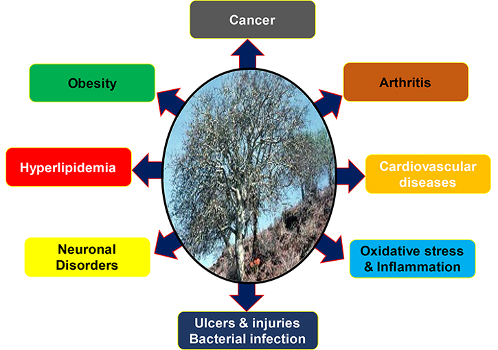
    It is already well established that the gum resin of C. wightii has been used traditionally for centuries against various chronic diseases like arthritis, obesity, diabetes and cardiovascular diseases (Figure 1) [83]. Recent studies have reported that the resin extracts from C. wightii lower the levels of low-density lipoprotein (LDL) and cholesterol in different experimental settings [58, 84, 85]. The C. wightii was shown to decrease LDL, very-low-density lipoprotein (VLDL), and cholesterol and increase high-density lipoprotein (HDL). It was also shown to reduce high fat-induced obesity in rat models [86]. Additionally, C. wightii was reported to improve oxidative stress, inflammation, edema, and necrosis in an ischemic rat model, thereby displaying its cardioprotective effects [80]. In another study, C. wightii was also shown to prevent lipid layer damage by inhibiting lipid peroxidation [87]. Further, this plant was shown to inhibit diabetes by decreasing the expression of aspartate aminotransferase, alanine aminotransferase, and oxidative markers, i.e. lipid peroxidation and protein oxidation; and inducing nuclear receptors such as peroxisome proliferator-activated receptor alpha (PPARα), peroxisome proliferator-activated receptor-gamma (PPARγ) and liver X receptor [88, 89]. Besides, another constituent, dehydroabietic acid, from Commiphora sp. was found to enhance the diabetic wound healing via reversing TNF-α-induced activation of forkhead box protein O1 (FOXO1) and the transforming growth factor β (TGF-β)1/Smad3 signaling [90].

    Biological activities of Commiphora wightii (Mark W. Skinner/www.discoverlife.org)

    Further, guggulipid could act as an antinociceptive agent by reducing neural responses and hyperalgesic activities in an in vivo model of neuropathic pain [91]. Studies have also advocated the neuroprotective properties of C. wightii by modulating various markers of oxidative stress such as thiobarbituric acid reactive substances, nitric oxide (NO), TNF-α, glutathione (GSH), superoxide dismutase (SOD), and catalase levels [92], thus indicating its potential against neuroinflammation-related disorders [93]. The anticancer property of C. wightii has also been highlighted in several studies where it was found to inhibit cancer cell proliferation by inducing cell cycle arrest and apoptosis in prostate cancer (PC) cells [94, 95]. Similar anticancer activities were also reported by different compounds isolated from C. wightii in various cancers such as breast cancer, lung cancer, colon cancer, and melanoma [96]. Furthermore, C. wightii was shown to induce anti-bacterial properties by inhibiting the growth of gram-positive and gram-negative bacteria [97]. Additional findings also suggest the use of this plant and its extracts against gastric ulcer, skin injury [98], dementia [99], arthritic inflammation [100], and blood clots [101].

    Chemical nature of GS

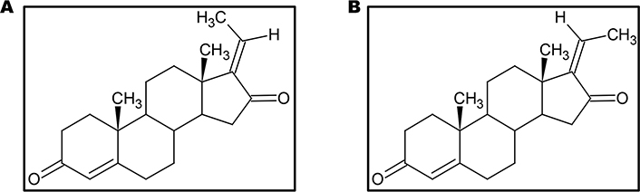
    GS, also known as 4, 17(20)-pregnadiene-3, 16-dione (C21H28O2), is the key component of the guggulu resin from C. wightii, which exists in E- and Z-isomers (Figure 2) and are represented as its cis- and trans-forms respectively [72, 77, 102, 103]. These E- and Z-GS are steroidal isomers and are inter-convertible in 3D space. They differ in the arrangement of CH3 molecule at C20 position and a distorted rotation of C-C double bond present at C17 and C20 positions is observed [104].

    Structures of the isomers of GS. (A) E-GS; (B) Z-GS

    Molecular targets of GS

    The multi-targeted compound, GS, is basically identified as an FXR inhibitor. FXR is a nuclear hormone receptor that regulates bile acid production and transport [79]. This protein might also plausibly regulate apoptosis in the cancer cells [105]. The activation of FXR is known to promote TGF-β-induced epithelial-mesenchymal transition (EMT), which is a hallmark of cancer [106]. FXR has also been identified as a marker of breast cancer-associated bone metastasis, and Z-GS has been found to efficiently inhibit FXR and its target associated bone proteins such as osteopontin, osteocalcin, and bone sialoprotein [107]. Besides FXR, GS is also known to alter the expression of various proteins in the cell and thus regulate different cellular processes such as cell growth, cell metabolism, cell survival, invasion, EMT and metastasis. Numerous preclinical studies on different cancer models have reported the chemoprotective effect of GS via modulation of multiple factors that are involved in cell growth and proliferation: COX-2, receptor activator of nuclear factor-κB ligand (RANKL); cell cycle proteins: c-Myc, cyclin D, cdc2, p21, p27; VEGF, MMP-9; proteins contributing to EMT: N-cadherin, TGF-β; molecules involved in apoptosis: caspases, survivin, Bcl-2, IAP1, XIAP, Bfl-1/A1, Bcl-2-associated X protein (Bax) and Bcl-2 homologous antagonist/killer (Bak); enzymes: IκBα kinase, Src homology 2 domain-containing protein tyrosine phosphatase 1 (SHP-1); markers involved in cell development: homeobox 2, caudal type homeobox 2 (CDX2) [65, 94, 106, 108122]. Most of these targets constitute the key components of the major pathways regulated by GS in cancer cells such as the NF-κB pathway [123, 124]; the intrinsic mitochondrial apoptotic pathway [125, 126]; the JAK/STAT pathway [67] and the STAT3 pathway [123]. GS also regulates other pathways like the Akt pathway [66, 112]; the c-Jun N-terminal kinase (JNK) pathway [112, 127]; the extracellular signal-regulated kinase (ERK) pathway [127]; steroid Src/FAK signaling [67]; β-catenin signaling [128]; and the mitogen-activated protein kinase (MAPK) pathway [124]. Further, the proteomic profiling of GS treated cancer cells have revealed that GS-induced downregulation of several factors such as proteins contributing to cell growth: methionine adenosyltransferase 2A (MAT2A) and U1 small nuclear ribonucleoprotein A (SNRPA); proteins involved in cell growth and migration: F-box only protein 2 (FBXO2), high mobility group box 3 (HMGB3), and Ras-related protein Rab-21; proteins contributing to tumorigenesis: caveolin-1, importin karyopherin subunit alpha 2 (KPNA2), and protein arginine N-methyltransferase 5 (PRMT5); proteins involved in purine metabolism: purine nucleoside phosphorylase (PNP); proteins involved in DNA replication: DNA replication licensing factor minichromosome maintenance complex component 3 (MCM3), and replication protein A 70 kDa DNA-binding subunit (RPA1); heat shock proteins (HSP): heme oxygenase-2 (HO-2), Hsp70 and Hsp27 [126]. GS also induced upregulation of annexin A7, which is involved in exocytosis/tumor suppression and the cytoskeletal protein, filamin B that regulates cell shape and migration [126]. In concordance with other studies, this study also demonstrated that GS-mediated suppression of colorectal cancer (CRC) involved modulation of the prime cellular pathway, the TNF-α/NF-κB signaling pathway [126]. GS is also known to induce reactive oxygen species (ROS)-dependent apoptosis in cancer cells via modulation of JNK [129131]. Furthermore, 14-3-3 zeta, which is involved in cancer recurrence and therapeutic resistance, has also been identified as a molecular target of GS [132, 133]. In addition, GS is also known to inhibit expression of P-glycoprotein (P-gp) in cancer cells, thereby chemosensitizing these cells to standard chemotherapeutic drugs [134136]. For example, GS sensitized hepatocellular carcinoma (HCC) cells to doxorubicin (DOX) by modulation of COX-2/P-gp dependent pathway [137], MCF-7/DOX cells to DOX by reducing the levels of Bcl-2 and P-gp [138], gall bladder cancer cells to gemcitabine through suppression of NF-κB [139], and glioblastoma cells to sonic hedgehog inhibitor SANT-1 via inflection of Ras/NF-κB pathway [140]. Furthermore, the combination of GS with bexarotene decreased DOX resistance in breast cancer cells by stimulating the secretion of exosome-associated breast cancer resistance protein (BCRP) [141]. GS has also been reported to induce CCAAT/enhancer-binding protein homologous protein (CHOP)-dependent death receptor (DR)-5 expressions through modulation of ROS-dependent endoplasmic reticulum (ER)-stress, and thus sensitize liver cancer cells to TNF-related apoptosis inducing ligand (TRAIL)-induced apoptosis [142]. Thus, GS acts on a diverse range of molecular targets and prevent the development and progression of cancer (Figure 3).

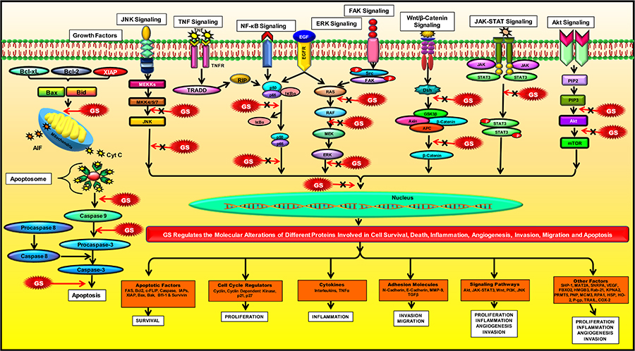
    Modulation of molecular pathways by GS. GS modulates several signaling pathways and regulate the expression of various proteins involved in inflammation, apoptosis, cell cycle, angiogenesis, invasion, metastases, and chemoresistance

    The tumor microenvironment (TME) comprises of cancer cells, cancer stem cells, immune cells, factors such as growth factors, cytokines, enzymes, chemotherapeutic drugs, the components of the extracellular matrix (ECM), fibroblasts, inflammatory cells, blood vessels, and signaling molecules [143146]. These components regulate tumorigenesis and various associated processes, such as oxidative stress, EMT, metastasis and autophagy [147153]. The TME is immunosuppressive; therefore, identification of new and effective compounds that would restore the immune response in tumor cells and inhibit the progression of tumors is critical for the prevention and treatment of cancer [154, 155]. The natural compound, GS, plays a potential role in remodeling of TME by regulating oxidative stress, autophagy, and expression of ECM proteins. For instance, GS was reported to protect PC12 cells from hydrogen peroxide-induced oxidative stress by decreasing the levels of extracellular lactate dehydrogenase, NO and ROS, and preventing the loss of mitochondrial membrane potential (ΔΨm) [156]. The antioxidant activity of GS was further evinced through GS-induced reduction of plasma trimethylamine-N-oxide expression and stimulation of Nrf2/HO-1 signaling that protects the cells from ROS-induced oxidative stress [157, 158]. GS, being an FXR antagonist, was also found to be involved in the regulation of autophagy [159, 160]. This compound has also been reportedly associated with FXR-mediated differentiation of bone marrow stromal cells into osteoblasts or adipocytes [161]. A particular study also evinced GS-induced modulation of TME components like TGFβ-induced EMT markers and COX-2 in inflammatory cells [162]. Furthermore, GS was reported to regulate the survival and stimulation of hepatic stellate cells through the regulation of NF-κB and apoptosis [114]. Thus, GS shows efficacy in the regulation of TME remodeling and exerts a significant anticancer effect.

    Biological activities of GS

    Congregate evidence suggests that GS has enormous potential in the prevention and treatment of various chronic diseases in humans owing to its multi-targeted properties. These include Alzheimer’s disease, arthritis, asthma, cancer, dermatitis, diabetes, gingivitis, inflammatory bowel disease, infectious diseases, intestinal metaplasia, otitis media, respiratory diseases, pancreatitis and psoriasis [79]. However, in the current review, we focus on its anti-cancer properties.

    Effect of GS in different cancers

    Surfeit number of preclinical evidences unveiled the cancer chemopreventive and therapeutic properties of GS against a wide range of cancers, including cancers of the brain, breast, colon, head and neck, liver, pancreas and prostate, and hematological malignancies like leukemia and lymphoma.

    These studies proved that GS has enormous potential for both the prevention and treatment of different cancers (Table 1) and are briefly summarized below.

    Potential of GS and its derivatives in the prevention and treatment of cancer

    CancerModelCombinationMechanismReferences
    Brain tumorIn vitrorBMECs-↓P-gp, ↑P-gp ATPase[134]
    In vitroA172, U87MG,
    T98G
    SANT-1↑Caspase-3, -9, ↑cytochrome c, ↑Bax, ↑IκBα,
    ↓pIκKα/β, ↓NF-κB, ↓Ras, ↓p-STAT3, ↑ERK,
    ↓c-Myc
    [140]
    Breast cancerIn vitroMCF-7IR↓NF-κB, ↓ERα, ↓IGF-1Rβ, ↑p21
    ↑Radiosensitivity
    [172]
    In vitroMCF-7/DOXDOX↑Apoptosis, ↓drug-transport activity,
    ↓MRP1
    [174]
    In vitroMDA-MB-231,
    MCF-7
    -↑Apoptosis, ↑Caspase-3, ↓β-Catenin, ↓TCF,
    ↓c-Myc, ↓Cyclin D1, ↓survivin
    [128]
    In vitroMCF-7-↓MMP-9, ↓p65/p50, ↓IκBα, ↓IKKαβ,
    ↓IKK/IκB/NF-κB axisA
    [117]
    In vitroMCF-7-↓MMP-9, ↓AP-1, ↓MAPK, ↓ERK, ↓JNKB[117]
    In vivoBALB/c miceDOX↓Tumor growth, ↓Bcl-2, ↓P-gp[138]
    In vitroMDA-MB-231Bex & DOX↑Apoptosis, ↓BCRP, ↓MDR proteins[141]
    CRCIn vitroHT-29-↓STAT3, ↓ARNT, ↓VEGF, ↓MMP-2, -9[110]
    In vitroHT-29-↓Procaspase-9, -3, ↓Bcl-2, ↓cIAP-1
    ↑Fas, ↑caspase-8, ↑p-JNK, ↑p-cJun, ↑tBid
    [72]
    In vivonu/nu mice-↓Tumor growth[72]
    In vitroHT-29IR↓IGF-1Rβ, ↓NF-κB, ↑p21, ↑γH2AX[172]
    In vitroH508, SNU-C4,
    HT-29
    -↓FXR, ↑EGFR, ↑Src, ↑ERK-1/2[177]
    In vitroHCT-116-↑p-53, ↓NF-κB, ↓Bcl-2, ↓cIAP-1, ↓survivin[126]
    CCAIn vitroSk-ChA-1,
    Mz-ChA-1
    -↑Caspase-9, -3, -8, ↑PARP, ↓Bcl-2,
    ↓survivin
    [111]
    In vitroHuCC-T1, RBE-↑Caspase-9, -3, -8, ↑DR5, ↑tBid, ↓Bid,
    ↑p-JNK1/2, ↑p-p38, ↑p-ERK1/2, ↑P-eIF2α, ↑BiP, ↑GRP78, ↑CHOP
    [130]
    Esophageal cancerIn vitroBE-derived cells-↑Apoptosis, ↑Caspase-3[105]
    In vitroBic-1-↓CDX2[113]
    In vitroTE-3-↓Cell proliferation[179]
    TE-12, SKGT-4,
    SKGT-5
    -↓FXR, ↓RAR-β2, ↓COX-2, ↓MMP-9,
    ↑Caspase-9, -3, -8
    In vivonu/nu mice-↓Tumor growth[179]
    In vitroSKGT-4, SKGT-5,
    TE-3, TE-12
    Amiloride↓Cell viability, ↑apoptosis[180]
    In vivonu/nu nude miceAmiloride↓Tumor formation, ↓growth[180]
    In vitroOE33, OE19-↓IκBα, ↓COX-2, ↓CDX-2, ↓PGE2[115]
    GBCIn vitroTGBC1, TGBC2-↓NF-κB p65, ↓MMP-2, ↓VEGF-C[139]
    In vitroTGBC1, TGBC2Gemcitabine↓NF-κB p65[139]
    Haematological malignanciesIn vitroKBM-5-↓NF-κB, ↑Caspase, ↑PARP cleavage
    ↑TNF-induced apoptosis
    [65]
    In vitroU937-↓ΔΨm, ↓p-ERK, ↑ROS, ↑HO-1, ↓GSH[202]
    In vitroU937-↓Cyclin D1, ↓cdc2, ↓Bfl-1, ↓XIAP, ↓cFLIP,
    ↓survivin, ↓Bcl-xL, ↓Bcl-2, ↓COX-2, ↓c-Myc,
    ↓IL-6, ↓IL-1β, ↓TNF,↑p21, ↑p27, ↑caspase-8, -9, -3,↑PARP, ↑BiD, ↑Bax, ↑cytochrome c, ↑JNK, ↓PI3K/Akt
    [112]
    In vitroU266-↓STAT3, ↓c-Src, ↓p-JAK2, ↑SHP-1,
    ↓Bcl-2, ↓Bcl-xL, ↓Mcl-1, ↓cyclin D1,
    ↑Caspase-3, ↑PARP
    [109]
    In vitroMM.1S-↓STAT3[109]
    In vitroK562/DOX-↓P-gp, ↓MDR[135]
    HCCIn vitroHep3B-↑CHOP, ↑DR5, ↑ROS, ↑BiP, ↑p-IRE1,
    ↑p-JNK, ↑p-PERK, ↑eIF-2α, ↑ATF4
    [142]
    In vitroHep3BTRAIL↓mtTMPt, ↑ Caspase-9, -3, -8, ↑PARP, ↓Bcl-2[142]
    In vitroHepG2-↓Bcl-2, ↑Bax, ↓TGF-β1, ↓VEGF, ↑TNF-α[125]
    In vitroHepG2R-↓COX-2, ↓P-gp[137]
    In vitroPLC/PRF/5RDOX↓COX-2, ↓P-gp, ↓PGE2, ↓MDR[137]
    In vitroHuH-7-↓EMT, ↓NR0B2, ↓CDH2 (N-cadherin)[106]
    HNCIn vitroSCC4-↓STAT3[109]
    In vitroPCI-37a,
    UM-22b,
    1483
    -↓STAT-3, ↓HIF-1α[108]
    In vivoNude mice-↑Apoptosis, ↓STAT-3[108]
    In vitroSCC4-↓Cyclin-D1, ↓XIAP, ↓Mcl-1, ↓c-Myc,
    ↓Survivin, ↑caspase-9, -3, -8, ↑Fas/CD95, ↑Bax/Bcl2,
    [133]
    In vitroSCC4, HSC2-↓PI3K/Akt, ↓GSK3β, ↓PDK1, ↓p-Raf, ↓pS6,
    ↓p-Bax, ↓p-Bad
    [66]
    In vitroSCC4, HSC2-↓p-IκBα, ↓NF-κB p65, ↓COX-2, ↓IL-6,
    ↓p-STAT-3, ↓VEGF
    [123]
    Lung cancerIn vitroH1299-↓NF-κB, ↓IκBα, ↓IKK, ↓COX-2, ↓MMP-9,
    ↓VEGF, ↓Cyclin D1, ↓c-Myc, ↓cIAP1, ↓XIAP,
    ↓Bfl-1, ↓Bcl-2, ↓TRAF1, ↓cFLIP, ↓survivin
    [65]
    Pancreatic cancerIn vitroMIAPaCa-2,
    Panc-1
    Gemcitabine↓Bcl-2, ↓p-Akt, ↓NF-κB, ↑Bax, ↑p-JNK[211]
    In vivoBALB/c
    Nude Mice
    Gemcitabine↓NF-κB, ↓Akt, ↓Bcl-2, ↑JNK[211]
    In vitroCD18/HPAF,
    Capan1
    -↓XIAP, ↓Bcl2, ↓Cyclin D1, ↑BAD, ↑Bax,
    ↓MUC4, ↑Caspase-3, ↓JAK/STAT,
    ↓Src/FAK
    [67]
    In vitroMIAPaCa2,
    PANC-1
    -↓FXR[209]
    In vitroPC-swIR↓IGF-1Rβ, ↓NF-κB[172]
    In vitroPANC-1-↓Aktc[210]
    PCIn vitroPC-3-↓Bcl-2, ↓Bcl-xL, ↑Bax, ↑Bak, ↑caspase-9, -3, -8[94]
    In vitroPC-3, LNCaP-↑JNK1/2, ↑p38 MAPK, ↑ERK1/2[131]
    In vitroDU145,
    HUVEC
    -↓VEGF, ↓G-CSF, ↓IL-17, ↓MMP-2,
    ↓p-Akt, ↓VEGF-R2,
    [218]
    In vivoDU145 cells
    implanted
    nude mice
    -↓VEGF-R2, ↓factor VIII, ↓CD31[218]
    Skin cancerIn vivoSENCAR mice-↓Skin edema, ↓hyperplasia, ↓ODC activity,
    ↓COX-2, ↓iNOS, ↓MAPKs, ↓NF-κB, ↓IKKα, ↓IκBα
    [124]
    In vitroB16/F10 mouse
    melanoma
    -↓Melanogenesis, ↓tyrosinase, ↓TRP-1
    ↓TRP-2, ↓MITF
    [219]
    Display full size

    A Cis-GS; B Trans-GS; C GS derivatives, GSD1 & GSD7; AP-1: activator protein 1; Bex: Bexarotene; BiP: binding immunoglobulin protein; cIAP-1: cellular inhibitor of apoptosis protein 1; HIF-1α: hypoxia-inducible factor 1alpha; IL-6: interleukin-6; iNOS: inducible nitric oxide synthase; MM: multiple myeloma; MMP-2: matrix metalloproteinase-2; mtTMPt: mitochondrial transmembrane potential; ODC: ornithine decarboxylase; STAT-3: signal transducer and activator of transcription 3; tBid: truncated Bid

    Brain cancer

    Brain cancer consists of more than 120 types that cover 2% of the entire global cancer incidence. It can be defined as a primary and secondary tumor depending on its tumor development status, type of tissue, and nature of its malignancy [163, 164]. Few studies reported that GS exhibited significant anticancer properties against brain cancer cells in preclinical settings. For example, GS was shown to decrease the expression of P-gp and increase the activity of P-gp ATPase dose-dependently in rat brain microvessel endothelial cells (rBMECs), thus overcoming the low accumulation of the drug and resistance in brain cancer [134]. Furthermore, GS reduced expression of Ras and NF-κB in glioblastoma cells resulting in the sensitization of these cells to SANT-1 (which is a Gli1 protein inhibitor). It also increased the expression of caspases-3 and -9, Bax, and cytochrome c, which ultimately leads to the intrinsic pathway of apoptosis [140].

    Breast cancer

    Breast cancer is the most commonly diagnosed cancer among females, and triple-negative breast cancer is the most aggressive form which is more frequently diagnosed in younger females with poor prognosis and a high recurrence rate [165171]. Metastasis of breast cancer cells is common which is mostly seen in the bone, liver, lungs, and brain tissues [149] and contributes to an increased mortality rate due to this disease [146]. Several studies have well-documented the prospective of GS in inhibiting the growth of breast cancer. For example, a study showed that GS induced radiosensitization in breast cancer cells and reduced the growth of estrogen-positive tumors resistant to tamoxifen, through the suppression of NF-κB activation and IGF1-Rβ, and ERα [172]. Another study on the effect of GS isomers against breast cancer cells demonstrated that cis-GS repressed TPA-induced MMP expression by blocking IKK/NF-κB signaling, whereas trans-GS blocked the MAPK/AP-1 signaling. Moreover, the combination of these isomers exhibited an additive effect on the inhibition of invasion of MCF-7 cells [117]. Besides, the treatment of monocytes with GS reduced RANKL-activated NF-κB activation, which correlated with inhibition of IKK and phosphorylation and degradation of IκBα. In addition, GS also suppressed the differentiation of monocytes to osteoclasts that were induced by co-incubating breast cancer cells, MDA-MB-468, or human multiple myeloma cells, U266 with monocytes [116]. A study conducted by Silva et al. [173], revealed that the release of deoxycholate from osteoblast-like cells, MG63 and bone tissues trigger the survival and migration of metastatic breast cancer cells, MDA-MB231, which was inhibited upon treatment with GS through the induction of apoptosis and modulation of the expression of urokinase-type plasminogen activator (uPA).

    Further, the guggulipid that contains GS has shown immense potential in the reduction of breast cancer cell growth and stimulation of apoptosis through the induction of cytoplasmic histone-associated DNA fragmentation, activation of caspase-3, and suppression of T-cell factor 4 and Wnt/β-Catenin pathway via suppression of its targets proteins such as cyclin D1, c-Myc, and survivin [128]. Additionally, the combined treatment of GS and DOX increased the sensitivity of the MCF-7/DOX cells to DOX by increasing the population of the apoptotic cells compared to DOX. This effect was plausibly mediated via the suppression of multi-drug resistance (MDR) protein, MRP1 [174]. Further, the combination of GS and DOX also inhibited tumor growth in BALB/c mice model by suppressing the expression of Bcl-2 and P-gp [138]. Similarly, the combination of GS and bexarotene increased DOX retention in breast cancer cells and enhanced cell death through increased secretion of exosome-associated BCRP/ABCG2 and reduced MDR levels [141].

    CRC

    CRC is the third most common cancer occurring globally [175, 176]. Copious investigations have found the enormous potential of GS in combating this disease. For example, An et al. [72], found that treatment of HT29 colon cancer cells with GS elevated apoptosis by activating caspases-3 and -8. It has also been reported that GS increased the expression of truncated BH3 interacting domain death agonist (Bid), Fas, p-JNK, and p-c-Jun levels in vitro and decreased the expression of cIAP-1, cIAP-2, and Bcl-2, and suppressed tumor growth in an in vivo mouse model. Further, GS induced apoptosome and apoptosis in HCT 116 colon cancer cells via activation of caspase-3/7, through modulating the expression of Bcl-2, and releasing cytochrome c from mitochondria. The same study also showed that GS increased the levels of p53, and suppressed the expression of NF-κB and its regulated molecules, survivin, Bcl-2, and cIAP-1 [126]. Moreover, GS suppressed viability, angiogenesis, and metastasis of colon cancer cells by inhibiting the expression of VEGF, aryl hydrocarbon receptor nuclear translocator (ARNT), STAT3 proteins, and the activity of MMP-2 and -9 [110]. Besides, GS also increased the radiation sensitivity of colon cancer cells through the suppression of IGF1-Rβ and NF-κB [172]. Contrasting to other studies, Peng et al. [177], reported that over-expression of FXR suppressed the proliferation of H508, SNU-C4, and HT-29 colon cancer cells, whereas the inhibition of FXR by GS resulted in increased proliferation of the colon cancer cells through the upregulation of Src, EGFR, and ERK-1/2.

    Esophageal cancer (EC)

    EC ranks eighth among all cancers globally and it is the sixth most common cause of cancer-related mortality with a 5-year survival rate of 15–25%. It originates in the GE junction and cardia and represents two major histological subtypes, i.e. esophageal squamous cell carcinoma and esophageal adenocarcinoma (EAC) [178]. Many studies have proven the efficacy of GS against EC. It was reported that overexpression of FXR in EAC is linked with increased tumor grade, tumor size, and high lymph node metastasis and inverse expression of retinoic acid receptor-β2 (RAR-β2), therefore knockdown of FXR yielded in decreased cell growth in vitro and decreased tumor size in the mouse model. This study suggests that GS holds enormous potential in the treatment of EC through the reduction of cell viability and induction of apoptosis via FXR suppression [179]. Similarly, the treatment of FXR overexpressed Barrett’s esophagus (BE)-derived cells with GS induced caspase-3 activity and resulted in apoptosis [105]. Further, GS treatment blocked IκBα phosphorylation induced by deoxycholic acid and decreased CDX2, COX-2, and prostaglandin E2 (PGE2) expressions in EAC cells, and also caused increased sub-G1 phase apoptosis in BE and EAC cells [115]. Besides, GS in combination with amiloride suppressed the viability of EC cells, induced apoptosis, and inhibited tumor growth in a mouse model [180].

    Head and neck cancer (HNC)

    HNC includes a range of cancers among which more than 90% is represented by head and neck squamous cell carcinoma (HNSCC) [181187]. HNSCC has a widespread global incidence rate with poor prognosis and a 50% occurrence in the oropharynx region mostly observed in the palatine tonsil and bottom of the tongue [188]. Several studies have demonstrated the potential of GS in inhibiting HNC. For instance, the SCC4 cells treated with GS were found to exhibit apoptosis via decreased expression of cyclin D1, phosphorylated Bcl-2-associated death promoter (BAD), XIAP, myeloid cell leukemia 1 (Mcl-1), c-Myc, and survivin, and increased expression of p21 and p27, Bax/Bcl2 ratio, caspases-9, -3, -8 and Fas/CD95 proteins [133]. Further, the pre-treatment of HNC cells with GS resulted in inhibition of smokeless tobacco/nicotine-induced expression of PI3K, phosphoinositide-dependent kinase-1 (PDK1), Akt, Raf, Glycogen synthase kinase 3 beta (GSK3β), and pS6, thereby blocking the PI3K/Akt pathway. It also suppressed Akt-associated Bax and BAD phosphorylation in HNC cells [66]. Similarly, in another study, GS was shown to suppress NF-κB and pSTAT3 and their target proteins COX-2 and VEGF, and also reduced the expression of phosphorylated IκBα and IL-6, induced by smokeless tobacco/nicotine in HNC cells [123]. Further, GS inhibited the growth of HNC cells by decreasing the expression of STAT3 [109]. The inhibition of STAT3 by GS was enhanced when combined with erlotinib or cetuximab in vitro and in vivo. Besides, GS also reduced the invasion of HNC cells by suppressing the expression of HIF-1α [108]. Additionally, it was also shown that the proteasome inhibitor bortezomib causes an increase in total STAT3, pSTAT3, and cellular STAT3 levels in HNC cells and the combined treatment of GS (natural STAT3 inhibitor) with bortezomib could induce synergistic death of cancer cells [189].

    HCC

    HCC or liver cancer is one of the highest occurring malignancies in the world [2, 53, 190197]. Despite the advancement in therapeutic strategies, there is a lack of efficacious and non-toxic drugs for the treatment of HCC. In pursuit of alternative medicine for the treatment of HCC, GS was investigated in several studies. For instance, treatment with GS significantly reduced the proliferation, induced G0/G1 phase arrest and apoptosis in HepG2 cells via regulating the expression of Bax, Bcl-2, TGF-β1, TNF-α, and VEGF [125]. Further, GS was also found to inhibit TGF-β-induced EMT in HCC cells [106]. In this study, GS was found to decrease the mRNA expression of CDH2, which codes for N-cadherin, a marker of EMT, and nuclear receptor subfamily 0, group B, member 2 (NR0B2), which is an FXR target gene [106]. Additionally, the treatment of GS sensitized DOX-resistant PLC/PRF/5R cells to DOX via suppression of the expression of COX-2, P-gp, and PGE2 [137]. In another study, the combination of GS with TRAIL was found to induce apoptosis in HCC cells through the reduction of mitochondrial transmembrane potential and caspase activation [142]. The receptor for TRAIL, DR5, was also found to be upregulated along with activation of proteins associated with ER stress and apoptosis such as eukaryotic initiation factor-2α (eIF2α) and CHOP in GS treated cells [142].

    Hematological malignancies

    Hematological malignancies such as leukemia, lymphoma, and multiple myeloma are serious health issues worldwide [2, 198201]. Most of the hematological malignancies are a result of genetic alterations, as an example, the mutation of FMS-like tyrosine kinase3-internal tandem duplication (FLT3-ITD), which is common in acute myeloid leukemia (AML) [27, 29]. Therefore, the therapeutic potential of GS was explored for the treatment of hematological malignancies. GS was shown to potentiate the TNF-induced apoptosis of leukemia cells by inhibiting NF-κB and increasing caspase-mediated cleavage of PARP protein [65]. This plant sterol was also reported to reduce the proliferation of U937 leukemia cells by inhibiting DNA synthesis, inducing G1/S phase arrest, and decreasing the levels of cyclin D1 and cdc2 and upregulating CDK inhibitors such as p21 and p27. Further, GS was shown to induce apoptosis in these cells through activation of caspases-3, -8, and -9, the release of cytochrome c, Bid and PARP cleavage, and by decreasing anti-apoptotic proteins such as Bfl-1, XIAP, cFLIP, Bcl-xL, Bcl-2, survivin, COX-2, c-Myc, IL-6, IL-1β and TNF levels. In addition, GS also activated JNK and suppressed Akt activity in these cells [112]. Moreover, the cis- and trans-GS was reported to cause apoptosis in AML cells, HL60 and U937, through phosphatidylserine externalization and loss of ΔΨm. The trans-GS was also reported to play a remarkable role in the differentiation of these cells as evidenced through elevated levels of surface proteins such as CD11b and CD14, while cis-GS reduced intracellular GSH levels and promoted oxidation of cardiolipin, which is known to be involved in mitochondrial function and prevention of apoptosis [202]. Moreover, the combined treatment of GS and DOX reversed MDR by inhibiting P-gp and increasing cellular accumulation of DOX in K562/DOX cells [135]. Additionally, GS inhibited the proliferation of multiple myeloma cells by decreasing the expression of STAT3 via activation of SHP-1, which leads to the suppression of c-Src and p-JAK2 proteins. Further, GS was also shown to inhibit Bcl-2, Bcl-xL, Mcl-1, and cyclin D1 gene expressions in these cells [109].

    Pancreatic cancer (PaCa)

    PaCa is one of the most fatal malignancies in the world that ranks seventh in both males and females worldwide [2]. Studies have shown the efficacy of natural compounds like resveratrol, curcumin, γ-tocotrienol, food supplement like ZyflamendTM, and small molecules like protein kinase D (PKD) inhibitor, CRT0066101 against PaCa [203208]. Similarly, GS has also been reported to exhibit antineoplastic and chemosensitizing potential in PaCa models. In a particular study, GS was found to suppress proliferation and survival and induce apoptosis in PaCa cells, Capan1 and CD18/HPAF, through enhanced activation of caspase-3, altered BAD phosphorylation, decreased cyclin D1, and reduced level of anti-apoptotic proteins, Bcl-2 and XIAP. This compound also suppressed invasion and metastasis in PaCa cells by dysregulating the cytoskeletal organization, inhibiting the activation of FAK and Src signaling, and reducing MMP-9 levels and JAK/STAT pathway-mediated mucin4 (MUC4) expressions [67]. Similarly, GS-mediated FXR inhibition was found to remarkably suppress migration and invasion in PaCa cells [209]. Besides, the derivatives of GS, GSD-1, and GSD-7 were also reported to induce morphological changes and reduce cell survival in PANC-1 cells by inhibiting Akt protein [210]. Further, GS was also evinced to induce radiosensitization in PC-Sw cells through reduced levels of NF-κB and IGF1-Rβ [172]. In addition, GS administration enhanced gemcitabine-mediated growth suppression and apoptosis in PaCa cells via suppression of NF-κB activity, levels of Akt and Bcl-2, and activation of c-JNK and Bax [211].

    PC

    In terms of incidence, PC is the second most common malignancy in males worldwide [2, 212215]. The factors like unhealthy diet and lifestyles were reported to be associated with the development of PC [216, 217]. Multiple lines of evidence indicate that GS has immense potential in the prevention and treatment of PC. For example, GS induced apoptosis in PC-3 and LNCaP cells by triggering ROI-dependent JNK activation [131]. In LNCaP cells, GS was also found to reduce the expression and promoter activity of the androgen receptor [131]. Besides, GS treatment was also reported to induce apoptosis in PC-3 cells by inducing DNA fragmentation and upregulating the expression of Bax, Bak, and caspases -3, -8, and -9 [94]. Further, the administration of Z-GS was reported to inhibit tube formation of HUVEC cells and migration of DU145 and HUVEC cells via suppression of VEGF, granulocyte colony-stimulating factor (G-CSF), VEGF receptor (VEGF-R2), and Akt. The anti-angiogenic effect of GS was also evident in the nude mice model, where the oral administration of GS was found to suppress tumor growth and decrease levels of angiogenic markers, factor VIII, CD31, and VEGF-R2 [218].

    Other cancers

    Apart from the aforementioned cancers, the anticancer potential of GS was documented in other cancers such as cholangiocarcinoma (CCA), gallbladder cancer, melanoma, and lung cancer. For example, GS was shown to reduce the growth of CCA cells (Sk-ChA-1 and Mz-ChA-1) and induce apoptosis via enhancement of caspases -3, -8, and -9 expression and PARP cleavage, and suppression of survivin and Bcl-2 [111]. Further, GS treatment was reported to induce apoptosis in HuCC-T1 and RBE CCA cells through the regulation of the ROS/JNK pathway [130]. In addition, treatment with GS was shown to inhibit melanogenesis in B16 murine melanoma cells through the reduction of tyrosinase, microphthalmia-associated transcription factor (MITF), and tyrosinase-related protein (TRP-1 and TRP-2). This compound was also found to suppress melanogenesis induced by α-melanocyte-stimulating hormone and forskolin [219]. Another study revealed that GS lowered skin tumor incidence in SENCAR mice by inhibiting various inflammation and tumor-associated markers such as COX-2, iNOS, MAPKs, IKKα, IκBα, and NF-κB [124]. Furthermore, the activity of GS was also studied in H1299 lung cancer cells where it inhibited NF-κB activation induced by various agents such as TNF, IL-1β, and carcinogens; and also inhibited IκBα and IKK. Besides, GS suppressed the activities of other proteins such as COX2, MMP9, VEGF, cell cycle proteins, and anti-apoptotic proteins [65]. In another study, the potential of GS on gall bladder cancer (GBC) was investigated. This study showed that GS suppressed the proliferation and invasion of TGBC1 and TGBC2, GBC cells via inhibition of NF-κB p65, VEGF-C, and MMP-2. In addition, the combination of GS and gemcitabine significantly inhibited the growth of GBC, thereby exerting its chemosensitizing potential [139]. Thus, GS has immense potential as a drug candidate for cancer treatment as proved by preclinical studies. However, the pharmacokinetics (Pk) and pharmacodynamics of this compound should be studied for its safe and effective use as a clinical drug.

    Pk and pharmacodynamics of GS

    A limited number of studies have reported the Pk and pharmacodynamics of GS to date. In 1998, Verma et al. [220], performed HPLC of Z-GS, E-GS, drug-free rat serum, GS-spiked serum, serum from GS (50 mg/kg)-dosed rats (at 4 h), and serum from GS-dosed rats (at 24 h). The recovery percentage of Z-GS and E-GS from spiked serum samples was more than 90% at all concentrations of spiking (25, 50, 250, 2, 500 ng/mL) while the chromatogram analysis suggested that under in vivo conditions, GS is plausibly metabolized from Z- to E-form upon administration and is retained in the same form in the body. In another study, the effect of both oral and intravenous administration on various Pk parameters of Z-GS and E-GS were analysed; however, no statistically significant difference was observed. It was reported that the Pk parameters like terminal half-life, systemic clearance, area under the curve, and volume of distribution were 4.48 and 3.56 h, 1.76 and 2.24 L/h, 5.95 and 4.75 μgh/mL, and 11.36 and 10.76 L, respectively. These results demonstrated that the absorption of Z-GS was rapid from the gastrointestinal tract, leading to maximum concentration (Cmax) in the serum 2 h after oral administration. Moreover, the bioavailability of orally administered Z-GS relative to the intravenous administration was found to be 42.9% [221]. In another study, Bhatta et al. [222], developed a liquid chromatography-tandem mass spectrometry (LC-MS/MS) method for the simultaneous determination of both Z- and E-GS in rabbit plasma. However, this method faced a limitation of the long run time of 20 min. Subsequently, in 2015, Chhonker et al. [223], developed and validated an extremely specific and sensitive LC-MS/MS method for the estimation of E- and Z-GS in rat plasma within a run time of 6 min. Using this method, the ADME properties of GS such as metabolic stability, pH-dependent stability, plasma protein binding, solubility and Pk of GS isomers administered orally in rats were evaluated. Moreover, E- and Z-GS were reported to be soluble up to 50 μM (1, 561 ng/mL) under physiological conditions. The findings also suggested that E- and Z-GS were stable in the gastrointestinal fluid and were not subjected to enzymatic degradation; their plasma protein binding was high and independent of the concentration of GS. Further, both the stereoisomers exhibited low bioavailability of GS owing to wide first pass metabolism through high clearance and short half-life in rats [223].

    The efficacy, toxicity, and pharmacodynamics of a drug depend hugely on its metabolic fate. In 2012, Yang et al. [224], reported a basic metabolic profile of Z-GS. However, to gain an in-depth understanding of the Pk and metabolism of GS, in 2018, Chhonker et al. [225], conducted another metabolic investigation where GS was found to metabolize to produce nineteen metabolites in human liver microsomes, and S9 fractions and hydroxylation was identified as the prime metabolic pathway. Also, the binding efficiency of GS with human serum albumin ranged from low to moderate. Further, CYP profiling and inhibition studies revealed that GS is a substrate for various CYPs, majorly CYP3A4, and it inhibited CYP2C19 [225]. Recently, another study was performed to detect electrophilic reactive metabolites of GS isomers. It was hypothesized that these metabolites are responsible for the toxic reactions of GS. The results showed that hydroxylated metabolites of GS isomers formed adducts with the trapping agents, GSH and N-acetylcysteine [226]. Overall, these studies form the background for further structural modification of GS, enhancement in the stability, and designing of its analogs.

    Discussion and conclusion

    The increasing rate of cancer cases and deaths pose a huge concern worldwide [227231]. The currently available drugs are expensive, induce severe adverse side-effects, and are not very effective [6, 38, 232]. Thus, alternative drugs are sought for the efficient management of cancer, and natural compounds have shown promising results [233245]. Among these compounds, GS, the FXR antagonist, has exhibited immense potential as an anticancer agent against various cancer types. This multi-targeted agent has shown to affect different cellular processes such as inflammation, cell proliferation, survival, angiogenesis, invasion, metastasis, EMT, and apoptosis in cancer cells in various preclinical models. In this process, GS has been reported to inflect multiple pathways like NF-κB, STAT-3, ERK/MAPK, JAK/STAT, ROS/JNK and PI3K/Akt, and several genes and proteins such as COX-2, MMP-9, p38, PGE2, HIF-1α, VEGF, interleukins, cyclin D1, survivin, p21, p27, p53, PARP, Bid and caspases. This active metabolite of guggulipid has also been known to regulate cellular stress and ΔΨm in several in vitro models. Further, GS also inhibited tumor growth remarkably in in vivo models of cancer. Not only this, but GS could also induce sensitization of cancer cells to standard chemotherapeutic drugs like DOX and gemcitabine in various cancer models. It has also been reported to sensitize the cancer cells to TRAIL and enhance the efficacy of radiation in cancer models, thereby leading to cancer cell death. These studies show that GS is a potential therapeutic agent for the prevention and treatment of various cancers. However, the preclinical findings need to be validated in clinical settings with a detailed investigation of its safety, toxicity, and bioavailability. Studies have identified two prime stereoisoforms of GS, Z-GS and E-GS, along with 19 metabolites. GS is characterized by physiological solubility upto 50 μM, high plasma protein binding, and limited bioavailability. Hence, studies have been carried out to design analogs of this pharmacophore with enhanced bioavailability. Thus, modulation of the pharmacodynamics and Pk properties of this compound may lead to the development of analogs and derivatives of GS for better management of cancer.

    Abbreviations

    AML:

    acute myeloid leukemia

    ARNT:

    aryl hydrocarbon receptor nuclear translocator

    BAD:

    Bcl-2-associated death promoter

    Bak:

    Bcl-2 homologous antagonist/killer

    Bax:

    Bcl-2-associated X protein

    Bcl-2:

    B-cell lymphoma 2

    BCRP:

    breast cancer resistance protein

    BE:

    Barrett’s esophagus

    Bfl-1/A1:

    Bcl-2-related protein A1

    Bid:

    BH3 interacting domain death agonist

    C. wightii:

    Commiphora wightii

    CCA:

    cholangiocarcinoma

    CDX2:

    caudal type homeobox 2

    cFLIP:

    cellular FLICE (FADD-like IL-1β-converting enzyme)-inhibitory protein

    CHOP:

    CCAAT/enhancer-binding protein homologous protein

    COX-2:

    cyclooxygenase 2

    CRC:

    colorectal cancer

    DOX:

    doxorubicin

    DR:

    death receptor

    EAC:

    esophageal adenocarcinoma

    EC:

    esophageal cancer

    ECM:

    extracellular matrix

    eIF2α:

    eukaryotic initiation factor-2α

    EMT:

    epithelial-mesenchymal transition

    ER:

    endoplasmic reticulum

    ERK:

    extracellular signal-regulated kinase

    FAK:

    focal adhesion kinase

    FXR:

    farnesoid X receptor

    GBC:

    gall bladder cancer

    GS:

    guggulsterone

    GSH:

    glutathione

    GSK3β:

    Glycogen synthase kinase 3 beta

    HCC:

    hepatocellular carcinoma

    HNC:

    head and neck cancer

    HNSCC:

    head and neck squamous cell carcinoma

    IAP1:

    inhibitor of apoptosis protein 1

    IKK:

    IkappaB kinase

    IκBα:

    IkappaB alpha

    JAK:

    Janus kinase

    JNK:

    Jun N-terminal kinase

    LC-MS/MS:

    liquid chromatography-tandem mass spectrometry

    LDL:

    low-density lipoprotein

    MAPK:

    mitogen-activated protein kinase

    Mcl-1:

    myeloid cell leukemia 1

    MDR:

    multi-drug resistance

    MITF:

    microphthalmia-associated transcription factor

    MMP:

    matrix metalloproteinase

    MUC4:

    mucin 4

    NF-κB:

    nuclear factor kappa B

    NO:

    nitric oxide

    NR0B2:

    nuclear receptor subfamily 0, group B, member 2

    PaCa:

    pancreatic cancer

    PC:

    prostate cancer

    PDK1:

    phosphoinositide-dependent kinase-1

    PGE2:

    prostaglandin E2

    P-gp:

    P-glycoprotein

    PI3K:

    phosphoinositide 3-kinase

    Pk:

    pharmacokinetics

    PKD:

    protein kinase D

    PPARγ:

    peroxisome proliferator-activated receptor-gamma

    QOL:

    quality of life

    RANKL:

    receptor activator of nuclear factor-κB ligand

    rBMECs:

    rat brain microvessel endothelial cells

    ROS:

    reactive oxygen species

    SHP-1:

    Src homology 2 domain-containing protein tyrosine phosphatase 1

    Src:

    steroid receptor coactivator

    STAT:

    signal transducers and activators of transcription

    TGF-β:

    transforming growth factor β

    TME:

    tumor microenvironment

    TNF:

    tumor necrosis factor

    TNFR:

    tumor necrosis factor receptor

    TRAIL:

    TNF-related apoptosis-inducing ligand

    TRP:

    tyrosinase-related protein

    VEGF:

    vascular endothelial growth factor

    XIAP:

    X-linked inhibitor of apoptosis protein

    ΔΨm:

    mitochondrial membrane potential

    Declarations

    Acknowledgments

    The author Kishore Banik acknowledges UGC, New Delhi, India, for providing him the fellowship.

    Author contributions

    ABK contributed to the study design, conceptualisation, supervision and review editing. SG, CH, and KB performed bibliographic search. SG and CH contributed to original manuscript. DP contributed to table preparation and proofreading. KB and DP performed artwork. CH, KB and DP contributed to review editing. SG, CH and KB contributed to proofreading.

    Conflicts of interest

    The authors declare that they have no conflicts of interest.

    Ethical approval

    Not applicable.

    Consent to participate

    Not applicable.

    Consent to publication

    Not applicable.

    Availability of data and materials

    Not applicable.

    Funding

    This project was supported by NCD/NER/4/2018-19 awarded to ABK by Indian Council of Medical Research (ICMR), Government of India. The funder had no role in study design, data collection and analysis, decision to publish, or preparation of the manuscript.

    Copyright

    © The Author(s) 2020.

    References

    Hassanpour SH, Dehghani M. Review of cancer from perspective of molecular. J Cancer Res Pract. 2017;4:1279. [DOI]
    Bray F, Ferlay J, Soerjomataram I, Siegel RL, Torre LA, Jemal A. Global cancer statistics 2018: GLOBOCAN estimates of incidence and mortality worldwide for 36 cancers in 185 countries. CA Cancer J Clin. 2018;68:394424. [DOI] [PubMed]
    Ferlay J, Colombet M, Soerjomataram I, Mathers C, Parkin DM, Piñeros M, et al. Estimating the global cancer incidence and mortality in 2018: GLOBOCAN sources and methods. Int J Cancer. 2019;144:194153. [DOI] [PubMed]
    Girisa S, Shabnam B, Monisha J, Fan L, Halim CE, Arfuso F, et al. Potential of zerumbone as an anti-cancer agent. Molecules. 2019;24:734. [DOI]
    Henamayee S, Banik K, Sailo BL, Shabnam B, Harsha C, Srilakshmi S, et al. Therapeutic emergence of rhein as a potential anticancer drug: a review of its molecular targets and anticancer properties. Molecules. 2020;25:2278. [DOI]
    Khatoon E, Banik K, Harsha C, Sailo BL, Thakur KK, Khwairakpam AD, et al. Phytochemicals in cancer cell chemosensitization: current knowledge and future perspectives. Semin Cancer Biol. 2020;[Epub ahead of print]. [DOI]
    Anand P, Kunnumakkara AB, Sundaram C, Harikumar KB, Tharakan ST, Lai OS, et al. Cancer is a preventable disease that requires major lifestyle changes. Pharm Res. 2008;25:2097116. [DOI] [PubMed] [PMC]
    Kunnumakkara AB, Anand P, Aggarwal BB. Curcumin inhibits proliferation, invasion, angiogenesis and metastasis of different cancers through interaction with multiple cell signaling proteins. Cancer Lett. 2008;269:199225. [DOI] [PubMed]
    Kunnumakkara AB, Bordoloi D, Harsha C, Banik K, Gupta SC, Aggarwal BB. Curcumin mediates anticancer effects by modulating multiple cell signaling pathways. Clin Sci (Lond). 2017;131:178199. [DOI] [PubMed]
    Roy NK, Bordoloi D, Monisha J, Padmavathi G, Kotoky J, Golla R, et al. Specific targeting of Akt kinase isoforms: taking the precise path for prevention and treatment of cancer. Curr Drug Targets. 2017;18:42135. [DOI] [PubMed]
    Shabnam B, Padmavathi G, Banik K, Girisa S, Monisha J, Sethi G, et al. Sorcin a potential molecular target for cancer therapy. Transl Oncol. 2018;11:137989. [DOI] [PubMed] [PMC]
    Banik K, Ranaware AM, Deshpande V, Nalawade SP, Padmavathi G, Bordoloi D, et al. Honokiol for cancer therapeutics: a traditional medicine that can modulate multiple oncogenic targets. Pharmacol Res. 2019;144:192209. [DOI] [PubMed]
    Kunnumakkara AB, Shabnam B, Girisa S, Harsha C, Banik K, Devi TB, et al. Inflammation, NF-κB, and chronic diseases: how are they linked? Crit Rev Immunol. 2020;40:139. [DOI] [PubMed]
    Shanmugam MK, Warrier S, Kumar AP, Sethi G, Arfuso F. Potential role of natural compounds as anti-angiogenic agents in cancer. Curr Vasc Pharmacol. 2017;15:50319. [DOI] [PubMed]
    Hsieh YS, Yang SF, Sethi G, Hu DN. Natural bioactives in cancer treatment and prevention. Biomed Res Int. 2015;2015:182835. [DOI] [PubMed] [PMC]
    Dai X, Zhang J, Arfuso F, Chinnathambi A, Zayed ME, Alharbi SA, et al. Targeting TNF-related apoptosis-inducing ligand (TRAIL) receptor by natural products as a potential therapeutic approach for cancer therapy. Exp Biol Med (Maywood). 2015;240:76073. [DOI] [PubMed] [PMC]
    Khwairakpam AD, Shyamananda MS, Sailo BL, Rathnakaram SR, Padmavathi G, Kotoky J, et al. ATP citrate lyase (ACLY): a promising target for cancer prevention and treatment. Curr Drug Targets. 2015;16:15663. [DOI] [PubMed]
    Sailo BL, Banik K, Girisa S, Bordoloi D, Fan L, Halim CE, et al. FBXW7 in cancer: what has been unraveled thus far? Cancers (Basel). 2019;11:246. [DOI]
    Aggarwal BB, Kunnumakkara AB, Harikumar KB, Tharakan ST, Sung B, Anand P. Potential of spice-derived phytochemicals for cancer prevention. Planta Med. 2008;74:15609. [DOI] [PubMed]
    Guruvayoorappan C, Sakthivel KM, Padmavathi G, Bakliwal V, Monisha J, Kunnumakkara AB. Cancer preventive and therapeutic properties of fruits and vegetables: an overview. In: Kunnumakkara AB, editor. Anticancer properties of fruits and vegetable: Scientific Review. Singapore: World Scientific; 2015. pp. 152.
    Roy PS, Saikia BJ. Cancer and cure: a critical analysis. Indian J Cancer. 2016;53:4412. [DOI] [PubMed]
    Chakraborty C, Sharma AR, Sharma G, Sarkar BK, Lee SS. The novel strategies for next-generation cancer treatment: miRNA combined with chemotherapeutic agents for the treatment of cancer. Oncotarget. 2018;9:1016474. [DOI] [PubMed] [PMC]
    Ranaware AM, Banik K, Deshpande V, Padmavathi G, Roy NK, Sethi G, et al. Magnolol: a neolignan from the magnolia family for the prevention and treatment of cancer. Int J Mol Sci. 2018;19:2362. [DOI]
    Arruebo M, Vilaboa N, Sáez-Gutierrez B, Lambea J, Tres A, Valladares M, et al. Assessment of the evolution of cancer treatment therapies. Cancers (Basel). 2011;3:3279330. [DOI] [PubMed] [PMC]
    Kanojia D, Garg M, Martinez J, M TA, Luty SB, Doan NB, et al. Kinase profiling of liposarcomas using RNAi and drug screening assays identified druggable targets. J Hematol Oncol. 2017;10:173. [DOI] [PubMed] [PMC]
    Warrier VU, Makandar AI, Garg M, Sethi G, Kant R, Pal JK, et al. Engineering anti-cancer nanovaccine based on antigen cross-presentation. Biosci Rep. 2019;39:BSR20193220. [DOI] [PubMed] [PMC]
    Garg M, Nagata Y, Kanojia D, Mayakonda A, Yoshida K, Haridas Keloth S, et al. Profiling of somatic mutations in acute myeloid leukemia with FLT3-ITD at diagnosis and relapse. Blood. 2015;126:2491501. [DOI] [PubMed] [PMC]
    Kirtonia A, Gala K, Fernandes SG, Pandya G, Pandey AK, Sethi G, et al. Repurposing of drugs: an attractive pharmacological strategy for cancer therapeutics. Semin Cancer Biol. 2020;[Epub ahead of print]. [DOI]
    Kirtonia A, Pandya G, Sethi G, Pandey AK, Das BC, Garg M. A comprehensive review of genetic alterations and molecular targeted therapies for the implementation of personalized medicine in acute myeloid leukemia. J Mol Med (Berl). 2020;98:106991. [DOI] [PubMed]
    Monisha J, Padmavathi G, Roy NK, Deka A, Bordoloi D, Anip A, et al. NF-κB blockers gifted by mother nature: prospectives in cancer cell chemosensitization. Curr Pharm Des. 2016;22:4173200. [DOI] [PubMed]
    Roy NK, Deka A, Bordoloi D, Mishra S, Kumar AP, Sethi G, et al. The potential role of boswellic acids in cancer prevention and treatment. Cancer Lett. 2016;377:7486. [DOI] [PubMed]
    Khwairakpam AD, Bordoloi D, Thakur KK, Monisha J, Arfuso F, Sethi G, et al. Possible use of Punica granatum (Pomegranate) in cancer therapy. Pharmacol Res. 2018;133:5364. [DOI] [PubMed]
    Monisha J, Jaiswal A, Banik K, Choudhary H, Singh AK, Bordoloi D, et al. Cancer cell chemoresistance: a prime obstacle in cancer therapy. In: Kunnumakkara AB, Bordoloi D, Monisha J, editors. Cancer cell chemoresistance and chemosensitization. Sinapore: World Scientific; 2018. pp. 1549.
    Banik K, Ranaware AM, Harsha C, Nitesh T, Girisa S, Deshpande V, et al. Piceatannol: a natural stilbene for the prevention and treatment of cancer. Pharmacol Res. 2020;153:104635. [DOI] [PubMed]
    Kunnumakkara AB, Nair AS, Ahn KS, Pandey MK, Yi Z, Liu M, et al. Gossypin, a pentahydroxy glucosyl flavone, inhibits the transforming growth factor beta-activated kinase-1-mediated NF-kappaB activation pathway, leading to potentiation of apoptosis, suppression of invasion, and abrogation of osteoclastogenesis. Blood. 2007;109:511221. [DOI] [PubMed] [PMC]
    Bordoloi D, Roy NK, Monisha J, Padmavathi G, Kunnumakkara AB. Multi-targeted agents in cancer cell chemosensitization: what we learnt from curcumin thus far. Recent Pat Anticancer Drug Discov. 2016;11:6797. [DOI] [PubMed]
    Sailo BL, Banik K, Padmavathi G, Javadi M, Bordoloi D, Kunnumakkara AB. Tocotrienols: the promising analogues of vitamin E for cancer therapeutics. Pharmacol Res. 2018;130:25972. [DOI] [PubMed]
    Kunnumakkara AB, Bordoloi D, Sailo BL, Roy NK, Thakur KK, Banik K, et al. Cancer drug development: the missing links. Exp Biol Med (Maywood). 2019;244:66389. [DOI] [PubMed] [PMC]
    Thomas D, Govindhan S, Baiju EC, Padmavathi G, Kunnumakkara AB, Padikkala J. Cyperus rotundus L. prevents non-steroidal anti-inflammatory drug-induced gastric mucosal damage by inhibiting oxidative stress. J Basic Clin Physiol Pharmacol. 2015;26:48590. [DOI] [PubMed]
    Deng S, Shanmugam MK, Kumar AP, Yap CT, Sethi G, Bishayee A. Targeting autophagy using natural compounds for cancer prevention and therapy. Cancer. 2019;125:122846. [DOI] [PubMed]
    Tewari D, Nabavi SF, Nabavi SM, Sureda A, Farooqi AA, Atanasov AG, et al. Targeting activator protein 1 signaling pathway by bioactive natural agents: possible therapeutic strategy for cancer prevention and intervention. Pharmacol Res. 2018;128:36675. [DOI] [PubMed]
    Mishra S, Verma SS, Rai V, Awasthee N, Chava S, Hui KM, et al. Long non-coding RNAs are emerging targets of phytochemicals for cancer and other chronic diseases. Cell Mol Life Sci. 2019;76:194766. [DOI] [PubMed]
    Pandey MK, Sung B, Ahn KS, Kunnumakkara AB, Chaturvedi MM, Aggarwal BB. Gambogic acid, a novel ligand for transferrin receptor, potentiates TNF-induced apoptosis through modulation of the nuclear factor-kappaB signaling pathway. Blood. 2007;110:351725. [DOI] [PubMed] [PMC]
    Khwairakpam AD, Monisha J, Roy NK, Bordoloi D, Padmavathi G, Banik K, et al. Vietnamese coriander inhibits cell proliferation, survival and migration via suppression of Akt/mTOR pathway in oral squamous cell carcinoma. J Basic Clin Physiol Pharmacol. 2019;31:/j/jbcpp.2020.31.issue-3/jbcpp-2019-0162/jbcpp-2019-0162.xml. [DOI]
    Padmavathi G, Rathnakaram SR, Monisha J, Bordoloi D, Roy NK, Kunnumakkara AB. Potential of butein, a tetrahydroxychalcone to obliterate cancer. Phytomedicine. 2015;22:116371. [DOI] [PubMed]
    Padmavathi G, Roy NK, Bordoloi D, Arfuso F, Mishra S, Sethi G, et al. Butein in health and disease: a comprehensive review. Phytomedicine. 2017;25:11827. [DOI] [PubMed]
    Banik K, Harsha C, Bordoloi D, Lalduhsaki Sailo B, Sethi G, Leong HC, et al. Therapeutic potential of gambogic acid, a caged xanthone, to target cancer. Cancer Lett. 2018;416:7586. [DOI] [PubMed]
    Khwairakpam AD, Damayenti YD, Deka A, Monisha J, Roy NK, Padmavathi G, et al. Acorus calamus: a bio-reserve of medicinal values. J Basic Clin Physiol Pharmacol. 2018;29:10722. [DOI] [PubMed]
    Bordoloi D, Monisha J, Roy NK, Padmavathi G, Banik K, Harsha C, et al. An investigation on the therapeutic potential of butein, a tretrahydroxychalcone against human oral squamous cell carcinoma. Asian Pac J Cancer Prev. 2019;20:343746. [DOI] [PubMed] [PMC]
    Roy NK, Parama D, Banik K, Bordoloi D, Devi AK, Thakur KK, et al. An update on pharmacological potential of boswellic acids against chronic diseases. Int J Mol Sci. 2019;20:4101. [DOI]
    Singh YP, Girisa S, Banik K, Ghosh S, Swathi P, Deka M, et al. Potential application of zerumbone in the prevention and therapy of chronic human diseases. J Funct Foods. 2019;53:24858. [DOI]
    Prasannan R, Kalesh KA, Shanmugam MK, Nachiyappan A, Ramachandran L, Nguyen AH, et al. Key cell signaling pathways modulated by zerumbone: role in the prevention and treatment of cancer. Biochem Pharmacol. 2012;84:126876. [DOI] [PubMed]
    Siveen KS, Ahn KS, Ong TH, Shanmugam MK, Li F, Yap WN, et al. Y-tocotrienol inhibits angiogenesis-dependent growth of human hepatocellular carcinoma through abrogation of AKT/mTOR pathway in an orthotopic mouse model. Oncotarget. 2014;5:1897911. [DOI] [PubMed] [PMC]
    Ramachandran L, Manu KA, Shanmugam MK, Li F, Siveen KS, Vali S, et al. Isorhamnetin inhibits proliferation and invasion and induces apoptosis through the modulation of peroxisome proliferator-activated receptor γ activation pathway in gastric cancer. J Biol Chem. 2012;287:3802840. [DOI] [PubMed] [PMC]
    Manu KA, Shanmugam MK, Ramachandran L, Li F, Siveen KS, Chinnathambi A, et al. Isorhamnetin augments the anti-tumor effect of capecitabine through the negative regulation of NF-κB signaling cascade in gastric cancer. Cancer Lett. 2015;363:2836. [DOI] [PubMed]
    Lee JH, Chiang SY, Nam D, Chung WS, Lee J, Na YS, et al. Capillarisin inhibits constitutive and inducible STAT3 activation through induction of SHP-1 and SHP-2 tyrosine phosphatases. Cancer Lett. 2014;345:1408. [DOI] [PubMed]
    Yamada T, Sugimoto K. Guggulsterone and its role in chronic diseases. Adv Exp Med Biol. 2016;929:32961. [DOI] [PubMed]
    Urizar NL, Moore DD. GUGULIPID: a natural cholesterol-lowering agent. Annu Rev Nutr. 2003;23:30313. [DOI] [PubMed]
    Soni V, Swarnkar PL. Conservation strategies for Commiphora wightii. An important medicinal plant species. Medicinal Plant Conservation. 2006;12:402.
    Deng R. Therapeutic effects of guggul and its constituent guggulsterone: cardiovascular benefits. Cardiovasc Drug Rev. 2007;25:37590. [DOI] [PubMed]
    Ghritlahare SK, Satapathy T, Panda PK, Mishra G. Ethnopharmacological story of guggul sterones: an overview. Res J Pharmacognosy and Phytochem. 2017;9:1828. [DOI]
    Jain N, Nadgauda R. Commiphora wightii (Arnott) Bhandari-a natural source of guggulsterone: facing a high risk of extinction in its natural habitat. Am J Plant Sci. 2013;04:5768. [DOI]
    Almazari I, Surh YJ. Cancer chemopreventive and therapeutic potential of guggulsterone. Top Curr Chem. 2013;329:3560. [DOI] [PubMed]
    Shishodia S, Azu N, Rosenzweig JA, Jackson DA. Guggulsterone for chemoprevention of cancer. Curr Pharm Des. 2016;22:294306. [DOI] [PubMed]
    Shishodia S, Aggarwal BB. Guggulsterone inhibits NF-kappaB and IkappaBalpha kinase activation, suppresses expression of anti-apoptotic gene products, and enhances apoptosis. J Biol Chem. 2004;279:4714858. [DOI] [PubMed]
    Macha MA, Matta A, Chauhan SS, Siu KW, Ralhan R. Guggulsterone targets smokeless tobacco induced PI3K/Akt pathway in head and neck cancer cells. PLoS One. 2011;6:e14728. [DOI] [PubMed] [PMC]
    Macha MA, Rachagani S, Gupta S, Pai P, Ponnusamy MP, Batra SK, et al. Guggulsterone decreases proliferation and metastatic behavior of pancreatic cancer cells by modulating JAK/STAT and Src/FAK signaling. Cancer Lett. 2013;341:16677. [DOI] [PubMed] [PMC]
    Liu X, Guo GL, Kong B, Hilburn DB, Hubchak SC, Park S, et al. Farnesoid X receptor signaling activates the hepatic X-box binding protein 1 pathway in vitro and in mice. Hepatology. 2018;68:30416. [DOI] [PubMed] [PMC]
    Owsley E, Chiang JY. Guggulsterone antagonizes farnesoid X receptor induction of bile salt export pump but activates pregnane X receptor to inhibit cholesterol 7alpha-hydroxylase gene. Biochem Biophys Res Commun. 2003;304:1915. [DOI] [PubMed]
    Kuzu OF, Noory MA, Robertson GP. The role of cholesterol in cancer. Cancer Res. 2016;76:206370. [DOI] [PubMed] [PMC]
    Buchwald H. Cholesterol inhibition, cancer, and chemotherapy. Lancet. 1992;339:11546. [DOI] [PubMed]
    An MJ, Cheon JH, Kim SW, Kim ES, Kim TI, Kim WH. Guggulsterone induces apoptosis in colon cancer cells and inhibits tumor growth in murine colorectal cancer xenografts. Cancer Lett. 2009;279:93100. [DOI] [PubMed]
    Witkamp R. Biologically active compounds in food products and their effects on obesity and diabetes. Comprehensive natural products II: Chemistry and biology. Oxford: Elsevier; 2010. pp. 50945.
    Jasuja ND, Choudhary J, Sharama P, Sharma N, Joshi SC. A review on bioactive compounds and medicinal uses of Commiphora mukul. J Plant Sci. 2012;7:11337. [DOI]
    Shah R, Gulati V, Palombo EA. Pharmacological properties of guggulsterones, the major active components of gum guggul. Phytother Res. 2012;26:1594605. [DOI] [PubMed]
    Shishodia S, Harikumar KB, Dass S, Ramawat KG, Aggarwal BB. The guggul for chronic diseases: ancient medicine, modern targets. Anticancer Res. 2008;28:364764. [PubMed]
    Bajaj AG, Dev S. Chemistry of ayurvedic crude drugs-V: Guggulu (resin from commiphora mukul)-5 some new steroidal components and, stereochemistry of guggulsterol-I at C-20 and C-22. Tetrahedron. 1982;38:294954. [DOI]
    Shen T, Li GH, Wang XN, Lou HX. The genus Commiphora: a review of its traditional uses, phytochemistry and pharmacology. J Ethnopharmacol. 2012;142:31930. [DOI] [PubMed]
    Kunnumakkara AB, Banik K, Bordoloi D, Harsha C, Sailo BL, Padmavathi G, et al. Googling the Guggul (Commiphora and Boswellia) for prevention of chronic diseases. Front Pharmacol. 2018;9:686. [DOI] [PubMed] [PMC]
    Ojha S, Bhatia J, Arora S, Golechha M, Kumari S, Arya DS. Cardioprotective effects of Commiphora mukul against isoprenaline-induced cardiotoxicity: a biochemical and histopathological evaluation. J Environ Biol. 2011;32:7318. [PubMed]
    Rupani R, Chavez A. Medicinal plants with traditional use: ethnobotany in the Indian subcontinent. Clin Dermatol. 2018;36:3069. [DOI] [PubMed]
    Sharma A, Kumar A. Traditional uses of herbal medicinal plants of Rajashthan: Guggal. Int J Life Sci Pharma Res. 2012;2:7782.
    Baliga MS, Nandhini J, Emma F, Venkataranganna MV, Venkatesh P, Fayad R. Indian medicinal plants and spices in the prevention and treatment of ulcerative colitis. In: Watson R, Preedy V, editors. Bioactive food as dietary interventions for liver and gastrointestinal disease. San Diego: Academic Press; 2013. pp. 17385.
    Urizar NL, Liverman AB, Dodds DT, Silva FV, Ordentlich P, Yan Y, et al. A natural product that lowers cholesterol as an antagonist ligand for FXR. Science. 2002;296:17036. [DOI] [PubMed]
    Agrawal P, Vegda R, Laddha K. Simultaneous estimation of Withaferin A and Z-Guggulsterone in marketed formulation by RP-HPLC. J Chromatogr Sci. 2015;53:9404. [DOI] [PubMed]
    Nadeem S. Synergistic effect of Commiphora mukul (gum resin) and Lagenaria siceraria (fruit) extracts in high fat diet induced obese rats. Asian Pac J Trop Dis. 2012;2:S8836. [DOI]
    Rehman NU, Al-Riyami SA, Hussain H, Ali A, Khan AL, Al-Harrasi A. Secondary metabolites from the resins of Aloe vera and Commiphora mukul mitigate lipid peroxidation. Acta Pharm. 2019;69:43341. [DOI] [PubMed]
    Ramesh B, Karuna R, Sreenivasa RS, Haritha K, Sai MD, Sasi BR, et al. Effect of Commiphora mukul gum resin on hepatic marker enzymes, lipid peroxidation and antioxidants status in pancreas and heart of streptozotocin induced diabetic rats. Asian Pac J Trop Biomed. 2012;2:895900. [DOI] [PubMed] [PMC]
    Cornick CL, Strongitharm BH, Sassano G, Rawlins C, Mayes AE, Joseph AN, et al. Identification of a novel agonist of peroxisome proliferator-activated receptors alpha and gamma that may contribute to the anti-diabetic activity of guggulipid in Lep(ob)/Lep(ob) mice. J Nutr Biochem. 2009;20:80615. [DOI] [PubMed]
    Wang XW, Yu Y, Gu L. Dehydroabietic acid reverses TNF-α-induced the activation of FOXO1 and suppression of TGF-β1/Smad signaling in human adult dermal fibroblasts. Int J Clin Exp Pathol. 2014;7:861626. [PubMed] [PMC]
    Goyal S, Khilnani G, Singhvi I, Singla S, Khilnani AK. Guggulipid of Commiphora mukul, with antiallodynic and antihyperalgesic activities in both sciatic nerve and spinal nerve ligation models of neuropathic pain. Pharm Biol. 2013;51:148798. [DOI] [PubMed]
    Ahmad MA, Najmi AK, Mujeeb M, Akhtar M. Protective effect of guggulipid in high fat diet and middle cerebral artery occlusion (MCAO) induced ischemic cerebral injury in rats. Drug Res (Stuttg). 2016;66:40714. [DOI] [PubMed]
    Niranjan R, Nath C, Shukla R. Guggulipid and nimesulide differentially regulated inflammatory genes mRNA expressions via inhibition of NF-kB and CHOP activation in LPS-stimulated rat astrocytoma cells, C6. Cell Mol Neurobiol. 2011;31:75564. [DOI] [PubMed]
    Singh SV, Zeng Y, Xiao D, Vogel VG, Nelson JB, Dhir R, et al. Caspase-dependent apoptosis induction by guggulsterone, a constituent of Ayurvedic medicinal plant Commiphora mukul, in PC-3 human prostate cancer cells is mediated by Bax and Bak. Mol Cancer Ther. 2005;4:174754. [DOI] [PubMed]
    Shen T, Zhang L, Wang YY, Fan PH, Wang XN, Lin ZM, et al. Steroids from Commiphora mukul display antiproliferative effect against human prostate cancer PC3 cells via induction of apoptosis. Bioorg Med Chem Lett. 2012;22:48016. [DOI] [PubMed]
    Mallavadhani UV, Chandrashekhar M, Nayak VL, Ramakrishna S. Synthesis and anticancer activity of novel fused pyrimidine hybrids of myrrhanone C, a bicyclic triterpene of Commiphora mukul gum resin. Mol Divers. 2015;19:74557. [DOI] [PubMed]
    Saeed MA, Sabir AW. Antibacterial activities of some constituents from oleo-gum-resin of Commiphora mukul. Fitoterapia. 2004;75:2048. [DOI] [PubMed]
    Haffor AS. Effect of myrrh (Commiphora molmol) on leukocyte levels before and during healing from gastric ulcer or skin injury. J Immunotoxicol. 2010;7:6875. [DOI] [PubMed]
    Saxena G, Singh SP, Pal R, Singh S, Pratap R, Nath C. Gugulipid, an extract of Commiphora whighitii with lipid-lowering properties, has protective effects against streptozotocin-induced memory deficits in mice. Pharmacol Biochem Behav. 2007;86:797805. [DOI] [PubMed]
    Gujral ML, Sareen K, Tangri KK, Amma MK, Roy AK. Antiarthritic and anti-inflammatory activity of gum guggul (Balsamodendron mukul Hook). Indian J Physiol Pharmacol. 1960;4:26773. [PubMed]
    Dini I. Spices and herbs as therapeutic foods. In: Grumezescu A, Holban AM, editors. Food quality: balancing health and disease. San Diego: Academic Press; 2018. pp. 43369.
    Chander R, Rizvi F, Khanna AK, Pratap R. Cardioprotective activity of synthetic guggulsterone (E and Z-isomers) in isoproterenol induced myocardial ischemia in rats: a comparative study. Indian J Clin Biochem. 2003;18:719. [DOI]
    Kalshetti PB, Thakurdesai P, Alluri R. A review on bioactive phytoconstituents and pharmacological uses of Commiphora mukul. J Curr Pharma Res. 2014;5:1392405.
    Kulhari A, Sheorayan A, Chaudhury A, Sarkar S, Kalia RK. Quantitative determination of guggulsterone in existing natural populations of Commiphora wightii (Arn.) Bhandari for identification of germplasm having higher guggulsterone content. Physiol Mol Biol Plants. 2015;21:7181. [DOI] [PubMed] [PMC]
    De Gottardi A, Dumonceau JM, Bruttin F, Vonlaufen A, Morard I, Spahr L, et al. Expression of the bile acid receptor FXR in Barrett’s esophagus and enhancement of apoptosis by guggulsterone in vitro. Mol Cancer. 2006;5:48. [DOI] [PubMed] [PMC]
    Kainuma M, Takada I, Makishima M, Sano K. Farnesoid X receptor activation enhances transforming growth factor β-induced epithelial-mesenchymal transition in hepatocellular carcinoma cells. Int J Mol Sci. 2018;19:1898. [DOI]
    Absil L, Journé F, Larsimont D, Body JJ, Tafforeau L, Nonclercq D. Farnesoid X receptor as marker of osteotropism of breast cancers through its role in the osteomimetism of tumor cells. BMC Cancer. 2020;20:640. [DOI] [PubMed] [PMC]
    Leeman-Neill RJ, Wheeler SE, Singh SV, Thomas SM, Seethala RR, Neill DB, et al. Guggulsterone enhances head and neck cancer therapies via inhibition of signal transducer and activator of transcription-3. Carcinogenesis. 2009;30:184856. [DOI] [PubMed] [PMC]
    Ahn KS, Sethi G, Sung B, Goel A, Ralhan R, Aggarwal BB. Guggulsterone, a farnesoid X receptor antagonist, inhibits constitutive and inducible STAT3 activation through induction of a protein tyrosine phosphatase SHP-1. Cancer Res. 2008;68:440615. Erratum in: Cancer Res. 2018;78:5184. [DOI] [PubMed]
    Kim ES, Hong SY, Lee HK, Kim SW, An MJ, Kim TI, et al. Guggulsterone inhibits angiogenesis by blocking STAT3 and VEGF expression in colon cancer cells. Oncol Rep. 2008;20:13217. [PubMed]
    Zhong F, Yang J, Tong ZT, Chen LL, Fan LL, Wang F, et al. Guggulsterone inhibits human cholangiocarcinoma Sk-ChA-1 and Mz-ChA-1 cell growth by inducing caspase-dependent apoptosis and downregulation of survivin and Bcl-2 expression. Oncol Lett. 2015;10:141622. [DOI] [PubMed] [PMC]
    Shishodia S, Sethi G, Ahn KS, Aggarwal BB. Guggulsterone inhibits tumor cell proliferation, induces S-phase arrest, and promotes apoptosis through activation of c-Jun N-terminal kinase, suppression of Akt pathway, and downregulation of antiapoptotic gene products. Biochem Pharmacol. 2007;74:11830. [DOI] [PubMed] [PMC]
    Yamada T, Osawa S, Hamaya Y, Furuta T, Hishida A, Kajimura M, et al. Guggulsterone suppresses bile acid-induced and constitutive caudal-related homeobox 2 expression in gut-derived adenocarcinoma cells. Anticancer Res. 2010;30:195360. [PubMed]
    Kim BH, Yoon JH, Yang JI, Myung SJ, Lee JH, Jung EU, et al. Guggulsterone attenuates activation and survival of hepatic stellate cell by inhibiting nuclear factor kappa B activation and inducing apoptosis. J Gastroenterol Hepatol. 2013;28:185968. [DOI] [PubMed]
    Yamada T, Osawa S, Ikuma M, Kajimura M, Sugimoto M, Furuta T, et al. Guggulsterone, a plant-derived inhibitor of NF-TB, suppresses CDX2 and COX-2 expression and reduces the viability of esophageal adenocarcinoma cells. Digestion. 2014;90:20817. [DOI] [PubMed]
    Ichikawa H, Aggarwal BB. Guggulsterone inhibits osteoclastogenesis induced by receptor activator of nuclear factor-kappaB ligand and by tumor cells by suppressing nuclear factor-kappaB activation. Clin Cancer Res. 2006;12:6628. Erratum in: Clin Cancer Res. 2018;24:4347. [DOI] [PubMed]
    Noh EM, Chung EY, Youn HJ, Jung SH, Hur H, Lee YR, et al. Cis-guggulsterone inhibits the IKK/NF-κB pathway, whereas trans-guggulsterone inhibits MAPK/AP-1 in MCF-7 breast cancer cells: guggulsterone regulates MMP-9 expression in an isomer-specific manner. Int J Mol Med. 2013;31:3939. [DOI] [PubMed]
    Puar YR, Shanmugam MK, Fan L, Arfuso F, Sethi G, Tergaonkar V. Evidence for the involvement of the master transcription factor NF-κB in cancer initiation and progression. Biomedicines. 2018;6:82. [DOI]
    Shin EM, Hay HS, Lee MH, Goh JN, Tan TZ, Sen YP, et al. DEAD-box helicase DP103 defines metastatic potential of human breast cancers. J Clin Invest. 2014;124:380724. [DOI] [PubMed] [PMC]
    Ahn KS, Sethi G, Chaturvedi MM, Aggarwal BB. Simvastatin, 3-hydroxy-3-methylglutaryl coenzyme A reductase inhibitor, suppresses osteoclastogenesis induced by receptor activator of nuclear factor-kappaB ligand through modulation of NF-kappaB pathway. Int J Cancer. 2008;123:173340. [DOI] [PubMed]
    Sethi G, Ahn KS, Sung B, Aggarwal BB. Pinitol targets nuclear factor-kappaB activation pathway leading to inhibition of gene products associated with proliferation, apoptosis, invasion, and angiogenesis. Mol Cancer Ther. 2008;7:160414. [DOI] [PubMed]
    Ahn KS, Sethi G, Jain AK, Jaiswal AK, Aggarwal BB. Genetic deletion of NAD(P)H: quinone oxidoreductase 1 abrogates activation of nuclear factor-kappaB, IkappaBalpha kinase, c-Jun N-terminal kinase, Akt, p38, and p44/42 mitogen-activated protein kinases and potentiates apoptosis. J Biol Chem. 2006;281:19798808. [DOI] [PubMed]
    Macha MA, Matta A, Chauhan SS, Siu KW, Ralhan R. Guggulsterone (GS) inhibits smokeless tobacco and nicotine-induced NF-κB and STAT3 pathways in head and neck cancer cells. Carcinogenesis. 2011;32:36880. [DOI] [PubMed]
    Sarfaraz S, Siddiqui IA, Syed DN, Afaq F, Mukhtar H. Guggulsterone modulates MAPK and NF-kappaB pathways and inhibits skin tumorigenesis in SENCAR mice. Carcinogenesis. 2008;29:20118. [DOI] [PubMed] [PMC]
    Shi JJ, Jia XL, Li M, Yang N, Li YP, Zhang X, et al. Guggulsterone induces apoptosis of human hepatocellular carcinoma cells through intrinsic mitochondrial pathway. World J Gastroenterol. 2015;21:1327787. [DOI] [PubMed] [PMC]
    Leo R, Therachiyil L, Siveen SK, Uddin S, Kulinski M, Buddenkotte J, et al. Protein expression profiling identifies key proteins and pathways involved in growth inhibitory effects exerted by guggulsterone in human colorectal cancer cells. Cancers (Basel). 2019;11:1478. [DOI]
    Kim DG, Bae GS, Choi SB, Jo IJ, Shin JY, Lee SK, et al. Guggulsterone attenuates cerulein-induced acute pancreatitis via inhibition of ERK and JNK activation. Int Immunopharmacol. 2015;26:194202. [DOI] [PubMed]
    Jiang G, Xiao X, Zeng Y, Nagabhushanam K, Majeed M, Xiao D. Targeting beta-catenin signaling to induce apoptosis in human breast cancer cells by z-guggulsterone and Gugulipid extract of Ayurvedic medicine plant Commiphora mukul. BMC Complement Altern Med. 2013;13:203. [DOI] [PubMed] [PMC]
    Xiao D, Zeng Y, Prakash L, Badmaev V, Majeed M, Singh SV. Reactive oxygen species-dependent apoptosis by gugulipid extract of Ayurvedic medicine plant Commiphora mukul in human prostate cancer cells is regulated by c-Jun N-terminal kinase. Mol Pharmacol. 2011;79:499507. [DOI] [PubMed] [PMC]
    Zhong F, Tong ZT, Fan LL, Zha LX, Wang F, Yao MQ, et al. Guggulsterone-induced apoptosis in cholangiocarcinoma cells through ROS/JNK signaling pathway. Am J Cancer Res. 2016;6:22637. [PubMed] [PMC]
    Singh SV, Choi S, Zeng Y, Hahm ER, Xiao D. Guggulsterone-induced apoptosis in human prostate cancer cells is caused by reactive oxygen intermediate dependent activation of c-Jun NH2-terminal kinase. Cancer Res. 2007;67:743949. [DOI] [PubMed]
    Neal CL, Yu D. 14-3-3 ζ as a prognostic marker and therapeutic target for cancer. Expert Opin Ther Targets. 2010;14:134354. [DOI] [PubMed] [PMC]
    Macha MA, Matta A, Chauhan S, Siu KM, Ralhan R. 14-3-3 zeta is a molecular target in guggulsterone induced apoptosis in head and neck cancer cells. BMC Cancer. 2010;10:655. [DOI] [PubMed] [PMC]
    Chen XZ, Xu HB, Xu LZ, Mao XP, Li L. Guggulsterone regulates the function and expression of P-glycoprotein in rat brain microvessel endothelial cells. Eur J Pharmacol. 2013;718:249. [DOI] [PubMed]
    Xu HB, Li L, Liu GQ. Reversal of P-glycoprotein-mediated multidrug resistance by guggulsterone in doxorubicin-resistant human myelogenous leukemia (K562/DOX) cells. Pharmazie. 2009;64:6605. [PubMed]
    Xu HB, Xu LZ, Li L, Fu J, Mao XP. Reversion of P-glycoprotein-mediated multidrug resistance by guggulsterone in multidrug-resistant human cancer cell lines. Eur J Pharmacol. 2012;694:3944. [DOI] [PubMed]
    Xu HB, Fu J, Huang F, Yu J. Guggulsterone sensitized drug-resistant human hepatocarcinoma cells to doxorubicin through a Cox-2/P-gp dependent pathway. Eur J Pharmacol. 2017;803:5764. [DOI] [PubMed]
    Xu HB, Shen ZL, Fu J, Xu LZ. Reversal of doxorubicin resistance by guggulsterone of Commiphora mukul in vivo. Phytomedicine. 2014;21:12219. [DOI] [PubMed]
    Yang MH, Lee KT, Yang S, Lee JK, Lee KH, Moon IH, et al. Guggulsterone enhances antitumor activity of gemcitabine in gallbladder cancer cells through suppression of NF-κB. J Cancer Res Clin Oncol. 2012;138:174351. [DOI] [PubMed]
    Dixit D, Ghildiyal R, Anto NP, Ghosh S, Sharma V, Sen E. Guggulsterone sensitizes glioblastoma cells to Sonic hedgehog inhibitor SANT-1 induced apoptosis in a Ras/NFκB dependent manner. Cancer Lett. 2013;336:34758. [DOI] [PubMed]
    Kong JN, He Q, Wang G, Dasgupta S, Dinkins MB, Zhu G, et al. Guggulsterone and bexarotene induce secretion of exosome-associated breast cancer resistance protein and reduce doxorubicin resistance in MDA-MB-231 cells. Int J Cancer. 2015;137:161020. [DOI] [PubMed] [PMC]
    Moon DO, Park SY, Choi YH, Ahn JS, Kim GY. Guggulsterone sensitizes hepatoma cells to TRAIL-induced apoptosis through the induction of CHOP-dependent DR5: involvement of ROS-dependent ER-stress. Biochem Pharmacol. 2011;82:164150. [DOI] [PubMed]
    Wang M, Zhao J, Zhang L, Wei F, Lian Y, Wu Y, et al. Role of tumor microenvironment in tumorigenesis. J Cancer. 2017;8:76173. [DOI] [PubMed] [PMC]
    Denton AE, Roberts EW, Fearon DT. Stromal cells in the tumor microenvironment. Adv Exp Med Biol. 2018;1060:99114. [DOI] [PubMed]
    Arneth B. Tumor microenvironment. Medicina (Kaunas). 2019;56:15. [DOI]
    Libring S, Shinde A, Chanda MK, Nuru M, George H, Saleh AM, et al. The dynamic relationship of breast cancer cells and fibroblasts in fibronectin accumulation at primary and metastatic tumor sites. Cancers (Basel). 2020;12:1270. [DOI]
    Hardy SD, Shinde A, Wang WH, Wendt MK, Geahlen RL. Regulation of epithelial-mesenchymal transition and metastasis by TGF-β, P-bodies, and autophagy. Oncotarget. 2017;8:10330214. [DOI] [PubMed] [PMC]
    Shinde A, Libring S, Alpsoy A, Abdullah A, Schaber JA, Solorio L, et al. Autocrine fibronectin inhibits breast cancer metastasis. Mol Cancer Res. 2018;16:157989. [DOI] [PubMed] [PMC]
    Shinde A, Wilmanski T, Chen H, Teegarden D, Wendt MK. Pyruvate carboxylase supports the pulmonary tropism of metastatic breast cancer. Breast Cancer Res. 2018;20:76. [DOI] [PubMed] [PMC]
    Shinde A, Hardy SD, Kim D, Akhand SS, Jolly MK, Wang WH, et al. Spleen tyrosine kinase-mediated autophagy is required for epithelial-mesenchymal plasticity and metastasis in breast cancer. Cancer Res. 2019;79:183143. [DOI] [PubMed] [PMC]
    Shinde A, Paez JS, Libring S, Hopkins K, Solorio L, Wendt MK. Transglutaminase-2 facilitates extracellular vesicle-mediated establishment of the metastatic niche. Oncogenesis. 2020;9:16. [DOI] [PubMed] [PMC]
    Uzunalli G, Dieterly AM, Kemet CM, Weng HY, Soepriatna AH, Goergen CJ, et al. Dynamic transition of the blood-brain barrier in the development of non-small cell lung cancer brain metastases. Oncotarget. 2019;10:633448. [DOI] [PubMed] [PMC]
    Wilmanski T, Zhou X, Zheng W, Shinde A, Donkin SS, Wendt M, et al. Inhibition of pyruvate carboxylase by 1α,25-dihydroxyvitamin D promotes oxidative stress in early breast cancer progression. Cancer Lett. 2017;411:17181. [DOI] [PubMed] [PMC]
    Wu AA, Drake V, Huang HS, Chiu S, Zheng L. Reprogramming the tumor microenvironment: tumor-induced immunosuppressive factors paralyze T cells. Oncoimmunology. 2015;4:e1016700. [DOI] [PubMed] [PMC]
    Tormoen GW, Crittenden MR, Gough MJ. Role of the immunosuppressive microenvironment in immunotherapy. Adv Radiat Oncol. 2018;3:5206. [DOI] [PubMed] [PMC]
    Xu HB, Li L, Liu GQ. Protection against hydrogen peroxide-induced cytotoxicity in PC12 cells by guggulsterone. Yao Xue Xue Bao. 2008;43:11907. [PubMed]
    Gautam A, Paudel YN, Abidin S, Bhandari U. Guggulsterone, a farnesoid X receptor antagonist lowers plasma trimethylamine-N-oxide levels: an evidence from in vitro and in vivo studies. Hum Exp Toxicol. 2019;38:35670. [DOI] [PubMed]
    Xu Y, Guan J, Xu J, Chen S, Sun G. Z-Guggulsterone attenuates glucocorticoid-induced osteoporosis through activation of Nrf2/HO-1 signaling. Life Sci. 2019;224:5866. [DOI] [PubMed]
    Wu K, Zhao T, Hogstrand C, Xu YC, Ling SC, Chen GH, et al. FXR-mediated inhibition of autophagy contributes to FA-induced TG accumulation and accordingly reduces FA-induced lipotoxicity. Cell Commun Signal. 2020;18:47. [DOI] [PubMed] [PMC]
    Yang R, Hu Z, Zhang P, Wu S, Song Z, Shen X, et al. Probucol ameliorates hepatic stellate cell activation and autophagy is associated with farnesoid X receptor. J Pharmacol Sci. 2019;139:1208. [DOI] [PubMed]
    Id Boufker H, Lagneaux L, Fayyad-Kazan H, Badran B, Najar M, Wiedig M, et al. Role of farnesoid X receptor (FXR) in the process of differentiation of bone marrow stromal cells into osteoblasts. Bone. 2011;49:121931. [DOI] [PubMed]
    Chen B, You WJ, Xue S, Qin H, Zhao XJ, Zhang M, et al. Overexpression of farnesoid X receptor in small airways contributes to epithelial to mesenchymal transition and COX-2 expression in chronic obstructive pulmonary disease. J Thorac Dis. 2016;8:306374. [DOI] [PubMed] [PMC]
    Khwairakpam AD, Monisha J, Banik K, Choudhary H, Sharma A, Bordoloi D, et al. Chemoresistance in brain cancer and different chemosensitization approaches. In: Kunnumakkara AB, Bordoloi D, Monisha J, editors. Cancer cell chemoresistance and chemosensitization. Singapore: World Scientific; 2018. pp. 10727.
    Bhuvanalakshmi G, Gamit N, Patil M, Arfuso F, Sethi G, Dharmarajan A, et al. Stemness, pluripotentiality, and Wnt antagonism: sFRP4, a Wnt antagonist mediates pluripotency and stemness in glioblastoma. Cancers (Basel). 2018;11:25. [DOI]
    Thakur KK, Bordoloi D, Kunnumakkara AB. Alarming burden of triple-negative breast cancer in India. Clin Breast Cancer. 2018;18:e3939. [DOI] [PubMed]
    Shanmugam MK, Ahn KS, Hsu A, Woo CC, Yuan Y, Tan KHB, et al. Thymoquinone inhibits bone metastasis of breast cancer cells through abrogation of the CXCR4 signaling axis. Front Pharmacol. 2018;9:1294. [DOI] [PubMed] [PMC]
    Liu L, Ahn KS, Shanmugam MK, Wang H, Shen H, Arfuso F, et al. Oleuropein induces apoptosis via abrogating NF-κB activation cascade in estrogen receptor-negative breast cancer cells. J Cell Biochem. 2019;120:450413. [DOI] [PubMed]
    Wang C, Kar S, Lai X, Cai W, Arfuso F, Sethi G, et al. Triple negative breast cancer in Asia: an insider’s view. Cancer Treat Rev. 2018;62:2938. [DOI] [PubMed]
    Bhuvanalakshmi G, BasappaRangappa KS, Dharmarajan A, Sethi G, Kumar AP, et al. Breast cancer stem-like cells are inhibited by Diosgenin, a steroidal saponin, by the attenuation of the Wnt β-Catenin signaling via the Wnt antagonist secreted frizzled related protein-4. Front Pharmacol. 2017;8:124. [DOI] [PubMed] [PMC]
    Mohan CD, Srinivasa V, Rangappa S, Mervin L, Mohan S, Paricharak S, et al. Trisubstituted-imidazoles induce apoptosis in human breast cancer cells by targeting the oncogenic PI3K/Akt/mTOR signaling pathway. PLoS One. 2016;11:e0153155. [DOI] [PubMed] [PMC]
    Jia LY, Shanmugam MK, Sethi G, Bishayee A. Potential role of targeted therapies in the treatment of triple-negative breast cancer. Anticancer Drugs. 2016;27:14755. [DOI] [PubMed]
    Choudhuri R, Degraff W, Gamson J, Mitchell JB, Cook JA. Guggulsterone-mediated enhancement of radiosensitivity in human tumor cell lines. Front Oncol. 2011;1:19. [DOI] [PubMed] [PMC]
    Silva J, Dasgupta S, Wang G, Krishnamurthy K, Ritter E, Bieberich E. Lipids isolated from bone induce the migration of human breast cancer cells. J Lipid Res. 2006;47:72433. [DOI] [PubMed]
    Xu HB, Li L, Liu GQ. Reversal of multidrug resistance by guggulsterone in drug-resistant MCF-7 cell lines. Chemotherapy. 2011;57:6270. [DOI] [PubMed]
    Buhrmann C, Yazdi M, Popper B, Kunnumakkara AB, Aggarwal BB, Shakibaei M. Induction of the epithelial-to-mesenchymal transition of human colorectal cancer by human TNF-β (Lymphotoxin) and its reversal by resveratrol. Nutrients. 2019;11:704. [DOI]
    Buhrmann C, Kunnumakkara AB, Popper B, Majeed M, Aggarwal BB, Shakibaei M. Calebin A potentiates the effect of 5-FU and TNF-β (Lymphotoxin α) against human colorectal cancer cells: potential role of NF-κB. Int J Mol Sci. 2020;21:2393. [DOI]
    Peng Z, Raufman JP, Xie G. Src-mediated cross-talk between farnesoid X and epidermal growth factor receptors inhibits human intestinal cell proliferation and tumorigenesis. PLoS One. 2012;7:e48461. [DOI] [PubMed] [PMC]
    Bordoloi D, Banik K, Khwairakpam AD, Sharma A, Monisha J, Sailo BL, et al. Different approaches to overcome chemoresistance in esophageal cancer. In: Kunnumakkara AB, Bordoloi D, Monisha J, editors. Cancer cell chemoresistance and chemosensitization. Singapore: World Scientific; 2018. pp. 24166.
    Guan B, Li H, Yang Z, Hoque A, Xu X. Inhibition of farnesoid X receptor controls esophageal cancer cell growth in vitro and in nude mouse xenografts. Cancer. 2013;119:13219. [DOI] [PubMed] [PMC]
    Guan B, Hoque A, Xu X. Amiloride and guggulsterone suppression of esophageal cancer cell growth in vitro and in nude mouse xenografts. Front Biol (Beijing). 2014;9:7581. [DOI] [PubMed] [PMC]
    Elkashty OA, Ashry R, Tran SD. Head and neck cancer management and cancer stem cells implication. Saudi Dent J. 2019;31:395416. [DOI] [PubMed] [PMC]
    Sawhney M, Rohatgi N, Kaur J, Shishodia S, Sethi G, Gupta SD, et al. Expression of NF-kappaB parallels COX-2 expression in oral precancer and cancer: association with smokeless tobacco. Int J Cancer. 2007;120:254556. [DOI] [PubMed]
    Lee JH, Rangappa S, Mohan CD, BasappaSethi G, Lin ZX, Rangappa KS, Ahn KS. Brusatol, a Nrf2 inhibitor targets STAT3 signaling cascade in head and neck squamous cell carcinoma. Biomolecules. 2019;9:550. [DOI]
    Behera AK, Kumar M, Shanmugam MK, Bhattacharya A, Rao VJ, Bhat A, et al. Functional interplay between YY1 and CARM1 promotes oral carcinogenesis. Oncotarget. 2019;10:370924. [DOI] [PubMed] [PMC]
    Selvi RB, Swaminathan A, Chatterjee S, Shanmugam MK, Li F, Ramakrishnan GB, et al. Inhibition of p300 lysine acetyltransferase activity by luteolin reduces tumor growth in head and neck squamous cell carcinoma (HNSCC) xenograft mouse model. Oncotarget. 2015;6:4380618. [DOI] [PubMed] [PMC]
    Li F, Shanmugam MK, Siveen KS, Wang F, Ong TH, Loo SY, et al. Garcinol sensitizes human head and neck carcinoma to cisplatin in a xenograft mouse model despite downregulation of proliferative biomarkers. Oncotarget. 2015;6:514763. [DOI] [PubMed] [PMC]
    Li F, Shanmugam MK, Chen L, Chatterjee S, Basha J, Kumar AP, et al. Garcinol, a polyisoprenylated benzophenone modulates multiple proinflammatory signaling cascades leading to the suppression of growth and survival of head and neck carcinoma. Cancer Prev Res (Phila). 2013;6:84354. [DOI] [PubMed]
    Thakur KK, Bordoloi D, Prakash J, Monisha J, Roy NK, Kunnumakkara AB. Different chemosensitization approaches for the effective management of HNSCC. In: Kunnumakkara AB, Bordoloi D, Monisha J, editors. Cancer cell chemoresistance and chemosensitization. Singapore: World Scientific; 2018. pp. 399423.
    Li C, Zang Y, Sen M, Leeman-Neill RJ, Man DS, Grandis JR, et al. Bortezomib up-regulates activated signal transducer and activator of transcription-3 and synergizes with inhibitors of signal transducer and activator of transcription-3 to promote head and neck squamous cell carcinoma cell death. Mol Cancer Ther. 2009;8:221120. [DOI] [PubMed] [PMC]
    Singh AK, Roy NK, Anip A, Banik K, Monisha J, Bordoloi D, et al. Different methods to inhibit chemoresistance in hepatocellular carcinoma. In: Kunnumakkara AB, Bordoloi D, Monisha J, editors. Cancer cell chemoresistance and chemosensitization. Singapore: World Scientific; 2018. pp. 37398.
    Swamy SG, Kameshwar VH, Shubha PB, Looi CY, Shanmugam MK, Arfuso F, et al. Targeting multiple oncogenic pathways for the treatment of hepatocellular carcinoma. Target Oncol. 2017;12:110. [DOI] [PubMed]
    Sethi G, Chatterjee S, Rajendran P, Li F, Shanmugam MK, Wong KF, et al. Inhibition of STAT3 dimerization and acetylation by garcinol suppresses the growth of human hepatocellular carcinoma in vitro and in vivo. Mol Cancer. 2014;13:66. [DOI] [PubMed] [PMC]
    Mohan CD, Bharathkumar H, Bulusu KC, Pandey V, Rangappa S, Fuchs JE, et al. Development of a novel azaspirane that targets the Janus kinase-signal transducer and activator of transcription (STAT) pathway in hepatocellular carcinoma in vitro and in vivo. J Biol Chem. 2014;289:34296307. [DOI] [PubMed] [PMC]
    Dai X, Ahn KS, Kim C, Siveen KS, Ong TH, Shanmugam MK, et al. Ascochlorin, an isoprenoid antibiotic inhibits growth and invasion of hepatocellular carcinoma by targeting STAT3 signaling cascade through the induction of PIAS3. Mol Oncol. 2015;9:81833. [DOI] [PubMed] [PMC]
    Rajendran P, Li F, Shanmugam MK, Vali S, Abbasi T, Kapoor S, et al. Honokiol inhibits signal transducer and activator of transcription-3 signaling, proliferation, and survival of hepatocellular carcinoma cells via the protein tyrosine phosphatase SHP-1. J Cell Physiol. 2012;227:218495. [DOI] [PubMed]
    Rajendran P, Li F, Manu KA, Shanmugam MK, Loo SY, Kumar AP, et al. γ-Tocotrienol is a novel inhibitor of constitutive and inducible STAT3 signalling pathway in human hepatocellular carcinoma: potential role as an antiproliferative, pro-apoptotic and chemosensitizing agent. Br J Pharmacol. 2011;163:28398. [DOI] [PubMed] [PMC]
    Tan SM, Li F, Rajendran P, Kumar AP, Hui KM, Sethi G. Identification of beta-escin as a novel inhibitor of signal transducer and activator of transcription 3/Janus-activated kinase 2 signaling pathway that suppresses proliferation and induces apoptosis in human hepatocellular carcinoma cells. J Pharmacol Exp Ther. 2010;334:28593. [DOI] [PubMed]
    Bharti AC, Shishodia S, Reuben JM, Weber D, Alexanian R, Raj-Vadhan S, et al. Nuclear factor-kappaB and STAT3 are constitutively active in CD138+ cells derived from multiple myeloma patients, and suppression of these transcription factors leads to apoptosis. Blood. 2004;103:317584. [DOI] [PubMed]
    Shishodia S, Amin HM, Lai R, Aggarwal BB. Curcumin (diferuloylmethane) inhibits constitutive NF-kappaB activation, induces G1/S arrest, suppresses proliferation, and induces apoptosis in mantle cell lymphoma. Biochem Pharmacol. 2005;70:70013. [DOI] [PubMed]
    Siveen KS, Mustafa N, Li F, Kannaiyan R, Ahn KS, Kumar AP, et al. Thymoquinone overcomes chemoresistance and enhances the anticancer effects of bortezomib through abrogation of NF-κB regulated gene products in multiple myeloma xenograft mouse model. Oncotarget. 2014;5:63448. [DOI] [PubMed] [PMC]
    Arora L, Kumar AP, Arfuso F, Chng WJ, Sethi G. The role of signal transducer and activator of transcription 3 (STAT3) and its targeted inhibition in hematological malignancies. Cancers (Basel). 2018;10:327. [DOI]
    Samudio I, Konopleva M, Safe S, McQueen T, Andreeff M. Guggulsterones induce apoptosis and differentiation in acute myeloid leukemia: identification of isomer-specific antileukemic activities of the pregnadienedione structure. Mol Cancer Ther. 2005;4:198292. [DOI] [PubMed]
    Harikumar KB, Kunnumakkara AB, Sethi G, Diagaradjane P, Anand P, Pandey MK, et al. Resveratrol, a multitargeted agent, can enhance antitumor activity of gemcitabine in vitro and in orthotopic mouse model of human pancreatic cancer. Int J Cancer. 2010;127:25768. [DOI] [PubMed] [PMC]
    Kunnumakkara AB, Guha S, Krishnan S, Diagaradjane P, Gelovani J, Aggarwal BB. Curcumin potentiates antitumor activity of gemcitabine in an orthotopic model of pancreatic cancer through suppression of proliferation, angiogenesis, and inhibition of nuclear factor-kappaB-regulated gene products. Cancer Res. 2007;67:385361. [DOI] [PubMed]
    Dhillon N, Aggarwal BB, Newman RA, Wolff RA, Kunnumakkara AB, Abbruzzese JL, et al. Phase II trial of curcumin in patients with advanced pancreatic cancer. Clin Cancer Res. 2008;14:44919. [DOI] [PubMed]
    Kunnumakkara AB, Sung B, Ravindran J, Diagaradjane P, Deorukhkar A, Dey S, et al. {Gamma}-tocotrienol inhibits pancreatic tumors and sensitizes them to gemcitabine treatment by modulating the inflammatory microenvironment. Cancer Res. 2010;70:8695705. [DOI] [PubMed] [PMC]
    Kunnumakkara AB, Sung B, Ravindran J, Diagaradjane P, Deorukhkar A, Dey S, et al. Zyflamend suppresses growth and sensitizes human pancreatic tumors to gemcitabine in an orthotopic mouse model through modulation of multiple targets. Int J Cancer. 2012;131:E292303. [DOI] [PubMed] [PMC]
    Harikumar KB, Kunnumakkara AB, Ochi N, Tong Z, Deorukhkar A, Sung B, et al. A novel small-molecule inhibitor of protein kinase D blocks pancreatic cancer growth in vitro and in vivo. Mol Cancer Ther. 2010;9:113646. Erratum in: Mol Cancer Ther. 2010;9:2153. [DOI] [PubMed] [PMC]
    Lee JY, Lee KT, Lee JK, Lee KH, Jang KT, Heo JS, et al. Farnesoid X receptor, overexpressed in pancreatic cancer with lymph node metastasis promotes cell migration and invasion. Br J Cancer. 2011;104:102737. [DOI] [PubMed] [PMC]
    Kohyama A, Yokoyama R, Dibwe DF, El-Mekkawy S, Meselhy MR, Awale S, et al. Synthesis of guggulsterone derivatives as potential anti-austerity agents against PANC-1 human pancreatic cancer cells. Bioorg Med Chem Lett. 2020;30:126964. [DOI] [PubMed]
    Ahn DW, Seo JK, Lee SH, Hwang JH, Lee JK, Ryu JK, et al. Enhanced antitumor effect of combination therapy with gemcitabine and guggulsterone in pancreatic cancer. Pancreas. 2012;41:104857. [DOI] [PubMed]
    Zhang J, Sikka S, Siveen KS, Lee JH, Um JY, Kumar AP, et al. Cardamonin represses proliferation, invasion, and causes apoptosis through the modulation of signal transducer and activator of transcription 3 pathway in prostate cancer. Apoptosis. 2017;22:15868. [DOI] [PubMed]
    Lee JH, Kim C, Baek SH, Ko JH, Lee SG, Yang WM, et al. Capsazepine inhibits JAK/STAT3 signaling, tumor growth, and cell survival in prostate cancer. Oncotarget. 2017;8:1770011. [DOI] [PubMed] [PMC]
    Zhang J, Ahn KS, Kim C, Shanmugam MK, Siveen KS, Arfuso F, et al. Nimbolide-induced oxidative stress abrogates STAT3 signaling cascade and inhibits tumor growth in transgenic adenocarcinoma of mouse prostate model. Antioxid Redox Signal. 2016;24:57589. [DOI] [PubMed]
    Kim C, Cho SK, Kapoor S, Kumar A, Vali S, Abbasi T, et al. β-Caryophyllene oxide inhibits constitutive and inducible STAT3 signaling pathway through induction of the SHP-1 protein tyrosine phosphatase. Mol Carcinog. 2014;53:793806. [DOI] [PubMed]
    Muralimanoharan SB, Kunnumakkara AB, Shylesh B, Kulkarni KH, Haiyan X, Ming H, et al. Butanol fraction containing berberine or related compound from nexrutine inhibits NFkappaB signaling and induces apoptosis in prostate cancer cells. Prostate. 2009;69:494504. [DOI] [PubMed] [PMC]
    Heymach JV, Shackleford TJ, Tran HT, Yoo SY, Do KA, Wergin M, et al. Effect of low-fat diets on plasma levels of NF-κB-regulated inflammatory cytokines and angiogenic factors in men with prostate cancer. Cancer Prev Res (Phila). 2011;4:15908. [DOI] [PubMed] [PMC]
    Xiao D, Singh SV. z-Guggulsterone, a constituent of Ayurvedic medicinal plant Commiphora mukul, inhibits angiogenesis in vitro and in vivo. Mol Cancer Ther. 2008;7:17180. [DOI] [PubMed]
    Koo JH, Rhee KS, Koh HW, Jang HY, Park BH, Park JW. Guggulsterone inhibits melanogenesis in B16 murine melanoma cells by downregulating tyrosinase expression. Int J Mol Med. 2012;30:9748. [DOI] [PubMed]
    Verma N, Singh SK, Gupta RC. Simultaneous determination of the stereoisomers of guggulsterone in serum by high-performance liquid chromatography. J Chromatogr B Biomed Sci Appl. 1998;708:2438. [DOI] [PubMed]
    Verma N, Singh SK, Gupta RC. Pharmacokinetics of guggulsterone after intravenous and oral administration in rats. Pharm Pharmacol Comm. 1999;5:34954. [DOI]
    Bhatta RS, Kumar D, Chhonker YS, Jain GK. Simultaneous estimation of E- and Z-isomers of guggulsterone in rabbit plasma using liquid chromatography tandem mass spectrometry and its application to pharmacokinetic study. Biomed Chromatogr. 2011;25:105460. [DOI] [PubMed]
    Chhonker YS, Chandasana H, Mukkavilli R, Prasad YD, Laxman TS, Vangala S, et al. Assessment of in vitro metabolic stability, plasma protein binding, and pharmacokinetics of E- and Z-guggulsterone in rat. Drug Test Anal. 2016;8:96675. [DOI] [PubMed]
    Yang D, Yang J, Shi D, Xiao D, Chen YT, Black C, et al. Hypolipidemic agent Z-guggulsterone: metabolism interplays with induction of carboxylesterase and bile salt export pump. J Lipid Res. 2012;53:52939. [DOI] [PubMed] [PMC]
    Chhonker YS, Chandasana H, Bala V, Mukkavilli R, Kumar D, Vangala S, et al. In-vitro metabolism, CYP profiling and metabolite identification of E- and Z-guggulsterone, a potent hypolipidmic agent. J Pharm Biomed Anal. 2018;160:20211. [DOI] [PubMed]
    Balhara A, Ladumor M, Singh DK, Praneetha P, Preethi J, Pokharkar S, et al. In vitro evaluation of reactive nature of E- and Z-guggulsterones and their metabolites in human liver microsomes using UHPLC-Orbitrap mass spectrometer. J Pharm Biomed Anal. 2020;186:113275. [DOI] [PubMed]
    Singh AK, Roy NK, Bordoloi D, Padmavathi G, Banik K, Khwairakpam AD, et al. Orai-1 and Orai-2 regulate oral cancer cell migration and colonisation by suppressing Akt/mTOR/NF-κB signalling. Life Sci. 2020;261:118372. [DOI] [PubMed]
    Roy NK, Monisha J, Padmavathi G, Lalhruaitluanga H, Kumar NS, Singh AK, et al. Isoform-specific role of akt in oral squamous cell carcinoma. Biomolecules. 2019;9:253. [DOI]
    Parama D, Boruah M, Kumari Y, Rana V, Banik K, Harsha C, et al. Diosgenin, a steroidal saponin, and its analogues: effective therapies against different chronic diseases. Life Sci. 2020:118182. [DOI] [PubMed]
    Bordoloi D, Banik K, Padmavathi G, Vikkurthi R, Harsha C, Roy NK, et al. TIPE2 induced the proliferation, survival, and migration of lung cancer cells through modulation of Akt/mTOR/NF-κBsignaling cascade. Biomolecules. 2019;9:836. [DOI]
    Monisha J, Roy NK, Padmavathi G, Banik K, Bordoloi D, Khwairakpam AD, et al. NGAL is downregulated in oral squamous cell carcinoma and leads to increased survival, proliferation, migration and chemoresistance. Cancers (Basel). 2018;10:228. [DOI]
    Aggarwal BB, Sethi G, Ahn KS, Sandur SK, Pandey MK, Kunnumakkara AB, et al. Targeting signal-transducer-and-activator-of-transcription-3 for prevention and therapy of cancer: modern target but ancient solution. Ann N Y Acad Sci. 2006;1091:15169. [DOI] [PubMed]
    Goel A, Kunnumakkara AB, Aggarwal BB. Curcumin as “Curecumin”: from kitchen to clinic. Biochem Pharmacol. 2008;75:787809. [DOI] [PubMed]
    Anand P, Thomas SG, Kunnumakkara AB, Sundaram C, Harikumar KB, Sung B, et al. Biological activities of curcumin and its analogues (Congeners) made by man and Mother Nature. Biochem Pharmacol. 2008;76:1590611. [DOI] [PubMed]
    Anand P, Sundaram C, Jhurani S, Kunnumakkara AB, Aggarwal BB. Curcumin and cancer: an “old-age” disease with an “age-old” solution. Cancer Lett. 2008;267:13364. [DOI] [PubMed]
    Kunnumakkara AB, Bordoloi D, Padmavathi G, Monisha J, Roy NK, Prasad S, et al. Curcumin, the golden nutraceutical: multitargeting for multiple chronic diseases. Br J Pharmacol. 2017;174:132548. [DOI] [PubMed] [PMC]
    Kunnumakkara AB, Diagaradjane P, Guha S, Deorukhkar A, Shentu S, Aggarwal BB, et al. Curcumin sensitizes human colorectal cancer xenografts in nude mice to gamma-radiation by targeting nuclear factor-kappaB-regulated gene products. Clin Cancer Res. 2008;14:212836. [DOI] [PubMed]
    Bhutani M, Pathak AK, Nair AS, Kunnumakkara AB, Guha S, Sethi G, et al. Capsaicin is a novel blocker of constitutive and interleukin-6-inducible STAT3 activation. Clin Cancer Res. 2007;13:302432. [DOI] [PubMed]
    Pandey MK, Sandur SK, Sung B, Sethi G, Kunnumakkara AB, Aggarwal BB. Butein, a tetrahydroxychalcone, inhibits nuclear factor (NF)-kappaB and NF-kappaB-regulated gene expression through direct inhibition of IkappaBalpha kinase beta on cysteine 179 residue. J Biol Chem. 2007;282:1734050. [DOI] [PubMed]
    Kunnumakkara AB, Diagaradjane P, Anand P, Harikumar KB, Deorukhkar A, Gelovani J, et al. Curcumin sensitizes human colorectal cancer to capecitabine by modulation of cyclin D1, COX-2, MMP-9, VEGF and CXCR4 expression in an orthotopic mouse model. Int J Cancer. 2009;125:218797. [DOI] [PubMed]
    Pandey MK, Sung B, Kunnumakkara AB, Sethi G, Chaturvedi MM, Aggarwal BB. Berberine modifies cysteine 179 of IkappaBalpha kinase, suppresses nuclear factor-kappaB-regulated antiapoptotic gene products, and potentiates apoptosis. Cancer Res. 2008;68:53709. [DOI] [PubMed]
    Sung B, Kunnumakkara AB, Sethi G, Anand P, Guha S, Aggarwal BB. Curcumin circumvents chemoresistance in vitro and potentiates the effect of thalidomide and bortezomib against human multiple myeloma in nude mice model. Mol Cancer Ther. 2009;8:95970. [DOI] [PubMed] [PMC]
    Harikumar KB, Kunnumakkara AB, Ahn KS, Anand P, Krishnan S, Guha S, et al. Modification of the cysteine residues in IkappaB alpha kinase and NF-kappaB (p65) by xanthohumol leads to suppression of NF-kappaB-regulated gene products and potentiation of apoptosis in leukemia cells. Blood. 2009, 113:200313. [DOI] [PubMed] [PMC]
    Nair AS, Shishodia S, Ahn KS, Kunnumakkara AB, Sethi G, Aggarwal BB. Deguelin, an Akt inhibitor, suppresses IkappaBalpha kinase activation leading to suppression of NF-kappaB-regulated gene expression, potentiation of apoptosis, and inhibition of cellular invasion. J Immunol. 2006;177:561222. [DOI] [PubMed]
    Buhrmann C, Shayan P, Banik K, Kunnumakkara AB, Kubatka P, Koklesova L, et al. Targeting NF-κBsignaling by calebin A, a compound of turmeric, in multicellular tumor microenvironment: potential role of apoptosis induction in CRC cells. Biomedicines. 2020;8:236. [DOI]