 Mini Review


Affiliation:
1Centro de Biología Molecular Severo Ochoa, CSIC-UAM, 28049 Madrid, Spain
2Center for Networked Biomedical Research on Neurodegenerative Diseases (CIBERNED), Instituto de Salud Carlos III, 28029 Madrid, Spain
Email: javila@cbm.csic.es
ORCID: https://orcid.org/0000-0002-6288-0571


 ,
,
Affiliation:
1Centro de Biología Molecular Severo Ochoa, CSIC-UAM, 28049 Madrid, Spain
ORCID: https://orcid.org/0000-0001-8753-8249
Explor Neuroprot Ther. 2026;6:1004166 DOI: https://doi.org/10.37349/ent.2026.1004166
Received: March 26, 2026 Accepted: June 01, 2026 Published: August 05, 2026
Academic Editor: Thomas Müller, St. Joseph Hospital Berlin-Weissensee, Germany
The link between brain energy consumption and neuronal aging has been explored at the cellular and molecular levels. The brain is described as a “selfish organ”, as previously indicated in recent publications, demanding energy from peripheral organs to avoid a reduction of its energy expenditure. Aging is the main risk factor for neurodegenerative disorders, like tauopathies, related to neuronal energy changes. Thus, preventing, delaying, or reversing those changes should be a suitable way to carry out neuroprotection. Finally, possible therapies using as targets neuronal proteins like folate receptor alpha, GDF15 receptor, or tau protein, which are related to mitochondrial function and dysfunction, are proposed.
In this review, we examine recent studies highlighting aging as the main risk factor for neurological disorders such as tauopathies. We discuss how preventing or reversing neuronal aging could represent a promising strategy for neuroprotection against these disorders. Finally, we briefly consider potential therapeutic approaches targeting proteins predominantly expressed in neurons, including folate receptor alpha (FRα), receptor for growth differentiation factor 15 (GDF15), and tau protein.
A recent study [1], using plasma proteomics, reported a connection between brain aging, health span, and longevity. By analyzing aging patterns across different human organs, the authors showed that individuals with relatively youthful brain profiles tend to exhibit greater longevity.
Although many differences exist between the brain and peripheral organs, we will focus on one key aspect: energy consumption. The brain represents only about 2% of total body mass but consumes approximately 20% of the body’s total energy [2].
Neurons, due to their high energy demands, rely primarily on mitochondrial oxidative phosphorylation. They have limited glycolytic capacity and preferentially use astrocyte-derived lactate, converting it into pyruvate for mitochondrial oxidation to generate the ATP required for synaptic transmission. Beyond the lactate shuttle, astrocytes regulate the tripartite synapse by clearing excess glutamate to prevent neurotoxicity, regulating extracellular potassium, and modulating synaptic plasticity. Furthermore, their end-feet control the blood–brain barrier (BBB), proving that neurons cannot survive or process information without this constant astrocytic support [3, 4]. Neuronal high-energy demand largely reflects neuronal communication at synapses, particularly at presynaptic [5] and mainly at postsynaptic sites [6]. Synaptic activity requires significant ATP consumption to restore ion gradients, recycle neurotransmitters, and maintain the resting membrane potential. ATP production occurs in mitochondria through oxidative metabolism, whereby carbohydrates and lipids are metabolized to generate ATP [7].
At the cellular level, aging in both the brain and peripheral organs leads to the accumulation of senescent cells [8]. Cellular senescence is characterized by a metabolic shift from OXPHOS to glycolysis, along with a disruption of this homeostatic equilibrium [9, 10]. Astrocyte senescence disrupts the Astrocyte-Neuron Lactate Shuttle, inducing a critical neuronal energy deficit that drives neurodegeneration. When astrocytes enter a senescent state due to aging or cellular stress, they undergo metabolic reprogramming. This shift compromises their ability to synthesize and transport lactate to neurons, starving synaptic networks of their primary fuel [10]. Additionally, aging is often associated with elevated glucose levels (hyperglycemia), which can induce neuronal damage through mitochondrial dysfunction [11]. However, folate and its receptor, FRα—predominantly expressed in neurons—may help mitigate such damage [12].
Because the aging brain requires large amounts of energy, it may draw resources from the rest of the body. The brain has therefore been described as a “selfish organ”, prioritizing its own energy needs while contributing to aging in peripheral tissues. In this context, the Martin-Picard group proposed the brain-body energy conservation model [13, 14]. According to this model, the brain plays a central role in regulating systemic energy allocation, maintaining its own energy supply by suppressing energy expenditure in other organs [13].
One of the earliest cellular processes observed in the AD brain is cell cycle reentry in neurons, that cell cycle reentry may be abortive, triggering neuronal cell death [15, 16]. However, a more explored field and possible underlying mechanism for cerebral aging involves cellular senescence (Figure 1). During aging, some cells stop dividing and enter senescence. As indicated above for neurons, senescence is an energetically costly process and is associated with the secretion of inflammatory factors, including cytokines, in different cell types. One such cytokine, GDF15, signals increased energy demand to the brain [17]. GDF15 is secreted by several aging organs, whereas its receptor (GFRAL) [18, 19] is expressed almost exclusively in the brain [20]. GDF15 does not need to cross the BBB to exert its metabolic effects because its receptor, GFRAL, is located exclusively within the circumventricular organs of the hindbrain. GDF15 activates GFRAL-expressing neurons localized exclusively in the area postrema and nucleus tractus solitarius of the mouse brainstem. It then triggers the activation of neurons localized within the parabrachial nucleus and central amygdala that shape feeding responses to stressful conditions [20]. The brain monitors these energetically demanding processes and compensates by reducing energy expenditure in peripheral functions that are not essential for survival, such as maximal heart rate, muscle mass, bone mass, and hair pigmentation [14].
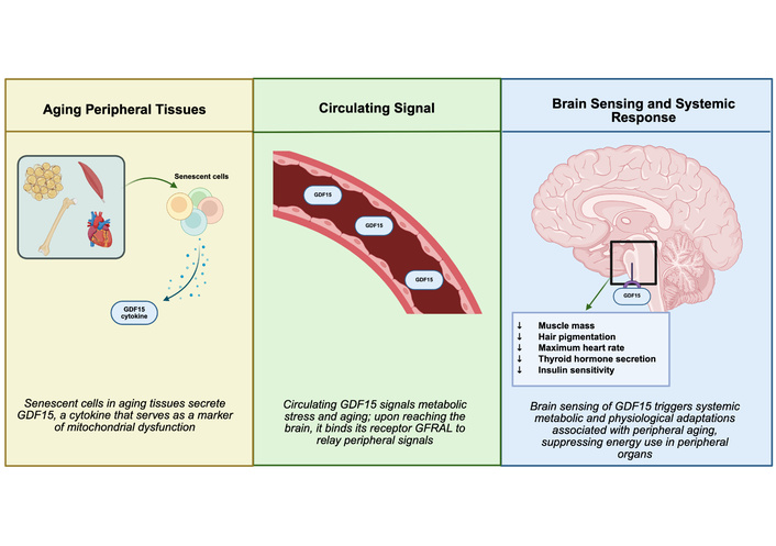
Senescent cell–brain communication through GDF15. Aging tissues accumulate senescent cells that secrete the cytokine GDF15 into the circulation. GDF15 reaches the brain, where its receptor is selectively expressed, allowing central sensing of peripheral metabolic stress. Activation of this signaling pathway triggers systemic physiological responses associated with peripheral aging, including reduced muscle mass, hair depigmentation, decreased maximal heart rate, altered thyroid hormone secretion, and changes in insulin sensitivity. GDF15: growth differentiation factor 15. Created in BioRender. Avila, J. (2026) https://BioRender.com/31x9vcu.
Consistent with this systemic regulation, recent findings show that aging neurons produce elevated levels of WD repeat and FYVE domain-containing protein 1 (WDFY1). This protein can be transported from the brain to bone via extracellular vesicles, contributing to bone–fat imbalance and osteoporosis [21].
GDF15 is a mitochondrial-stress-responsive cytokine [14] that acts as a biomarker for aging and neurodegenerative diseases, often correlating with phosphorylated tau (p-tau) in cerebrospinal fluid. Elevated GDF15 levels are associated with increased neuroinflammation, higher levels of Tau pathology, and cognitive impairment [17, 22]. Under chronic stress conditions, peripheral signals can activate the amygdala [7, 23], which in turn stimulates the hypothalamus–pituitary–adrenal (HPA) axis. This communication from the periphery involves the brainstem region known as the nucleus tractus solitarius [24], which may play a broader role in integrating signals between the brain and peripheral organs.
Not all brain regions consume energy equally. Studies measuring energy distribution across the functional connectome have shown that evolutionarily expanded regions of the human brain, particularly the cortex, require up to 67% more energy than regions mainly involved in sensory-motor processing, such as the brainstem [25]. In other words, if the brain is divided into cortex, limbic system, and brainstem, the frontal-parietal cortex is the most energy-demanding region [25].
Interestingly, tau protein—primarily expressed in neurons [26]—is also particularly abundant in cortical areas [27, 28]. This observation suggests a possible relationship between age-related changes in functional brain networks [29] and age-dependent alterations in tau levels that may contribute to neurodegenerative diseases known as tauopathies [30].
In aging neurons, tau accumulation may result from reduced energy availability for processes such as protein turnover. When tau degradation decreases, the protein accumulates and can eventually form pathological aggregates [31]. This abnormal accumulation is also associated with the presence of senescent neurons [32].
As discussed above, aging is the main risk factor for tauopathies. Therefore, effective neuroprotective strategies should aim to prevent, delay, or reverse neuronal aging.
A pioneering study by Yamanaka and Takahashi [33] demonstrated that differentiated peripheral cells can be reprogrammed into induced pluripotent stem cells (iPSCs) with embryonic-like characteristics through the expression of four transcription factors known as the Yamanaka factors (YF).
Subsequent work using mouse models showed that permanent expression of YF may promote tumor formation [34]. However, cyclic expression of YF at controlled levels promoted rejuvenation of peripheral tissues such as pancreas, spleen, skin, and muscle [35]. When neurons in culture were induced to express YF continuously, reprogramming into iPSCs did not occur [36]. However, cyclic expression of YF resulted in neuronal rejuvenation [37].
Despite these promising findings, such approaches rely on genetic manipulation in mouse models and are not easily translated into clinical therapies in humans. Therefore, efforts have been made to identify small molecules capable of reproducing similar rejuvenating effects.
In this context, the Izpisua-Belmonte laboratory described a cocktail of four compounds, including methionine (a one-carbon metabolism metabolite), that induced partial cellular reprogramming in non-neuronal cells [38]. Using a similar approach, rejuvenation of neural cells was later demonstrated [39]. However, an important improvement was achieved when methionine was replaced by folate [40].
Folate receptor is a glycosyl-phosphatidylinositol-bound membrane protein that mediates the cellular uptake of folate and the related coenzymes. Ligand binding to folate receptors stimulates their endocytic uptake by a clathrin-independent pathway [41]. Once internalized into endosomes, the acidic pH of the lysosome causes dissociation of folate; the receptor is transported to the cell nucleus [42], where it acts as a transcription factor regulating oncogenic genes and facilitating the expression of the so-called YF [43], and leading to neuronal rejuvenation [40, 42]. In addition, FRα functions via non-canonical signaling mechanisms: it physically associates with GP130 to activate the JAK-STAT3 pathway and triggers the ERK1/2 cascade [44].
Thus, neuronal rejuvenation may represent a promising strategy for neuroprotection against neurodegenerative diseases.
However, folate and its derivatives participate in many cellular pathways, and high doses may produce undesirable side effects. Consequently, researchers have searched for small molecules capable of mimicking folate activity through interaction with FRα.
A small FRα-binding peptide with functions similar to folate was first described some years ago [45]. More recently, a family of FRα-binding peptides with similar properties has been identified [46]. Importantly, these peptides can cross the BBB and may therefore be delivered through peripheral administration [46].
Figure 2 summarizes the mechanism by which these FRα-binding peptides promote neuronal rejuvenation.
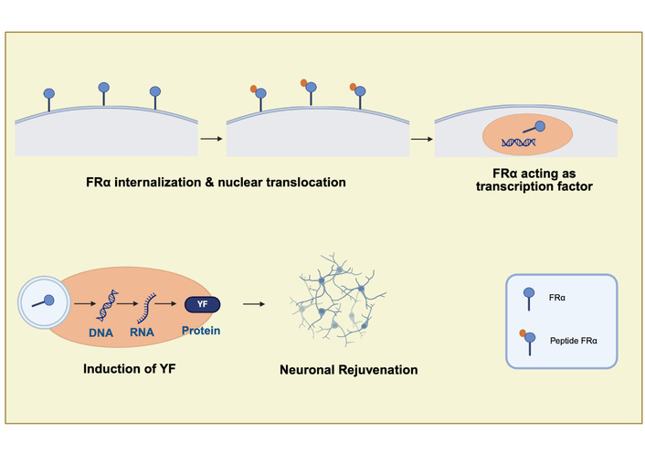
Mechanism of neuronal rejuvenation mediated by FRα-binding peptides. FRα-binding peptides interact with the FRα located at the neuronal cell membrane. Upon ligand binding, FRα undergoes internalization and translocation to the cell nucleus, where it functions as a transcription factor. This activity promotes the expression of YF, which are associated with neuronal rejuvenation and restoration of cellular youth functions. FRα: folate receptor alpha; YF: Yamanaka factors. Created in BioRender. Avila, J. (2026) https://BioRender.com/ghw2bdm.
Here we briefly discuss several therapeutic strategies currently being tested in cellular and animal models, with only a few approaches at a very early clinical stage.
As noted in the Introduction, aging is the main risk factor for neurological disorders and is strongly associated with decreased cellular energy levels due to mitochondrial dysfunction. Therefore, we will mainly focus on therapies aimed at preventing or reversing aging processes rather than lifestyle interventions such as exercise or dietary modifications [47], although such approaches may also delay aging—for example through the use of vitamin B cofactors involved in mitochondrial ATP production.
Nevertheless, it is worth briefly noting the role of lifestyle factors and circulating molecules that may regulate brain aging. Recent studies have shown that the liver-derived exercise factor GPLD1 (exerkine) targets glycosylphosphatidylinositol (GPI)-anchored proteins on the vasculature of the aging brain. Increased levels of GPLD1 have been associated with rejuvenation of the BBB and a reduction in pathological aging processes [48].
As mentioned above, GDF15 is a stress-induced cytokine that regulates metabolism, inflammation, and tissue repair, primarily by acting as a natural appetite suppressor via the GFRAL receptor. Elevated GDF15 levels are associated with various diseases, acting as a biomarker and therapeutic target for cancer cachexia, obesity, cardiovascular disease, and severe morning sickness. Thus, several clinical trials are actually ongoing (https://clinicaltrials.gov/search?term=GDF15). In addition, physical exercise promotes the secretion of irisin, a peptide derived from the FNDC5 protein, primarily released by skeletal muscle. Once in circulation, irisin can cross the BBB and has been shown to facilitate the clearance of damaged neuronal mitochondria through mitophagy. It may also induce the expression of brain-derived neurotrophic factor (BDNF). Together, these processes contribute to neuronal rejuvenation and improved brain function [49, 50].
In summary, based on these previous comments, potential therapeutic strategies include:
Maintaining cellular energy levels
Reducing tau protein accumulation (tau clearance)
Promoting neuronal rejuvenation using peptides that bind neuronal receptors such as FRα or the GDF15 receptor (GFRAL)
Eliminating senescent cells through senolytic agents
Given the rapid progress in this field, it is likely that additional strategies will emerge in the near future.
Electrical and magnetic stimulation techniques have been used for therapeutic brain modulation. Examples include electroconvulsive therapy (ECT) and transcranial magnetic stimulation (TMS), which are used to treat disorders such as depression [51, 52].
In addition, integrated brain–machine interface platforms are beginning to be applied clinically to restore sensory or motor function and to treat certain neurological disorders [53].
However, due to our limited expertise in electrophysiology and physics-based approaches, this mini-review will focus mainly on cellular and molecular therapeutic strategies.
At the molecular level, maintenance of neuronal energy homeostasis may involve intercellular mitochondrial transfer. Damaged neurons can restore their function by receiving mitochondria from neighboring glial cells through tunneling nanotubes [54].
In addition, mitochondrial transplantation—such as delivery via erythrocyte membrane encapsulation—has been successfully applied in mouse models of neurodegenerative disorders [55].
As mentioned earlier, mitochondrial dysfunction reduces cellular energy levels, impairing protein turnover and leading to the accumulation and aggregation, or even secretion, of neuronal proteins such as tau [56]. In addition, extracellular pathological tau may further exacerbate mitochondrial dysfunction [57].
Several strategies to reduce tau levels have been proposed [56]. More recent approaches include antisense oligonucleotide therapies [58], immunotherapies such as anti-tau vaccines [59], and tripartite motif containing 21 (TRIM21)-based degradation strategies [60].
Interestingly, heat exposure—resulting in elevated body temperature—has also been reported to promote tau clearance by enhancing proteostasis mechanisms [61].
Regarding tau clearance, specific cell types such as tanycytes have been proposed to mediate communication between the brain and the periphery (blood) for protein removal.
Tanycytes are specialized glial cells located in the median eminence, a region at the interface between the hypothalamus and the pituitary gland. In this strategic location, tanycytes regulate the transport of neuropeptides and hormones between the hypothalamus and the pituitary, thereby controlling their further release into the blood circulation and contributing to exchange processes between the brain (cerebrospinal fluid) and blood [62].
More recently, tanycytes have been implicated in the clearance of tau protein from the brain to the bloodstream [63]. In this context, tau can be taken up by tanycytes and subsequently transported to nearby blood vessels, facilitating its removal from the brain.
As discussed above, FRα-binding peptides capable of crossing the BBB and promoting neuronal rejuvenation may represent a promising therapeutic approach, although clinical translation will likely require further development (Figure 3).
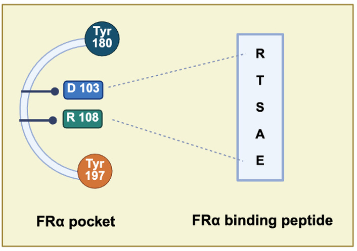
Molecular interaction between FRα and FRα-binding peptides. FRα-binding peptides, like the hexapeptide shown, interact with the FRα at the cell membrane. This interaction involves a defined molecular pocket spanning residues Tyr180 to Tyr197. Within this region, Asp103 of FRα is predicted to form electrostatic interactions with the arginine residue of the peptide, while Arg158 of FRα may interact with the glutamic acid residue of the peptide [46]. These interactions contribute to the specificity and stability of the peptide–receptor binding to facilitate the FRα transport from the cell membrane to the cell nucleus. FRα: folate receptor alpha. Created in BioRender. Avila, J. (2026) https://BioRender.com/s0472aa.
In addition, GDF15 is a cytokine associated with aging processes such as cellular senescence and mitochondrial dysfunction [14]. Although it is secreted by multiple organs, its receptor (GFRAL) is primarily expressed in the brain [20]. Identifying small molecules capable of modulating this receptor to delay brain aging represents an important future challenge.
Increasing the proportion of young versus senescent neurons may help prevent pathological brain aging.
Senescent cells are non-dividing cells characterized by DNA damage and expression of anti-apoptotic factors such as Bcl-2, PI3K, and PK13, which make them resistant to elimination. Their persistence is problematic because senescent cells secrete toxic and inflammatory factors, like GDF15, as indicated.
Senolytic drugs that selectively eliminate senescent cells—such as quercetin/dasatinib or navitoclax—have therefore been proposed as potential therapeutic agents [32].
In this review, we have briefly explored the role of aging in neurodegeneration, emphasizing the exceptionally high energy demands of both healthy and aging brains, particularly in the presence of accumulating senescent neurons.
Mitochondria play a central role in meeting these energy requirements. Notably, the three neuronal proteins highlighted in this mini-review—FRα [64], GDF15 [65] and tau [66–68]—are closely linked to mitochondrial function and dysfunction.
We propose that delaying neuronal aging or promoting the rejuvenation of adult neurons before they enter senescence may represent effective neuroprotective strategies. Finally, we have discussed several emerging therapeutic approaches aimed at preventing neuropathological aging and associated neuronal disorders.
BBB: blood–brain barrier
BDNF: brain-derived neurotrophic factor
ECT: electroconvulsive therapy
FRα: folate receptor alpha
GDF15: growth differentiation factor 15
GPI: glycosylphosphatidylinositol
HPA: hypothalamus–pituitary–adrenal
iPSCs: induced pluripotent stem cells
p-tau: phosphorylated tau
TMS: transcranial magnetic stimulation
TRIM21: tripartite motif containing 21
YF: Yamanaka factors
We acknowledge support from PTI NeurAging, Consejo Superior de Investigaciones Científicas (CSIC), Spain. We thank Nuria de la Torre for her generous assistance with the editing and preparation of the text.
JA: Conceptualization, Investigation, Writing—original draft, Writing—review & editing. FH: Conceptualization, Investigation, Writing—original draft, Writing—review & editing. Both authors read and approved the submitted version.
Jesús Avila, who is the Editorial Board Member of Exploration of Neuroprotective Therapy, had no involvement in the decision-making or the review process of this manuscript. Another author declares no conflicts of interest.
Not applicable.
Not applicable.
Not applicable.
Not applicable.
This work was supported by grants PID2024-1554470OB-I00 (JA) and PID2023-149460NB-I00 (FH) funded by MICIU/AEI/10.13039/501100011033/ and by FEDER, UE. Centro de Biología Molecular Severo Ochoa (CBM) receives an institutional grant from Fundación Ramón Areces and is a Severo Ochoa Center of Excellence (MICIN, Award CEX2021-001154-S). The funders had no role in study design, data collection and analysis, decision to publish, or preparation of the manuscript.
© The Author(s) 2026.
Open Exploration maintains a neutral stance on jurisdictional claims in published institutional affiliations and maps. All opinions expressed in this article are the personal views of the author(s) and do not represent the stance of the editorial team or the publisher.
Copyright: © The Author(s) 2026. This is an Open Access article licensed under a Creative Commons Attribution 4.0 International License (https://creativecommons.org/licenses/by/4.0/), which permits unrestricted use, sharing, adaptation, distribution and reproduction in any medium or format, for any purpose, even commercially, as long as you give appropriate credit to the original author(s) and the source, provide a link to the Creative Commons license, and indicate if changes were made.



View: 835
Download: 24
Times Cited: 0


