Review

Affiliation:
1Managed Care Department, NYC Health + Hospitals, New York, NY 10004, USA
Affiliation:
2Barts and the London School of Medicine and Dentistry, Queen Mary University of London, E1 2AD London, UK
Email: tuan.pham@qmul.ac.uk
ORCID: https://orcid.org/0000-0002-4255-5130
Explor Med. 2023;4:589–611 DOI: https://doi.org/10.37349/emed.2023.00163
Received: April 28, 2023 Accepted: June 07, 2023 Published: August 31, 2023
Academic Editor: Lindsay A. Farrer, Boston University School of Medicine, USA
Wound healing is a very dynamic and complex process as it involves the patient, wound-level parameters, as well as biological, environmental, and socioeconomic factors. Its process includes hemostasis, inflammation, proliferation, and remodeling. Evaluation of wound components such as angiogenesis, inflammation, restoration of connective tissue matrix, wound contraction, remodeling, and re-epithelization would detail the healing process. Understanding key mechanisms in the healing process is critical to wound research. Elucidating its healing complexity would enable control and optimize the processes for achieving faster healing, preventing wound complications, and undesired outcomes such as infection, periwound dermatitis and edema, hematomas, dehiscence, maceration, or scarring. Wound assessment is an essential step for selecting an appropriate treatment and evaluating the wound healing process. The use of artificial intelligence (AI) as advanced computer-assisted methods is promising for gaining insights into wound assessment and healing. As AI-based approaches have been explored for various applications in wound care and research, this paper provides an overview of recent studies exploring the application of AI and its technical developments and suitability for accurate wound assessment and prediction of wound healing. Several studies have been done across the globe, especially in North America, Europe, Oceania, and Asia. The results of these studies have shown that AI-based approaches are promising for wound assessment and prediction of wound healing. However, there are still some limitations and challenges that need to be addressed. This paper also discusses the challenges and limitations of AI-based approaches for wound assessment and prediction of wound healing. The paper concludes with a discussion of future research directions and recommendations for the use of AI-based approaches for wound assessment and prediction of wound healing.
A wound is caused by breakage or damage on the skin’s surface or some skin conditions including skin blistering disease (burn, eczema, psoriasis, or dystrophic epidermolysis bullosa). It is a result of a disruption of normal anatomic structures and function [1–3]. Wounds are classified depending on the cause, site, size, depth, mode of injury, exposure to external environment, degree of gross contamination, American Society of Anesthesiology score, severity and time frame of healing, and potential risk of infection [1, 2, 4–8]. Some common types are acute vs. chronic; open vs. closed; clean vs. contaminated; internal vs. external; superficial vs. deep; partial thickness vs. full thickness; surgical vs. traumatic; and pressure vs. diabetic ulcers [9–13].
A wound can be open or closed depending on the integrity of the skin overlying it. An injury that breaks the skin is called an open wound while a closed wound happens when there is damage to tissue under the intact skin. For example, abrasion, laceration, puncture, and avulsion are major types of open wounds while closed wounds consist of contusions, blisters, seromas, hematomas, and crush injuries.
According to the information given by the Centers for Disease Control and Prevention (CDC) in the United States [4, 7], there are four classes of wound statuses that represent the potential risk of infection: class 1 (clean), class 2 (clean-contaminated), class 3 (contaminated), and class 4 (dirty-infected). Clean wounds are those that are free from foreign materials such as dirt, feces, or blood. Contaminated wounds are those that are exposed to foreign materials. Dirty-infected wounds are those that are exposed to foreign materials and are infected. Infected wounds are those that are infected with microorganisms [14].
Internal wounds are those that are located inside the body. External wounds are those that are located outside the body. Wound site represents its location on the body. The depth of the wound determines the types of management, wound healing potential, and outcome. Its depth is classified into superficial, partial thickness, and full thickness [15]. Superficial wounds are those that affect only the epidermis. Partial thickness wounds affect the epidermis and dermis. Full thickness wounds affect the epidermis, dermis, and subcutaneous tissue.
Wound-level of severity is classified, depending on its types—penetrating wounds or blunt force trauma [16–18]. Penetrating wounds include punctured wounds; surgical wounds and incisions; thermal, chemical, or electric burns; bites and stings; gunshot wounds or other high velocity projectiles that can penetrate the body. Abrasions, lacerations, and skin tears are common examples of blunt force trauma.
Wound healing time is classified into acute, subacute, and chronic [1, 2, 19, 20]. Acute wounds heal within 3 months without complications. Subacute wounds heal within 3 to 6 months. Chronic wounds take more than 6 months to heal with some complications. Pressure injuries and diabetic ulcers are common types of chronic wounds. Wound healing time is also classified into primary, secondary, and tertiary [21]. Primary intention is the healing of a wound with minimal tissue loss. Secondary intention is the healing of a wound with significant tissue loss. Tertiary intention is the healing of a wound with delayed healing.
The body’s skin plays major roles in protection, sensation, thermoregulation, metabolism, excretion, and cosmetics [22]. Disruption to its integrity is problematic as it can interfere with its roles and functions [23, 24]. For example, a break in the skin can predispose the body to pain and infection. Inappropriate evaluation and management of wounds can have significant and direct on the patients as it can result in an emotional, physical and financial toll, bodily disfigure, psychological distress, decreased mobility, loss of productivity, severe disability, decreased quality of life, psychological distress, depression, reduced life expectancy and high morbidity, and high mortality [23–28]. Optimal wound management and healing outcomes require collaborative approach among interprofessional teams and ongoing multidisciplinary assessment, sound clinical decision-making, timely intervention, and comprehensive documentation of the wound’s progress and response to treatment [29]. Its healing process depends on the nature of the injury, etiologies, patient-specific characteristics, demographics, risk factors, prosthetic types, comorbidities, socioeconomic factors, and treatment modalities [8, 30].
The assessment of clinical wounds is challenging because of the current standard which focuses on wound itself and not its periwound area that is often accompanied by maceration, excoriation, dry skin, hyperkeratosis, dehydration, edema, undermining, rolled, callus, eczema, exudate, infection, tissue type, and wound dehiscence [31, 32]. These parameters are integral parts of wound healing. The initial clinical assessment determines the nature of the wound (i.e., acute vs. chronic wound). The process of wound healing could be anticipated well for acute wounds such as from trauma or surgery as the physiologically intact wound progresses through an orderly physiologic sequence of regular phases [33, 34]. Chronic wounds do not heal well, making it harder to predict healing processes and progress [35]. A successful clinical assessment of a wound requires a complete history, good review of systems, identification of possible risk factors and comorbidities, accurate inspection and visualization, good image analysis, and understanding of its underlying and periwound areas [36]. The heterogeneous etiology of chronic wounds and multiple factors that influence healing rates make wound healing prediction difficult and challenging [10, 30].
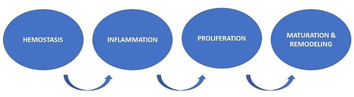
Wound healing is characterized by hemostasis, inflammatory, proliferative, maturation, and remodeling phases [37–41] (Figure 1). It is a complex process that involves both spatial and temporal synchronization of many special types of cells, cytokines, mediators, and the vascular system [4, 35]. The initial phase, hemostasis, starts immediately after the injury and lasts 5–10 min [39, 42]. Bleeding from the injury is slowed down and eventually stopped by the vasoconstriction of blood vessels. Released platelets go through a process of platelet aggregation and form a clot with fibrins that leak from injured blood vessels. The clot adheres to injured vessels and stops the bleeding and provides a scaffold for incoming inflammatory cells [43]. The inflammation phase lasts several days and includes hemostasis, chemotaxis, and increased vascular permeability [37, 40, 44]. Platelets from the initial phase release growth factors and cytokines (platelet-derived growth factors, transforming growth factor-beta, epidermal growth factor, and insulin growth factor) that are important cellular mediators that promote angiogenesis, thrombosis, and re-epithelialization [45]. The tissue inflammation activates and attracts inflammatory cells (neutrophils which are later replaced by macrophages) that assist in engulfing and digesting bacterial and cellular debris [46, 47]. Langerhans cells, dermal dendritic cells, and T cells are also activated to combat self and foreign antigens resulting in clearance of pathogens and debris and/or resolution of infection [48, 49]. Macrophages release growth factors that increase the level of fibroblasts that are essential for the next phase [50]. The proliferative phase lasts 6–21 days [41, 51]. During this phase, granulation tissue is formed and re-epithelialization and neovascularization occur. Fibroblasts released from the previous phase lay the foundation for new extracellular matrix for collagen and granulation tissue. New vessels formed within the granular tissue supply blood and nutrition to newly developed granular tissue. Fibroblasts stimulate the production of collagen giving tissue strength and structure. Myofibroblasts differentiated from some fibroblasts draw the wound margins together [52]. The dividing fibroblasts deposit extracellular matrix [53]. Wound edges and margins contract and the wound bed is covered with new epithelium resulting in a scar which serves as a rapid patch for the wound [54]. Maturation and remodeling phase is the final and longest phase of wound healing [55]. This phase lasts 1–2 years or even longer depending on a variety of mechanisms and etiologies. It is where the wound matures and achieves its maximal strength. As wound continues to contract and fibers are reorganized, remodeling occurs [19, 35]. Chronic wounds are commonly seen in immunocompromised and aging population and those with medical conditions like diabetes and vascular disease. These factors and conditions constitute the impairment in wound healing responses [56].
Image analysis enables the reconstruction of the wound and its underlying structures as well as periwound areas. The evaluation of wound healing processes can be made based on images. Artificial intelligence (AI) is an area of computer science, which has been developed since the 50s [57], aiming to mimic and automate the process of human thinking using computer algorithms. Artificial neural networks (ANNs) [58, 59] and fuzzy logic [60, 61] are among the main approaches for machine learning in AI. It is witnessed that AI has been pervading most aspects of modern technology and human life, where the development of the so-called “deep learning” [59, 62], which is a kind of ANNs, acts as a catalyst for the impact of AI.
An ANN architecture was inspired by the human brain, where brain cells (neurons) form an interconnected network to process complex information. An ANN consists of a few connected layers with artificial neurons, which are also called nodes, created by mathematical algorithms to learn on training data. A trained ANN is capable of making inference or prediction when it is encountered with similarly new information. A deep neural network or deep learning network has many connected layers with millions of artificial neurons. Such a depth of layers of a network empowers the computer algorithm to learn detailed and hidden information from training data. The deep learning model then becomes very useful for detecting objects or predicting outcomes that cannot be performed competitively by human experts.
AI-enabled image processing methods have been reportedly used as potential computerized tools for accurate wound assessment as their methods are objective and noninvasive [63]. In recent years, multiple predictive methods have been proposed, applying different data processing schemes and prediction algorithms, machine learning, and statistical models, which outperform human wound experts. Deep learning has gained popularity and led to breakthrough results in wound care. This article attempts to update reviews on developments and applications of AI methods for wound assessment and healing prediction.
Wounds have been called ‘the silent epidemic’ as they have a high prevalence and economic burden in the world. It has been shown in certain parts of the world, there are up to 4 people with at least one wound per 1,000 population [64–67]. Many of these wounds become chronic and are estimated to account for up to 5.5% of total health system costs in Wales [68]. The United States alone spends between $28.1 and $96.8 billion to manage chronic wounds and associated complications [69, 70]. Nonhealing wounds, surgical incisions, and trauma and burns cost the United States $50 billion, $12 billion, and $7.5 billion annually, respectively [71, 72]. Healing prediction has a direct impact on wound management and treatment and is very challenging due to the multifactorial complex nature of wounds including wound-, patient-, environmental- and socioeconomic-levels and etiologies. Implementing a complex computerized system thinking could revolutionize medicine by improving diagnostic accuracy, increasing efficiency, improving workflow, decreasing cost, improving treatment options, and shortening healing time.
Cross et al. [73] introduced an AI approach to risk profiling in the prevention and treatment of chronic wounds. The study primarily focuses on risk profiling, which involves identifying individuals at higher risk of developing chronic wounds and tailoring preventive and treatment strategies accordingly. The researchers employed various AI techniques including machine learning algorithms and data mining methods to develop the models which analyze demographic information, medical history, lifestyle factors, wound characteristics, and other relevant variables. The output of the model involves risk scores or probabilities indicating the likelihood of an individual developing chronic wounds. These scores would aid clinicians in identifying high-risk patients who may benefit from the targeted preventive measure.
The authors discussed the potential benefits of using AI in risk profiling for chronic wounds and provided examples of how AI could be used to identify patients who are at risk of developing chronic wounds or to predict the likelihood of wound healing. The authors concluded that AI which is a transformative technology could have immense potential in wound care. For example, the models may provide insights into the factors contributing to the risk of chronic wounds, assisting healthcare professionals in designing personalized interventions and treatment plans. There are several advantages in this study. First, it can enable earlier identification of patients who are at risk of developing chronic wounds, which can allow for timely intervention and potentially prevent the development and potential complications of these wounds. Second, it can enable more accurate predictions of wound healing, which can help clinicians tailor treatment plans to individual patients and improve outcomes. Third, it can help identify patterns and trends in wound healing that may not be apparent to human observers, which can help guide the development of new treatment strategies and modalities. Potential limitations and challenges include the need for large and diverse datasets to train and validate AI algorithms, the potential for bias in the data used to train the algorithms, and the need for careful validation and testing of the algorithms in clinical settings. Additionally, the use of AI in clinical decision-making raises ethical and legal concerns that will need to be addressed.
At the molecular level, composition of tissue types (epithelial, granulation, slough, and eschar) within a wound is a useful indicator of its healing progression. Accurate estimation of tissue composition allows clinicians to choose appropriate dressing, identify wounds at risk of not healing, timely refer patients to specialists, optimize wound care and healing outcomes, and tailor treatment to the patient’s condition. Clinicians depend on the naked eye to estimate wound tissue proportion exhibit considerable variability. Howell et al. [74] discussed the development of a method for the clinical evaluation of AI-based digital wound assessment tools. The researchers developed a framework for evaluating AI-based wound assessment tools using a combination of clinical and technical validation. The clinical validation included assessing the accuracy, consistency, and usability of the tool in a clinical setting, while the technical validation included assessing the performance of the AI algorithm and its ability to generalize to new data. The inputs of the AI models revolve around digital images of wounds. These images serve as the primary input data for the AI algorithms. Features extracted from the images may include parameters such as wound size, depth, color, texture, presence of necrotic tissue, granulation tissue, and signs of infection. These features are crucial for accurately assessing and diagnosing wounds. The AI models’ outputs would involve automated wound assessment based on the digital images which include classification of wound types, estimation of wound severity or stage, identification of complications, and tracking changes in wound healing progress over time. The outputs aim to provide clinicians with objective and standardized information to aid in treatment planning and monitoring. The authors showed in their study that AI-based wound annotation/assessment algorithm performed accurate assessment of wound area and percentage of granulation tissue (PGT) similarly to human wound specialists. The study has some potential advantages. First, it provided a standardized framework for evaluating AI-based wound assessment tools, which can help ensure that such tools are accurate, reliable, and usable in clinical settings. Second, the framework can help identify the strengths and limitations of different AI algorithms and guide the development of new algorithms that were better suited for wound assessment. Third, the framework can help accelerate the translation of AI-based wound assessment tools from research to clinical practice. However, it also has some limitations. First, the framework was still in the development stage and has not been fully validated in a clinical setting. Second, the framework focused on the evaluation of AI-based wound assessment tools and may not be applicable to other types of AI-based medical tools. Third, the framework did not address the ethical and regulatory issues associated with the use of AI in clinical practice, which would need to be addressed separately.
Chairat et al. [75] discussed the use of AI-assisted assessment of wound tissue using images taken with a smartphone. The authors collected a dataset of 31 wound images taken at different time points from 20 patients over 18 years of age with acute and chronic wounds who were attending the wound clinic of Songklanagarind Hospital. The dataset was used for training and testing the AI algorithm for segmenting a wound area, an epithelialization area, granulation tissue, and necrotic tissue. The wound area and composition on the images were annotated by three physicians with more than ten years of experience. Deep learning models were then developed to mimic what the physicians did on the images. The researchers examined two network variants, U-Net with EfficientNet and U-Net with MobileNetV2, on wound images with a size of 1,024 pixels × 1,024 pixels. The images were captured using a smartphone camera and included a variety of wound types, such as pressure ulcers, diabetic ulcers, and surgical wounds. AI techniques employed in the study may include computer vision techniques and image processing algorithms that can be used to extract features from the images and classify them into different categories. The inputs of the AI system consist of images captured with a smartphone. The study emphasizes the need for automatic color and measurement calibration, indicating that color correction and scaling are important components of input preprocessing. By calibrating the color and measurements, the AI system can account for potential variations caused by different smartphone cameras or lighting conditions, ensuring more accurate and standardized assessments. The outputs of the AI-assisted system would involve the assessment and analysis of wound tissue based on smartphone images. Outputs, which may include features such as wound size, color analysis, tissue characteristics (e.g., wound area, epithelialization area, necrotic tissue, granulation tissue), and other relevant wound parameters, aim to provide clinicians with objective and reliable information to support wound assessment and treatment decisions.
The author concluded that the use of an AI approach for automatic wound assessment resulted in efficient and accurate annotation of wound areas and compositions. This study has several potential advantages. First, it can enable quick and accurate assessment of wounds without the need for specialized equipment or expertise. Second, it can improve the consistency and reliability of wound assessments, which are important for tracking the progress of healing over time. Third, the use of a smartphone camera allows for remote monitoring of wounds, which can be especially useful for patients who are unable to travel to a healthcare facility for regular wound assessments. However, this study also has some limitations. First, the dataset used for training and testing the AI algorithm is relatively small, which may limit its generalizability to a larger population. Second, the study had mostly epithelialization area labels and granulation tissue labels and only a few necrotic tissue labels. This imbalance of class labels would affect the performance of the wound segmentation model. The model would have a small number of examples to learn so would not make accurate prediction. Third, the study did not assess the depth of the wound and the feasibility of using the AI algorithm in a clinical setting, which would require additional testing and validation.
Carrión et al. [76] developed a deep learning-based image analysis pipeline to evaluate and track wound size with high fidelity results on unseen data with minimal human intervention. The algorithm was trained and tested using a dataset of 256 wound images of wound inflicted lab mice. The study focuses on utilizing deep learning algorithms for the automatic detection and size estimation of wounds. The study aims to develop an AI-based system that can accurately identify wounds and estimate their size. The deep learning algorithms implemented in the study may include convolutional neural networks (CNNs) or similar architectures. Images containing wounds serve as the primary input data for the AI algorithms. The images were likely to be digital photographs or scans of wounds, capturing the visual appearance of the wound area. The outputs of the deep learning models would include two main components: wound detection and size estimation. The models would generate bounding boxes or segmentation masks to indicate the location and extent of the wound within the input images. Additionally, the size of the wound (i.e., length, width, or area) would be estimated. These outputs provide clinicians with objective measurements and visual representations of the wounds for accurate assessment and monitoring. The advantages of this study include the development of a reliable and accurate solution for wound detection and size estimation, which could potentially improve wound assessment and management. The study also demonstrated the feasibility and effectiveness of using deep learning algorithms for wound image analysis with minimal human intervention. The study provided critical information such as wound closure percentages and wound size for further evaluating the wounds. However, the study has several limitations, including the relatively small sample size and the limited diversity of wound types. Further validation with larger and more diverse datasets was needed to evaluate the generalizability and clinical usefulness of the algorithm. Additionally, there was a discrepancy between the wound size estimated by the algorithm and the actual wound size because human annotators considered the previous/next images when annotating the wound edge (temporal aspect) while the model did not. Human annotators were better at dealing with some of the challenges like occlusion and blur than the model. For future work, a network should be built to incorporate contextual information such as time and specific color/texture variations to predict surface area closer to what experts measure.
Ramachandram et al. [77] developed and evaluated a CNN, which is a deep learning network, for fully automated wound tissue segmentation using a mobile device. Digital images of wounds captured by mobile devices serve as the primary input data for AI algorithms. The images are likely to be obtained using smartphones or similar mobile devices, which makes the system more accessible and convenient for healthcare providers. The outputs of the deep learning models would involve the segmentation of wound tissue within the input images. The models would generate pixel-level masks that delineate different types of wound tissue, such as necrotic tissue, granulation tissue, and healthy tissue. These segmentation masks provide clinicians with detailed information about the spatial distribution and extent of different wound components, supporting accurate wound assessment and treatment planning. The study included a dataset of 58 anonymized wound images of various types of chronic wounds from Swift Medical’s Wound Database. Five wound clinicians labeled 4 different tissue types (epithelial, granulation, slough, and eschar) within the wound bed at 1-week intervals using a browser-based image annotation tool. Two deep CNN architectures were developed for wound segmentation and tissue segmentation and were used in sequence in the workflow. These models were trained using 465,187 and 17,000 image-label pairs, respectively. The algorithm provided objective wound tissue identification and measurement to assist clinicians in documenting the wound accurately. The advantages of this study include the development of a user-friendly, portable, and cost-effective solution for wound tissue segmentation that could potentially improve wound assessment and management. The study also demonstrated the feasibility and effectiveness of using deep learning algorithms on mobile devices for medical image analysis. The authors acknowledged challenges in labeling epithelialization within the wound bed and the distinction between epithelialization and epithelial tissue would lead to relatively poor performance of the model for epithelial tissue. There exist other possible limitations. First, the study’s cohort size and participant demographics were not specified, limiting the ability to assess the representativeness of the sample. A larger and more diverse cohort would have enhanced the study’s external validity and generalizability of the findings. Second, the dataset used to train the deep learning model may not have included a comprehensive representation of all possible wound types, sizes, and locations. This could limit the model’s ability to accurately segment wound tissues in scenarios that differ from those present in the training dataset. Consequently, the performance of the automated segmentation method may vary when applied to different types of wounds not included in the study. Third, the absence of a comparison with other existing methods or benchmarks for wound tissue segmentation would limit some understanding of the strengths and weaknesses of the proposed method in relation to existing approaches. While the study demonstrated the feasibility and effectiveness of the deep learning approach on mobile devices, it did not compare the performance of their method with alternative approaches or state-of-the-art techniques for wound tissue segmentation. Fourth, the study did not provide detailed information about potential biases in the dataset or the steps taken to mitigate them. Biases in the data used for training the deep learning model can influence its performance and generalizability. Addressing and reporting on data biases is crucial for ensuring the reliability and applicability of the automated segmentation method. Finally, the study also did not evaluate the impact of the algorithm on wound healing outcomes, which should be investigated in future research. Considering these limitations, further research and validation on larger and more diverse datasets are necessary to confirm the effectiveness and robustness of the proposed automated wound tissue segmentation method.
Barakat-Johnson et al. [78] studied the usability and effectiveness of an AI application for objective wound assessment and management during the COVID-19 pandemic. Virtual care has been quite convenient especially during the COVID-19 pandemic as it facilitated remote patient monitoring, increased patient adherence, enhanced efficiency, reduced patient travel time, allowed shared plans among providers, and resulted in optimal wound care. The study used retrospective data collected from patients with wounds who were assessed using the AI mobile application (app) compared to those who were assessed using traditional methods. The inputs for the AI app would be digital images of wounds taken by patients or healthcare providers using smartphones or other devices. The outputs of the AI app would involve the assessment and management recommendations for the wounds. The app, which utilizes the combination of computer vision techniques and machine learning algorithms, would analyze the uploaded images using AI algorithms to identify various wound characteristics, such as wound type, size, depth, color, and presence of necrotic or granulation tissue. Based on this analysis, the app would provide recommendations for appropriate wound management strategies, such as wound dressing, topical treatments, or referral to a specialist. These outputs aim to improve the accuracy and efficiency of wound assessment and guide healthcare providers in making informed treatment decisions. The study provided valuable evidence of the effectiveness and acceptability of an AI app in improving wound assessment and management during the COVID-19 pandemic. The study had some advantages. First, it demonstrated that the AI app improved the accuracy and consistency of wound assessment and management compared to traditional methods. Second, the study showed that the use of the AI app reduced the need for face-to-face consultations, which is particularly beneficial during the COVID-19 pandemic when remote consultations are encouraged. Third, the study provided evidence that the AI app was easy to use and was accepted by both patients and healthcare professionals. There are some disadvantages of this study. First, the study was conducted in a single center, which may limit the generalizability of the findings to other settings. Second, the study used retrospective data, which may be subject to bias and confounding. Third, the study did not evaluate the long-term outcomes of using the AI app, such as wound healing rates and patient satisfaction.
Berezo et al. [79] developed machine learning models to identify patients at risk of having wounds not healed within 4, 8, and 12 weeks from the start of treatment using electronic health record (EHR). The models were trained on three extensive datasets comprising a total of 1,220,576 wounds. These datasets included 187 factors that detailed patient information like demographics, underlying health conditions, and wound attributes. The information encompassed diverse patient characteristics, clinical data, and wound-related specifics, such as age, gender, wound size, duration, location, and type. Additionally, the dataset contained the healing duration for each wound. The machine learning models were fed inputs including pertinent features related to chronic wounds, like size, depth, location, infection presence, patient traits (age, gender, health conditions), and potentially other clinical factors. These features serve as input data for the machine learning models to learn patterns and relationships that can predict the healing time. The outputs of the machine learning models would involve the predicted healing time for a given chronic wound. The models would analyze the input features and generate an estimated timeframe for the wound to heal. The study demonstrates that accurately predicted chronic wound healing time by machine learning may improve treatment decisions and patient outcomes as the predicted healing time can assist healthcare providers in planning appropriate treatment strategies, setting expectations with patients, and allocating resources effectively. The authors acknowledge advantages and disadvantages of the study. This study offered a new perspective on the use of machine learning in wound care as it has several advantages. First, machine learning techniques can process large amounts of data quickly and accurately, making it possible to identify patterns and predict outcomes that might not be apparent to humans. Second, the study used a relatively large dataset, which increased the generalizability of the results and reduced the risk of overfitting. Third, predicting wound healing time can help healthcare providers plan treatment strategies and set realistic expectations for patients, potentially improving patient outcomes, and reducing costs. There are some potential disadvantages of this study. Even with a comprehensive dataset that included a wide range of patient demographics, clinical information, and wound-related characteristics, there might still be some unaccounted or unmeasured factors that could influence wound healing outcomes. These hidden factors could lead to incomplete or inaccurate predictions by the machine learning models, as they might not capture the full complexity of each individual case. The study also did not include any information on the type of wound dressing used, which may have an impact on wound healing time. The study did not include certain wound-related factors, such as wound depth and infection status, which may impact healing time. The study did not compare the machine learning model to traditional regression models, which would have helped to establish the superiority of the machine learning approach.
Cho et al. [80] developed a prediction model to predict healing rates of chronic wounds within 12 weeks, resulting in better quality of care. The authors analyzed electronic medical records (EMRs) for 620,356 chronic wounds of various etiologies in 261,398 patients from 532 wound care clinics in the United States. The dataset included various patient demographic, clinical, and wound-related characteristics, such as age, gender, comorbidities, wound size, duration, location, and type. The dataset also included the time it took for each wound to heal completely or reach 12 weeks without healing. Their study using real-world data from EMRs demonstrated that wound-level characteristics (location, area, depth, and etiology) were more powerful predictors than patient-level parameters such as demographics and comorbidities. Considering the objective of predicting wound healing, various machine learning algorithms including the logistic and classification tree models were utilized for the study. The inputs for the AI models would consist of relevant features associated with chronic wounds such as wound characteristics (size, depth, location, etiology), patient demographics (age, gender), comorbidities, wound treatments, and possibly other clinical variables. These features served as input data for the AI models to learn patterns and relationships that could predict the likelihood of wound healing within the specified 12-week timeframe. The outputs of the AI models would involve the predicted probability or likelihood of wound healing within 12 weeks. The models would analyze the input features and generate a probability score or a binary prediction indicating whether the wound was likely to heal within the specified timeframe. This provides important insights into the development of a predictive model for chronic wound healing as the accurate prediction can assist healthcare providers in assessing the potential outcome of a chronic wound and aid in treatment decision-making. The resulting severity adjustment model can become the basis for applications like quality measure development, research into clinical practice, and performance-based payment.
The study clearly had good advantages. First, a well-established statistical approach (Cox proportional hazards regression) was used to develop the predictive model, which has been widely used in medical research. Second, the study used a relatively large dataset, which increases the generalizability of the results and reduces the risk of overfitting. Third, the model was able to accurately predict wound healing within 12 weeks, which is a clinically relevant timeframe. The study also had several limitations. First, the study was based on retrospective data from a single center, which limits its generalizability to other settings and populations. Second, the study did not include certain wound-related factors, such as infection status, which may impact healing time. Third, the model may not be suitable for predicting healing beyond 12 weeks, as it was not validated for longer follow-up periods.
Ngo et al. [81] developed a computerized prediction model using thermal images to predict healing for venous leg ulcers (VLU) assessed in home settings. The researchers gathered data from 56 patients who had VLU over a span of 12 weeks. They examined 64 thermal images of these ulcers, where 17 wounds had healed by the end of the 12th week, while 47 wounds remained unhealed. The data set contained a variety of information about the patients, their health, and the characteristics of the wounds, including details like age, gender, and the size of the ulcers at the start of the study.
Thermal images of the wounds were taken in the patients’ homes and were marked as either healed or unhealed when checked after 12 weeks. The recordings were made in diverse environments and under different conditions. The dataset used in the study included sets of 19 texture features, which were obtained from the images. To simplify the data, they used a technique called principal component analysis (PCA).
The data were grouped based on the 12-week post-presentation healing label: healed and unhealed. The outputs of the AI models would involve the predicted probability or likelihood of healing for VLU. The models would analyze the input features and generate a probability score or a binary prediction indicating whether the ulcer was likely to heal. The top three textural features that provided the most significant differences were contrast, cluster prominence, and inverse different moment normalized. The authors demonstrated that the first three principal components of the textural features from one timepoint could be used as an input to a Bayesian neural network to discriminate between healed and unhealed wounds in the first assessment at week 0 accurately. The study used an optimal Bayesian neural network for the task of classification. This non-contact method, incorporating machine learning, can provide a computerized prediction of this delay in the first assessment (week 0) in participants’ homes compared to the current method that is able to do this in 3rd week and requires contact digital planimetry. This prediction can provide clinicians with valuable information to guide treatment decisions, set expectations with patients, and allocate appropriate resources. Wound management requires detailed evaluation and several follow-up visits due to complex and dynamic processes involved in wound healing. At each clinical encounter, a wound must be visualized and inspected carefully by a clinician to make diagnosis and prognosis, assess the effectiveness of treatment, and predict healing time and outcomes accurately. Virtual care has been made available due to the recent development of AI-based systems where many wound images could be captured, stored, and transmitted to wound specialists for evaluation and interpretation of the wound healing process. The study has several advantages. First, the study used a well-established machine learning approach to develop the predictive model, which has been widely used in medical research. Second, it was a noninvasive and noncontact method that would eliminate the risk of infection. It can support the early implementation of appropriate management to enable improved healing trajectories. Third, the study’s algorithm could result in a good area under the curve (AUC) value. The model was able to accurately predict wound healing, with a sensitivity and specificity of 78.57% and 60.00%, respectively, which is a clinically relevant outcome. The study also had some limitations due to its small number of subjects and lack of wound-related factors, such as wound depth and infection status, which may impact healing time. Controlled lighting and ambient temperature will be required for thermal image-based wound assessment if future studies involve the change of the images over time. The model may not be suitable for predicting healing beyond the studied period, as it was not validated for longer follow-up periods.
Anisuzzaman et al. [63] aimed to provide a systemic review of studies that have used imaged-based AI in wound assessment. The authors conducted a systematic review of studies that have used image-based AI in wound assessment. The studies included various wound types, such as pressure ulcers, diabetic foot ulcers, and VLU, and used various imaging techniques, such as digital photography, thermography, and hyperspectral imaging. The inputs for the AI techniques reviewed in this study would consist of digital images of wounds which serve as the primary input data for the AI algorithms used in image-based wound assessment. The specific details regarding the resolution, color channels, or preprocessing techniques applied to the input images would vary across the different studies reviewed. The outputs of the AI techniques vary depending on the specific techniques employed in the original studies reviewed by this study. However, in the context of wound assessment, the outputs typically involved the analysis and interpretation of the wound images such as wound segmentation, classification of wound types, measurement of wound dimensions, identification of wound characteristics, and prediction of wound healing outcomes. The review offered important insights into the current state of research on image-based AI in wound assessment. The authors concluded that AI-based digital platforms could play a significant role in delivering data-driven care to patients with debilitating chronic wounds. There are advantages of the study. First, the study provided a comprehensive overview of the current state of research on image-based AI in wound assessment, which can guide future research and clinical practice. Second, the study highlighted the potential of image-based AI in improving the accuracy and efficiency of wound assessment, which can lead to better wound management and outcomes. Third, the study identified various challenges and limitations of using image-based AI in wound assessment, such as the need for high-quality images, limited availability of training data, and potential bias in the AI algorithms. The authors clearly acknowledged the limitation of the study. First, the study was based on a systematic review of published studies, which may have variability in terms of quality and methodology, limiting its generalizability to other settings and populations. Second, the study did not provide any new data or insights into the use of image-based AI in wound assessment. Third, the study did not provide a meta-analysis of the results of the reviewed studies.
Tehsin et al. [82] presented a brief and impressive literature review on the potential contribution of AI in diabetic wound management to improve medical services for diabetic wound patients. The authors reviewed the diagnostic and treatment research under the umbrella of AI and computational science, for diabetic wound healing. The studies included various AI techniques, such as machine learning, deep learning, and computer vision, and used various data sources, such as patient data, wound images, and EHRs. This review is focused on existing and potential contribution of AI to improve medical services for diabetic wound patients. The article also discussed the future directions for the betterment of the field that can lead to facilitate both, clinicians, and patients. Their study offered important insights into the current state of research on and potential of AI in diabetic wound management.
They concluded that AI-based digital platforms could play a significant role in delivering data-driven care to patients with debilitating chronic wounds. There are advantages of this review. First, the review provided a comprehensive overview of the current state of research on the use of AI in diabetic wound management, which can guide future research and clinical practice. Second, the study highlighted the potential of AI in improving the accuracy and efficiency of diabetic wound detection, diagnosis, and management. Detecting wounds and its complications at early stage to prevent future amputation, predicting wound healing time, providing personalized wound care, improving quality of life, and saving healthcare costs are some examples that could be achieved by using AI. Third, the study identified various challenges and limitations of using AI in diabetic wound management, such as difficulty in acquiring medical data due to legal and privacy issues, the need for high-quality, clean, and reliable data, the risk of bias and overfitting in AI algorithms, and the need for validation in clinical practice. There are also some limitations in this study. First, the study was a mini-review and did not provide an in-depth analysis of any specific study or technique. Second, the study did not provide any new data or insights into the use of AI in diabetic wound management. Third, the study did not provide a quantitative analysis or meta-analysis of the results of the reviewed studies.
Robnik-Sikonja et al. [83] aimed to evaluate prognostic factors for wound healing and develop a prediction model using machine learning technique. The researchers analyzed the data concerning patients, wounds, and their treatment of a controlled clinical study of chronic wound healing acceleration as a result of electrical stimulation.
A total of 266 patients with 390 wounds were recorded in computer database but some of patient and wound data were missing so only 214 patients and 300 wound cases were finally used for the analysis. Among 300 wound cases, in only 174 cases, the observation period lasted until the completed wound closure and was shorter in 126 cases. The study involved four treatment groups: a conventional conservative treatment, sham treatment, biphasic pulsed current, and direct current electrical stimulation. Data was collected over 10 years and suffices for an analysis with machine learning methods. In the study, they investigated wound and patient attributes affecting chronic wound healing. They applied these attributes to attribute estimation algorithms to rank the prognostic factors to predict wound healing rate. The information gained from such algorithms helps formulate effective treatment decisions and needed resources for wound patients especially those with poor prognosis. Using the attribute estimation algorithms ReliefF and RReliefF, the researchers obtained a ranking of the prognostic factors which was comprehensible to experts. The specific AI techniques implemented in the study included machine learning algorithms, specifically regression and classification trees to build models for prediction of the wound healing rate. Decision trees are a type of supervised learning algorithm that can be used for classification and regression tasks. They created a tree-like model of decisions and their possible consequences based on input features. The inputs for the models in this study would consist of various factors or features related to wound healing. Factors such as wound characteristics (size, location, type), patient demographics (age, gender), comorbidities, and possibly other clinical variables served as input data for the decision tree models to learn the relationships between the input factors and the predicted wound healing outcomes. The outputs of the models would involve predictions or classifications related to wound healing. For example, the models could predict the probability or likelihood of wound healing within a specific timeframe or classify the wound healing outcome as successful or unsuccessful. The decision tree models aimed to provide comprehensible evaluations of the factors influencing wound healing, enabling clinicians to understand and interpret the decision-making process. The obtained results are encouraging and may form a basis for an expert system for the chronic wound healing rate prediction. If the wound healing rate is known, then the provided information can help to formulate the appropriate treatment decisions and orient resources toward individuals with poor prognosis.
There are some advantages of the study. First, the study used machine learning techniques to develop a prediction model for wound healing, which can improve the accuracy and efficiency of wound management. Second, the study identified several prognostic factors for wound healing, such as age, wound duration, and wound size, which can guide clinical decision-making. Third, the study used a comprehensible decision tree algorithm, which can provide interpretable and transparent predictions for clinicians. This study also had some limitations due to its small sample size of patients with chronic venous ulcers, which may limit generalizability of the findings. Secondly, the study did not include other types of wounds, such as pressure ulcers or diabetic foot ulcers, which may have different prognostic factors and wound healing patterns. Third, the study did not validate the prediction model on an independent dataset or in clinical practice, which may limit the reliability and effectiveness of the model.
Gupta et al. [84] developed an AI based prognostic model to objectively quantify wound healing and predict healing outcomes. The authors used deep learning-based objective features derived from wound images, pertaining to wound area and tissue amounts to train prognostic models that outperformed currently existing wound assessment tools such as Pressure Ulcer Scale for Healing (PUSH) and Bates-Jensen Wound Assessment Tool (BWAT). Their study included a dataset consisting of 2,151,185 wound evaluations and images derived from 201,463 wounds and 98,407 patients from 2,361 skilled nursing facilities (SNFs) and 141 home healthcare facilities (HHFs), spread across North America. The dataset focused on four frequently encountered wound types including pressure injuries, venous ulcers, diabetic wounds, and arterial ulcers. The inputs for the AI models in this study would involve various features and data points associated with the wounds being assessed. Subjective features included tissue type, exudate amount, exudate type, edges, edema, induration, wound area, normalized area, log of normalized area, wound location, wound type, clinical setting, age groups, and sex. These features served as input data for the AI models to learn patterns and relationships that can predict the quantification and progression of wound healing. Objective features were extracted directly from the images taken during wound evaluations. Deep learning-based models were employed for objective determination of wound region and quantification of different tissue types within the wound region. The segmented wound and tissue regions were subsequently used to objectively determine the wound area and relative amounts of different tissue types within the wound bed.
AutoTrace model which consisted of approximately 3.5 million parameters, was based on a deep convolutional encoder-decoder neural network architecture and was used for wound segmentation. The encoder block allowed the model to extract features, whereas the decoder block produced a wound segmentation mask from the learned features. AutoTrace model was trained using more than 400,000 image-label pairs with wound region labels determined by clinicians. The model was tested on 2,000 image-label pairs of four wound types. AutoTissue model, which used EfficientNetB0 architecture as the encoder, was used for wound tissue segmentation. The decoder was made up of 4 blocks, each consisting of a single two-dimensional (2-D) bilinear upsampling layer followed by 2 depth-wise convolutional layers. AutoTissue model consisted of approximately 3.8 million parameters and was trained using 17,000 anonymized wound images labeled by trained labelers and curated by wound clinicians. The model, which was running the objective feature extractor sequentially on a set of 2.1 million images using an instance based on a single central processing unit, segments detected wound regions into 5 separate tissue categories including epithelial, granulation, slough, eschar, and other, the latter of which included mostly healthy tissue and the HealX calibrant sticker.
The outputs of the AI models would involve objective measurements and predictions related to wound healing. This could include quantifying the rate of healing, estimating the time to complete healing, or predicting the likelihood of successful healing. The AI models aim to provide a quantitative assessment of wound healing, enabling healthcare providers to track progress, make informed decisions, and tailor treatment strategies based on the predicted outcomes. This study demonstrated the feasibility of developing prognostic tools based on the objective information derived from wound imaging. The feature importance investigation equips researchers with a guide to develop a more accurate, objective prognostic tool that can aid in accurate and faster detection of high-risk wounds, assisting clinicians in better decision-making and improving outcomes for patients.
The study had advantages due to a large and diverse dataset, including 98,407 patients with different types of chronic wounds, which can improve the generalizability and applicability of the model. Also, the study used high-resolution wound images, which can provide more accurate and objective measurements of wound size and healing progress. Lastly, the study used machine learning techniques, including CNNs and support vector regression, which can improve the accuracy and reliability of the prognostic model. There are also some limitations in the study. First, the study did not consider the impact of prognostic indices on clinical interventions, the study may fail to evaluate the effectiveness of different treatment strategies in relation to patient prognosis. This can limit the understanding of which interventions were most beneficial for specific prognostic groups, potentially leading to suboptimal treatment decisions. Second, neglecting the influence of these indices on clinical interventions poses a difficult in offering customized treatment guidance that matches each patient’s prognostic outlook. This limitation obstructs the optimization of treatment strategies and results for patients with varying prognostic attributes. Furthermore, overlooking the effects of these indices on clinical interventions can lead to missed chances to categorize patients by their prognosis. This might result in a uniform treatment approach that disregards risk differences, potentially causing extensive of inadequate treatment for specific patient subsets. Third, the study’s findings may have limited generalizability to real-world clinical practice. The lack of understanding of how prognostic indices influenced treatment decisions can undermine the applicability and relevance of the study’s results in broader healthcare contexts. In short, not considering the impact of prognostic indices on clinical interventions can result in incomplete treatment evaluations, lack of personalized recommendations, missed risk stratification opportunities, and limited generalizability of study findings. It is essential to integrate prognostic information with clinical interventions to fully understand the implications for patient management and optimize treatment outcomes.
Wang et al. [85] developed an immune profiling strategy for stratifying peri-implantitis patients with unique microbial colonization and clinical outcome by using a machine learning algorithm, namely Fast And Robust DEconvolution of Expression Profiles (FARDEEP), to analyze tissue samples from patients with peri-implantitis who were receiving reconstruction therapy. The study used data from 24 patients with at least one dental implant diagnosed with peri-implantitis, including demographic information, clinical parameters, microbial profiling data, and immune profiling data. The immune profiling data was generated using mass cytometry and included the expression levels of immune cell markers and cytokines. The algorithm helped them discover that immune signatures showed untapped potential in improving the risk-grading for peri-implantitis. It provides important insights into the use of machine learning and immune profiling for stratifying peri-implantitis patients with unique microbial colonization and clinical outcomes. The advantages of the study include the use of a machine learning algorithm to analyze immune profiling data, which can improve the accuracy and efficiency of immune profiling. The study used a multi-omics approach, including microbial and immune profiling data, which can provide a more comprehensive understanding of the pathogenesis of peri-implantitis. The researchers used a robust outlier-resistant machine learning algorithms for immune deconvolution which can improve the accuracy and reliability of the immune profiling strategy. The inputs for the machine learning models in this study would consist of immune profiling data obtained from peri-implantitis patients. Immune profiling data could include measurements of various immune markers, cytokines, chemokines, or other relevant immune-related parameters. These features served as input data for the machine learning models to learn the patterns and associations between immune profiles and clinical outcomes. The outputs of the machine learning models would involve the stratification of patients into distinct groups based on their immune profiles. This stratification aimed to identify subgroups of peri-implantitis patients with different microbial colonization patterns and clinical outcomes. By using machine learning techniques, the study sought to uncover hidden patterns or relationships within the immune profiling data that may be associated with specific clinical characteristics or treatment responses. The study has several advantages. First, it identified unique immune and microbial signatures associated with different clinical outcomes, which can facilitate personalized treatment strategies for peri-implantitis patients. Secondly, the study used a comprehensive immune profiling strategy, which can provide more accurate and objective measurements of immune cell markers and cytokines. There are also some limitations in the study. The study was conducted on a relatively small sample size of patients, which may limit the statistical power and generalizability of the findings. Secondly, the study did not validate the immune profiling strategy on an independent dataset or in clinical practice, which may limit the reliability and effectiveness of the strategy. Third, the study did not include other clinical variables, such as smoking, oral hygiene, comorbidities, or medications, which may influence the pathogenesis and clinical outcomes of peri-implantitis. Lastly, the study did not include other types of chronic wounds, such as diabetic foot ulcers or pressure ulcers, which may have different immune signatures and clinical outcomes.
Dabas et al. [86] conducted a scoping review of the literature on the application of AI methodologies to chronic wound care and management. The authors reviewed a total of 75 studies based on type of utilized AI methodology, wound type, medical record/database configuration, and research goal. These techniques could include machine learning algorithms, deep learning models, natural language processing (NLP), computer vision, or a combination of these approaches. The inputs for the AI models would vary depending on the specific studies reviewed. The input data could encompass various types of information related to chronic wound care and management. This may include patient data such as demographics, medical history, wound characteristics (e.g., size, location, severity), treatment modalities, imaging data, etc. The outputs of the AI models would likewise vary based on the specific studies reviewed. The outputs could include various outcomes related to chronic wound care and management. This may involve predictions or classifications of wound healing outcomes, risk assessments, treatment recommendations, wound monitoring or progression tracking, or other relevant information to support clinical decision-making and improve patient care.
The authors of this review article concluded that the implementation of machine learning algorithms in the diagnosis and management of hard-to-heal wounds was a promising approach for improving wound care delivered to hospitalized patients. The study had some advantages. First, the study provided a comprehensive overview of the current state of research on the use of AI methodologies in chronic wound care and management. Second, the study identified various AI techniques, including machine learning, deep learning, and NLP, that can be applied to chronic wound care and management. Third, the study highlighted the potential benefits of AI methodologies in chronic wound care and management, including improved accuracy, efficiency, and patient outcomes. The study had some limitations. First, the study did not conduct a meta-analysis or systematic review, which may limit the objectivity and generalizability of the findings. Second, the study did not evaluate the quality or rigor of the studies reviewed, which may limit the reliability of the conclusions drawn. Third, the study did not identify any specific gaps or areas for future research in the field of AI and chronic wound care and management.
In summary, Cross et al. [73] focused on risk profiling in chronic wound prevention and treatment using AI algorithms. Howell et al. [74] proposed a method for clinical evaluation of AI-based digital wound assessment tools. Chairat et al. [75] presented an AI-assisted approach for wound tissue assessment using smartphone images. Carrión et al. [76] developed deep learning algorithms for automatic wound detection and size estimation. Ramachandram et al. [77] introduced a fully automated wound tissue segmentation method using deep learning on mobile devices. Barakat-Johnson et al. [78] evaluated an AI app for wound assessment and management during the COVID-19 pandemic. Berezo et al. [79] and Cho et al. [80] employed machine learning techniques to predict chronic wound healing time within specified periods. Ngo et al. [81] utilized computerized prediction for VLU healing. The systematic review conducted by Anisuzzaman et al. [63] evaluated image-based AI techniques for wound assessment, highlighting their potential in improving wound care practices. Tehsin et al. [82] provided a mini-review on AI applications in diabetic wound management. Robnik-Sikonja et al. [83] explored comprehensible evaluation and prediction of wound healing using prognostic factors. Gupta et al. [84] proposed an AI-based objective prognostic model for quantifying wound healing. Wang et al. [85] discussed the application of machine learning for immune profiling in peri-implantitis patients. Finally, Dabas et al. [86] conducted a scoping review on the utilization of AI methodologies in chronic wound care and management. These studies collectively demonstrate the potential of AI in enhancing wound assessment, predicting healing outcomes, and improving clinical decision-making in wound care.
Studies on this review paper were performed around the globe including Australia, Canada, Israel, Pakistan, Slovenia, Thailand, the United Kingdom, and the United States of America from 2003 to 2023, mostly in 2022 and 2023 (Table 1). Authors of the studies obtained from VOSviewer (https://www.vosviewer.com/) are shown in Figure 2. Authors and AI methods for wound assessment and healing are summarized in Table 2.
Publications in AI for wound assessment and healing (in chronological order)
| Year | Location | Citation | Description of AI approach | Description of wound problem |
|---|---|---|---|---|
| 2003 | Trzaska, Ljubljana, Slovenia | [83] | Comprehensible evaluation of prognostic factors and prediction of wound healing | General |
| 2020 | Los Angeles, California, USA | [80] | Development of a model to predict healing of chronic wounds within 12 weeks | Chronic wounds |
| 2021 | Mineola, New York, USA | [74] | Development of a method for clinical evaluation of AI-based digital wound assessment tools | General |
| 2021 | Ann Arbor, Michigan, USA | [85] | Machine learning-assisted immune profiling stratifies peri-implantitis patients with unique microbial colonization and clinical outcomes | Peri-implantitis |
| 2022 | Santa Cruz, California, USA | [76] | Automatic wound detection and size estimation using deep learning algorithms | General |
| 2022 | Toronto, Ontario, Canada | [77] | Fully automated wound tissue segmentation using deep learning on mobile devices: cohort study | General |
| 2022 | Pittsburgh, Pennsylvania, USA | [79] | Predicting chronic wound healing time using machine learning | Chronic wounds |
| 2022 | Sydney, New South Wales, Australia | [78] | Evaluation of an AI app to improve wound assessment and management amid the COVID-19 pandemic | General |
| 2022 | Halifax, Nova Scotia, Canada | [73] | Risk profiling in the prevention and treatment of chronic wounds using AI | Chronic wounds |
| 2022 | Melbourne, Victoria, Australia | [81] | Computerised prediction of healing for VLU | VLU |
| 2022 | Milwaukee, Wisconsin, USA | [63] | Image-based AI in wound assessment: a systematic review | General |
| 2023 | Toronto, Ontario, Canada | [84] | Towards an AI-based objective prognostic model for quantifying wound healing | General |
| 2023 | Karachi, Sindh, Pakistan | [82] | Diabetic wounds and AI: a mini-review | Diabetic wounds |
| 2023 | Tel Aviv, Israel | [86] | Application of AI methodologies to chronic wound care and management: a scoping review | Chronic wounds |
| 2023 | Hat Yai, Songkhla, Thailand | [75] | AI-assisted assessment of wound tissue with automatic color and measurement calibration on images taken with a smartphone | General |
Authors and AI methods for wound assessment and healing
| Citation | Authors (Year) | Method used | Brief discussion | Limitation |
|---|---|---|---|---|
| [83] | Robnik-Sikonja M, et al. (2003) | Comprehensible evaluation of prognostic factors and prediction of wound healing | The authors propose a comprehensible evaluation method for prognostic factors and prediction of wound healing. The method involves analyzing various clinical and patient-related factors to develop predictive models for estimating wound healing outcomes. | Limitations may include the need for comprehensive datasets and accurate capture of relevant factors, potential challenges in quantifying and integrating diverse prognostic factors, and the dependence on accurate and consistent clinical assessments. |
| [80] | Cho SK, et al. (2020) | Model for predicting healing of chronic wounds within 12 weeks | The authors develop a predictive model for estimating wound healing within a 12-week timeframe. The model incorporates patient-related factors, wound characteristics, and clinical assessments to provide an objective prognosis for wound healing. | Limitations may include the need for validation in diverse patient populations, potential variations in healing outcomes for different wound types, and the dependence on accurate and consistent clinical assessments. |
| [74] | Howell RS, et al. (2021) | Clinical evaluation method for AI-based digital wound assessment tools | The authors describe the development of a method for the clinical evaluation of AI-based digital wound assessment tools. The method involves assessing the accuracy, reliability, and clinical utility of these tools in wound assessment. | Limitations may include the need for validation in larger and diverse patient populations, potential variations in tool performance across different wound types, and challenges in integrating the tools into clinical workflows. |
| [85] | Wang CW, et al. (2021) | Machine learning-assisted immune profiling for peri-implantitis patients | The authors utilize machine learning-assisted immune profiling to stratify peri-implantitis patients based on their microbial colonization and clinical outcomes. The method aims to provide personalized treatment strategies for improved management of peri-implantitis. | Limitations may include the need for comprehensive and representative datasets, potential variations in immune responses and microbial colonization across individuals, and the need for validation in larger patient populations. |
| [76] | Carrión H, et al. (2022) | Automatic wound detection and size estimation using deep learning | The authors employ deep learning algorithms for automatic wound detection and size estimation. The method utilizes CNNs to analyze images and accurately identify and measure wound areas. | Limitations may include the need for large and annotated datasets for training the deep learning algorithms, challenges in generalizing to different wound types, and potential difficulties in handling complex wound characteristics. |
| [77] | Ramachandram D, et al. (2022) | Fully automated wound tissue segmentation using deep learning on mobile devices | The authors present a fully automated method for wound tissue segmentation using deep learning algorithms deployed on mobile devices. The method aims to provide real-time and convenient wound assessment using readily available technology. | Limitations may include resource constraints of mobile devices that may limit the complexity of the deep learning models, potential challenges in generalizing to diverse wound types and characteristics, and the need for validation in larger and diverse clinical settings. |
| [79] | Berezo M, et al. (2022) | Machine learning for predicting chronic wound healing time | The authors develop a machine learning model for predicting the healing time of chronic wounds. The model utilizes various patient-related factors and wound characteristics to estimate the time required for wound healing. | Limitations may include the need for comprehensive and representative datasets, challenges in accurately capturing and quantifying wound characteristics, and potential variations in healing outcomes across different wound types. |
| [78] | Barakat-Johnson M, et al. (2022) | AI app for wound assessment and management | The authors evaluate an AI app designed to improve wound assessment and management, particularly during the COVID-19 pandemic. The app aims to provide remote wound monitoring and decision support for healthcare professionals. | Limitations may include the need for validation in larger patient populations, potential challenges in integrating the app into existing healthcare systems, and the dependence on reliable internet connectivity for remote monitoring. |
| [73] | Cross K, Harding K (2022) | Risk profiling using AI | The authors discuss the application of AI in risk profiling for the prevention and treatment of chronic wounds. AI techniques, such as machine learning, are utilized to develop predictive models for identifying individuals at higher risk of developing chronic wounds. | The limitations may include the need for high-quality and representative data, potential bias in the training data, and challenges in generalizability to diverse patient populations. |
| [81] | Ngo QC, et al. (2022) | Computerized prediction of healing for VLU | The authors propose a computerized prediction method for estimating healing outcomes of VLU. The method utilizes machine learning algorithms to analyze clinical and patient-related data and predict the likelihood of wound healing. | Limitations may include the need for comprehensive and well-curated datasets, potential challenges in accurately capturing and quantifying relevant clinical and patient-related factors, and the dependence on accurate and consistent data collection. |
| [63] | Anisuzzaman DM, et al. (2022) | Image-based AI | This systematic review explores the use of image-based AI in wound assessment. It provides an overview of various AI methods used in wound assessment, such as image classification, segmentation, and feature extraction. | The limitations of image-based AI in wound assessment include the need for large and diverse datasets, challenges in standardization, and potential biases in algorithm training. |
| [84] | Gupta R, et al. (2023) | AI-based objective prognostic model for wound healing | The authors work towards developing an AI-based objective prognostic model for quantifying wound healing. The model aims to provide accurate and reliable predictions of wound healing outcomes using various clinical and imaging data. | Limitations may include the need for large and diverse datasets, potential biases in the training data, challenges in capturing and quantifying relevant clinical and imaging factors, and the need for validation in diverse patient populations. |
| [82] | Tehsin S, et al. (2023) | AI for diabetic wound assessment | This mini-review discusses the application of AI in diabetic wound assessment. It provides an overview of various AI approaches used for diabetic wound analysis, including image processing, machine learning, and predictive modeling. | Limitations may include the need for large and diverse datasets, potential biases in the training data, and challenges in translating AI-based approaches into clinical practice. |
| [86] | Dabas M, et al. (2023) | AI in chronic wound care and management | This scoping review explores the application of AI methodologies in chronic wound care and management. It provides an overview of various AI approaches, including image analysis, machine learning, and predictive modeling, for improved wound assessment and treatment. | Limitations may include the need for validation and standardization of AI-based approaches, potential challenges in integrating AI technologies into clinical workflows, and the dependence on accurate and comprehensive data for training and validation. |
| [75] | Chairat S, et al. (2023) | AI-assisted assessment of wound tissue with smartphone images | The authors propose an AI-assisted method for wound tissue assessment using smartphone images. The method includes automatic color calibration and measurement calibration to enhance accuracy. | Limitations may include variations in image quality and lighting conditions captured by smartphones, potential challenges in accurately calibrating measurements, and the need for validation in diverse clinical settings. |
Although AI in medicine and health has been dramatically increasing and becoming one of the most impactful applications of AI, its role is still relatively not well explored for automated assessment of wounds and prediction of their healing. A major reason is the lack of public wound data that hinders the investigations of the AI research community. To facilitate standardized methods for accurate wound measurement and development of algorithms for predicting wound healing rates, some efforts have recently been initiated.
For such above reasons, the establishment of a national minimum data set (MDS) for generic wound assessment and monitoring wound healing progress has been proposed for being used across England to meet the standardization and variable assessment criteria [87]. Like the development of the MDS, a recording and communication tool for aiding wound-healing clinicians across China known as “WoundCareLog APP” has been introduced [88]. This tool allows the registration of digital images of chronic refractory wounds to allow for the acquisition of their morphological parameters needed for wound treatment and healing prediction, where the information provided by conventional medical history records is inadequate for the purposes. A new image database called “ComplexWoundDB” designed for the pixel-level classification of complex wounds into 5 categories (non-wound area, granulation, fibrinoid tissue, dry necrosis, and hematoma) has recently been available for public access [89]. However, the dataset is of a small size, which consists of 27 images obtained at the patients’ homes and labeled by four health professionals.
Furthermore, developed AI algorithms for wound assessment can be extended to automate the measurement of other wound features or adapted to evaluate other digital wound images and wound documentation on EMR systems. The incorporation of machine learning and other computational methods for further feature extraction from deep learning features at different levels of a CNN would be worth investigating for improving predictive AI models for wound healing. The development of AI-based systems for evidence-based decision making in wound management would be useful for advancing the state-of-the-art wound research. It will be important to rigorously validate the AI-based performance in helping clinicians obtain accurate wound assessments to guide clinical care.
Based on the literature review, there are many opportunities for the applications of AI for automating complex wound measurement and wound-healing prediction. These areas of research are relatively under-explored and need closer collaborations between wound and AI researchers to perform translational medicine for improving human health and reducing hospital costing. The rationale is that the accuracy of wound healing prediction would potentially translate into good clinical practice and higher performance-based payment.
AI: artificial intelligence
ANN: artificial neural network
app: mobile application
CNN: convolutional neural network
EMR: electronic medical record
VLU: venous leg ulcers
DTPL: Writing—original draft. TDP: Conceptualization, Writing—original draft. Both authors read and approved the submitted version.
The authors declare that they have no conflicts of interest.
Not applicable.
Not applicable.
Not applicable.
Not applicable.
Not applicable.
© The Author(s) 2023.
Copyright: © The Author(s) 2023. This is an Open Access article licensed under a Creative Commons Attribution 4.0 International License (https://creativecommons.org/licenses/by/4.0/), which permits unrestricted use, sharing, adaptation, distribution and reproduction in any medium or format, for any purpose, even commercially, as long as you give appropriate credit to the original author(s) and the source, provide a link to the Creative Commons license, and indicate if changes were made.