 Review


Affiliation:
1Ruhr Universität Bochum, 44801 Bochum, Germany
2RVZ Steglitz, 12163 Berlin, Germany
Email: j.braun@rvz-steglitz.de
ORCID: https://orcid.org/0000-0002-9156-5095


Explor Musculoskeletal Dis. 2026;4:1007139 DOI: https://doi.org/10.37349/emd.2026.1007139
Received: April 08, 2026 Accepted: September 06, 2026 Published: October 18, 2026
Academic Editor: Ann K. Rosenthal, Medical College of Wisconsin, USA
Joint hypermobility (JHM) arises from multifactorial influences including age, gender, trauma, conditioning, infection, inflammation, and genetic factors. It frequently co-occurs with skin overstretching seen in connective tissue disorders such as Ehlers-Danlos syndrome (EDS). The 2017 international classification updated definitions to distinguish primarily between hypermobile EDS (hEDS) and generalized hypermobility spectrum disorder (g-HSD). Primary diagnostic tools remain the Beighton score and a validated 5-part questionnaire (5PQ) instrument. As in the general population, hypermobility is observed frequently with inflammatory rheumatic musculoskeletal diseases (RMD) and in patients presenting with arthralgia without overt inflammation. However, the extent to which hypermobility influences the clinical course or treatment outcomes remains unclear and warrants further research.
The term hypermobility is defined as increased range of motion (ROM) in the joints, considering age, gender, and ethnicity [1]. Generalized joint hypermobility (JHM) is defined as hypermobility affecting multiple joints, ideally involving the limbs and the axial skeleton. Generalized JHM is a common condition found in 2–57% of the population. Of those subjects, about 10% report accompanying physical and/or psychological symptoms [2]. This variation is in part due to different problems in measuring hypermobility. Primary diagnostic tools remain the Beighton score (Figure 1) [3, 4] and a validated 5-part questionnaire (5PQ, Table 1) instrument [5, 6]. Age dependency of cutoffs of the Beighton score has been recently demonstrated [7]. Nevertheless, the reproducibility of scores and test results seems to be limited [8].
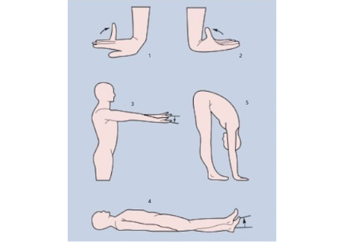
Beighton score for assessing hypermobility. (1) Extension of the little finger parallel to the forearm. (2) Adduction of the thumb towards the forearm. (3) Elbow hyperextension > 10°. (4) Knee hyperextension > 10°. (5) Flat hands on the floor in the forward bend. Total Beighton score = 9 points: Tests 1–4: 2 points each if bilateral; Test 5: 1 point. Generalised hypermobility in subjects < 25 years if score ≥ 6/9; Generalised hypermobility in subjects > 25–65 years if score ≥ 5/9; Generalised hypermobility in subjects > 65 years if score ≥ 4/9. Reprinted with permission from https://rheuma-schweiz.ch/krankheitsbilder/fachbereiche/weichteilrheumatismus/hyperlaxitaetssyndrom-2021/. Accessed April 7, 2026. © Rheuma Schweiz.
Hypermobility Questionnaire.
| Item | Question |
|---|---|
| The Hypermobility Questionnaire consists of 5 questions about current and past symptoms: | |
| 1 | Can you, or have you ever been able to, place your hands flat on the floor without bending your knees? |
| 2 | Can you, or have you ever been able to, bend your thumb towards your forearm? |
| 3 | As a child, did you amuse your friends by contorting your body into strange shapes, or were you able to do the splits? |
| 4 | As a child or teenager, did you dislocate your shoulder or kneecap more than once? |
| 5 | Do you consider yourself twice as flexible as other people? |
At least 2 positive answers are considered an indicator of JHS (sensitivity 80–85%) [5].
JHM arises from multifactorial influences including age, gender, trauma, conditioning, infection, inflammation, and genetic factors [9]. JHM often co-occurs with skin overstretching seen in connective tissue disorders such as the Ehlers-Danlos syndrome (EDS) [10]. In recent decades, JHM and related disorders have received growing research interest across multiple medical disciplines, albeit with variable emphasis in rheumatology [11]. JHM represents a complex phenotype arising from an interplay of genetic factors and environmental contributors such as age, gender, trauma, infection, and inflammation [12]. The 2017 international criteria replaced the older hypermobility syndrome nomenclature with two main entities: hypermobile EDS (hEDS) and generalized hypermobility spectrum disorder (g-HSD) [13, 14]. g-HSD is characterized by symptomatic hypermobility without clinical or genetic signs of EDS. This spectrum model acknowledges the fluid transition between benign hypermobility and pathologic syndromes with multisystem involvement [15].
This article summarizes current terminology, classification, and clinical relevance of hypermobility in rheumatology, aiming to raise awareness among clinicians.
Descriptions of hypermobility date to antiquity, with Hippocrates referencing joint laxity to enhance javelin throwing [16]. Scientific literature from the late 19th century, after the first description by Chernogubov in 1892 [17], onward documented hypermobility in contortionists and performers [18, 19]. The recognition that hypermobile joints often coincide with skin hyperextensibility laid the foundation for the identification of EDS—first described by Edvard Ehlers and Henri-Alexandre Danlos in the early 1900s [20]. EDS involves variable skin, vascular, musculoskeletal, and internal organ manifestations. Rheumatology traditionally focused on JHM syndrome, a descriptive concept before more precise genetic classifications were established [21–23]. The 2017 nosology unified and separated these classifications and terminology, enhancing clinical applicability [13–15].
Generalized hypermobility (GHM) is common globally and varies with age, sex, and ethnicity—more prevalent among women, children, and certain ethnic groups such as Inuit, Africans, and Asians [24]. In a Welsh population study, the diagnosed prevalence of GHM and EDS was approximately 194 per 100,000, with delayed diagnosis in females compared to males [25]. Background factors must be considered when interpreting hypermobility prevalence and clinical relevance.
JHM is defined by excessive joint ROM beyond normal physiological limits, determined by ligament laxity and connective tissue properties that are strongly genetically influenced [13–15]. Clinically, distinctions exist between hypermobility (objective excess motion), laxity (passive accessory movement), and functional instability (patient-perceived joint insecurity) [1, 9, 12]. The Beighton score remains the gold standard for quantifying hypermobility, assessing 9 joints through simple maneuvers [9, 12, 26]. Complementary screening uses the 5PQ, validated for adult populations [4, 27, 28] (Table 1). Although skin extensibility testing aids differentiation between hEDS and g-HSD, reproducibility remains limited [8, 29]. Comprehensive diagnosis requires training and experience due to overlaps with normal variability. Differential diagnosis depends on recognizing patterns of generalized, peripheral, localized, or historical hypermobility (see below). An overview is provided in Table 2 and Table 3.
Different types of Ehlers-Danlos syndrome [14].
| Types (n = 13) | Genes | Protein | Inheritance | Onset | Criteria |
|---|---|---|---|---|---|
| Classic | COL5A1, COL5A2, COL1A1 | Procollagen | AD | Childhood | Hyperextensibility with atrophic scarring, GHM |
| Classic-like (Type 1 ?) | TNXB | Tenascin-X | AR | Childhood and young adulthood | Skin hyperextensibility (velvet) without atrophic scarring, GHM, with/without recurrent dislocations, occurrence of haematomas |
| Cardiac and valvular involvement | COL1A2α2(I) | Procollagen | AR | Childhood, sometimes as early as the neonatal period | Severe progressive heart valve problems, skin hyperextensibility with atrophic scarring, thin skin, bruising, GHM |
| Vascular involvement | COL3A1, COL1A1α1(III) | Procollagen, α1(I) procollagen | AD | Childhood, sometimes as early as the neonatal period | - |
| Hypermobility | Not known | Not known | AD (?) | Any age | - |
| Arthrochalasia | COL1A1, COL1A2 | α1(I) procollagen, α2(I) procollagen | AR | Childhood, sometimes as early as the neonatal period | - |
| Dermatosparaxis | ADAMTS2 | Complement subcomponents 1r and 1s | AD | Childhood, sometimes as early as the neonatal period | Extreme, congenital skin fragility, characteristic craniofacial features, postnatal growth retardation, short limbs, redundant lax skin with excessive folds (especially palmar), severe bruising, with increased risk of subcutaneous haematomas and haemorrhages, umbilical hernias, perinatal complications due to tissue fragility |
| Kyphoscoliotic | PLOD1, FKBP14 | Lysyl hydroxylase 1, FKBP22 | AR | Childhood | Congenital muscular hypotonia, Early kypho-scoliosis, GHM with dislocations/subluxations |
| Corneal thinning | PRDM5, ZNF469 | PR-domain containing protein 5, zinc finger protein 469 | AR | Young children, some as early as the neonatal period | Thin cornea with or without rupture, early, progressive keratoconus, early progressive keratoglobus, blue sclera |
| Spondylodysplasia | B3GALT6, B4GALT7, SLC39A13 | Galactosyltransferase I and II, ZIP13 | AR | Neonatal period | Short stature, muscle hypotonia, GHM, curvature of the extremities |
| Muscle contractures | CHST14, DSE | Dermatan-4-sulfotransferase 1, dermatan sulfate epimerase 1 | AR | Childhood, sometimes as early as the neonatal period | Congenital contractures (particularly adducted thumbs, talipes equino-varus); characteristic facial features; hyperextensibility of the wrinkled skin (palmar) with atrophic scarring, haemorrhages |
| Myopathy | COL12A1α1(XII) | Procollagen | AD | Childhood, sometimes as early as the neonatal and prenatal periods | Congenital muscle hypotonia and/or atrophy, which improves with age, joint contractures (proximal), joint hypermobility (distal) |
| Periodontal involvement | C1R, C1S | Complement subcomponents 1r and 1s | - | Childhood | Early-onset, severe, untreatable periodontitis. Absence of gingival attachment, pretibial plaques, positive first-degree family history |
| Candidates for new clinical subtypes | |||||
| Classic-like (Type 2 ?) | AEBP1 | AE-binding protein 1 | AD | Childhood and early adulthood | Bilateral ptosis, webbed skin at the nape of the neck, low receding hairline, sagging cheeks, large ears, narrow high palate |
| Overlap associated with COL1 | COL1A1, COL1A2 | α1(I) procollagen, α2(II) procollagen | AD, AR | Childhood and early adulthood | Blue sclerae, flat feet with rearfoot valgus deformity, GHM, soft, doughy and/or hyperextensible skin |
n = 13 refers to the main clinical subtypes; two additional candidate subtypes are listed separately. AD: autosomal dominant; AE: adipocyte enhancer; AR: autosomal recessive; GHM: generalized hypermobility. ? It was left as is, following the reference.
Phenotypes belonging to the hypermobility spectrum.
| Phenotype | Symptoms | Beighton score | Musculoskeletal involvement |
|---|---|---|---|
| g-HM | Asymptomatic | Positive | No |
| p-HM | Asymptomatic | Negative | No |
| l-HM | Asymptomatic | Usually negative | No |
| g-HSD | Yes | Positive | Yes |
| p-HSD | Possible | Mostly negative | Yes |
| l-HSD | Possible | Negative | Yes |
| h-HSD* | Possible | Negative | Yes |
| hEDS** | Yes | Positive | Possible |
*History of joint hypermobility (e.g., positive 5-item questionnaire); ** plus a positive family history (first-degree relatives) and/or specific systemic manifestations (see new criteria). All these phenotypes require careful exclusion of other acquired and hereditary disorders, some of which overlap. (a) Peripheral joint hypermobility is typically limited to the hands and/or feet; the Beighton score is usually negative. (b) Localised joint hypermobility is restricted to individual joints or body parts; the Beighton score is negative. (c) See the new criteria for the operational definition of ‘generalised joint hypermobility’ as a mandatory feature/criterion of hypermobile Ehlers-Danlos syndrome. g-HM: generalised hypermobility disorder; g-HSD: generalised hypermobility spectrum disorder; hEDS: hypermobile Ehlers-Danlos syndrome; l-HSD: localised hypermobility spectrum disorder; p-HM: peripheral joint hypermobility; p-HSD: peripheral hypermobility spectrum disorder.
EDS encompasses a heterogeneous group [10] of 13 genetically defined, mostly rare [30] connective tissue disorders, characterized mainly by GHM, skin fragility, abnormal scarring, and bruising [13–15]. Most subtypes possess identified causative gene variants impacting the extracellular matrix (ECM), except hEDS and g-HSD, where molecular markers are lacking [13–15]. Diagnosing EDS involves ROM assessments, skin examination [29], molecular genetic testing where available, and exclusion of differential diagnoses (see below). The 2017 International Classification redefined EDS into 13 subtypes and introduced HSD as a separate diagnostic framework for symptomatic hypermobility not fulfilling hEDS criteria. Inclusion of g-HSD as part of the EDS spectrum has increased diagnostic rates of g-HSD in some settings [12].
Numerous genetic syndromes manifest generalized or peripheral hypermobility due to pathogenic variants in genes encoding ECM proteins such as collagen and tenascin-X [13–15, 31]. Collagen types I, II, IX, XI, and TNX are critical for ligament structure and function [32, 33]; mutations disrupt tissue homeostasis, leading to symptoms across multiple organ systems. Both Mendelian inheritance patterns and multifactorial models incorporating lifestyle and developmental factors explain hypermobility phenotypes.
The 2017 international classification of EDS identifies 13 clinical subtypes caused by mutations in 19 genes [14]. Common subtypes include classic, vascular, and hEDS. hEDS diagnosis is clinical, based on strict criteria involving phenotype and history, supported by screening via the 5PQ [5, 6]. Severity of symptoms varies substantially [34]. Molecular testing is mandatory for other subtypes but not currently available for hEDS [13–15]. g-HSD covers symptomatic hypermobility without molecular confirmation. A large clinical cohort study showed g-HSD accounting for 66.5% of hypermobility diagnoses, with overlapping musculoskeletal and neurological symptoms reported in both g-HSD and hEDS patients [35].
A comprehensive differential diagnosis is essential when evaluating patients with generalized or localized hypermobility to avoid misclassification and ensure accurate management:
Marfan syndrome (MfS): Characterized by arachnodactyly (long, slender fingers), which also occurs in other diseases [36], dolichostenomelia (long limbs), skin elasticity, and chest deformities like pectus excavatum. It is caused by mutations in the fibrillin-1 gene leading to increased connective tissue elasticity [37]. MfS shares musculoskeletal and cardiovascular features with some forms but has distinct genetic and clinical criteria.
Osteogenesis imperfecta (OI): Known as “brittle bone disease”. OI is a rare hereditary condition primarily caused by mutations affecting type I collagen, resulting in bone fragility, blue sclera, hearing loss, and sometimes JHM [38]. Differentiation from EDS is critical due to differences in fracture risk and treatment.
Neuromuscular disorders: Certain inherited myopathies may present with JHM alongside muscle weakness and hypotonia. Recognition of muscle involvement helps distinguish these from connective tissue disorders [39].
Other hereditary connective tissue disorders: Conditions such as Loeys-Dietz syndrome and Stickler syndrome can demonstrate overlapping features with EDS and MfS, necessitating genetic testing and careful phenotyping.
Differentiating these disorders involves clinical examination, family history, molecular genetic testing when available, and consideration of systemic involvement. Awareness of these alternatives prevents diagnostic delays and inappropriate management [36–39].
GHM is linked to musculoskeletal complaints including joint pain, subluxations, and repeated microtrauma, especially in weight-bearing joints such as hips, knees, ankles, and shoulders [12, 24]. Pain is the most significant clinical issue relating to GHM, which reflects a complex interplay of biomechanical strain, altered proprioception, and neurogenic mechanisms arising through nociceptive, neuropathic, and central sensitization processes [12, 15, 40–47]. Thus, pain mechanisms include nociceptive injury from ligament laxity and neuropathic elements like small-fiber neuropathy and nerve entrapment [40, 41]. Many patients exhibit symptoms consistent with fibromyalgia syndrome (FM), with common comorbidities including headaches, migraines, and gastrointestinal disturbances [42–47]. These symptoms significantly impair quality of life and complicate management [48]. In a cohort of 733 patients from an EDS referral center, over half exhibited comorbid FM, with symptoms of joint pain, cognitive difficulties (“brain fog”), and atopy being common [46]. Patients with GHM alone had fewer comorbidities but increased risk of joint sprains and subluxations [49].
Several studies report hypermobility in 13–15% of patients referred to rheumatology, associated with higher incidence of soft tissue rheumatism, FM, osteoarthritis, and some autoimmune diseases [50–55]. A study in juvenile idiopathic arthritis revealed worse disease activity and outcomes in hypermobile children [56]. Limited data exist for adult rheumatoid arthritis and spondyloarthritis outcomes, though mechanical stress hypotheses point to plausible effects [53, 54]. The relationship between hypermobility and osteoarthritis remains controversial with mixed findings [57–66].
JHM and ROM were investigated in female dancers and control subjects aged 8–16 years [67]. The prevalence of JHM and joint ROM was significantly higher in female dancers, with age-dependent differences. Hypermobility (Beighton score > 4) was also diagnosed in two-thirds of Polish jazz dancers aged 18–25 years [68]. It seems that hypermobility could represent an advantage for some performers (e.g., dancers, musicians and acrobats).
The 2017 International Classification redefined EDS into 13 subtypes and introduced HSD as a separate diagnostic framework for symptomatic hypermobility not fulfilling hEDS criteria [13–15]. The criteria emphasize diagnostic specificity, accepting reduced sensitivity, especially for hEDS [69, 70]. The classification aligns with evolving trends in medicine by recognizing “spectrum” disorders, reflecting variable phenotypes and symptoms. Taken together, hEDS likely represents a complex/polygenic disorder with an incompletely understood molecular basis. Rheumatologists should vigilantly assess hypermobility in patients with musculoskeletal pain, including those diagnosed with FM or long COVID due to symptom overlap [71–73]. Molecular diagnostics confirm many EDS subtypes but not hEDS, warranting cautious clinical judgment and genetic consultation.
A discussion of treatment options for hypermobility syndromes is beyond the scope of this paper. However, a very recent survey in the USA [74] revealed considerable variability and a lack of standardisation in therapeutic interventions, raising questions about the quality of care.
While rheumatology is not the primary specialty for hypermobility syndrome diagnosis, it plays a key role in detection, classification, and appropriate referral. Simple clinical tools like the Beighton score and 5PQ screening facilitate this process in everyday practice. Enhanced awareness and research are needed to elucidate hypermobility’s impact on inflammatory rheumatic disease progression and treatment efficacy.
5PQ: 5-part questionnaire
ECM: extracellular matrix
EDS: Ehlers-Danlos syndrome
FM: fibromyalgia syndrome
GHM: generalized hypermobility
g-HSD: generalized hypermobility spectrum disorder
hEDS: hypermobile Ehlers-Danlos syndrome
JHM: joint hypermobility
MfS: Marfan syndrome
OI: osteogenesis imperfecta
ROM: range of motion
BJ: Methodology, Writing—review & editing, Writing—original draft. The author read and approved the submitted version.
Jürgen Braun, who is the Associate Editor of Exploration of Musculoskeletal Diseases, had no involvement in the decision-making or the review process of this manuscript. The author declares no other conflicts of interest.
Not applicable.
Not applicable.
Not applicable.
Not applicable.
Not applicable.
© The Author(s) 2026.
Open Exploration maintains a neutral stance on jurisdictional claims in published institutional affiliations and maps. All opinions expressed in this article are the personal views of the author(s) and do not represent the stance of the editorial team or the publisher.
Copyright: © The Author(s) 2026. This is an Open Access article licensed under a Creative Commons Attribution 4.0 International License (https://creativecommons.org/licenses/by/4.0/), which permits unrestricted use, sharing, adaptation, distribution and reproduction in any medium or format, for any purpose, even commercially, as long as you give appropriate credit to the original author(s) and the source, provide a link to the Creative Commons license, and indicate if changes were made.



View: 57
Download: 5
Times Cited: 0


