
 Previous
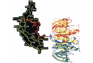
One of the most troubling developments of 2021 has been the number of fertile-age women who have been led to believe that mRNA vaccines against severe acute respiratory syndrome coronavirus-2 [SARS-CoV-2, coronavirus disease 2019 (COVID-19)] can cause infertility via cross-reactivity of immune response. Specifically, cross-reactivity of developed antibodies to syncytin-1, a protein found in human cell fusion, placentation and recently identified in the envelope gene of a human endogenous defective retrovirus, HERV-W (see “Syncytin is a captive retroviral envelope protein involved in human placental morphogenesis”. Nature. 2000;403:785–9. doi: 10.1038/35001608). The mechanism, evidence, and evaluation of the claim is presented concluding in a rejection due to lack of evidence.
One of the most troubling developments of 2021 has been the number of fertile-age women who have been led to believe that mRNA vaccines against severe acute respiratory syndrome coronavirus-2 [SARS-CoV-2, coronavirus disease 2019 (COVID-19)] can cause infertility via cross-reactivity of immune response. Specifically, cross-reactivity of developed antibodies to syncytin-1, a protein found in human cell fusion, placentation and recently identified in the envelope gene of a human endogenous defective retrovirus, HERV-W (see “Syncytin is a captive retroviral envelope protein involved in human placental morphogenesis”. Nature. 2000;403:785–9. doi: 10.1038/35001608). The mechanism, evidence, and evaluation of the claim is presented concluding in a rejection due to lack of evidence.
DOI: https://doi.org/10.37349/ei.2022.00032


A few pieces of research exist about the protective titer against severe acute respiratory syndrome (SARS) coronavirus 2 (CoV-2; SARS-CoV-2) in monkeys and humans in which the protection could be shown as dose-dependent. Early studies supposed that higher levels of pre-existing neutralizing antibodies (Nabs) against SARS-CoV-2 can potentially correlate with the protection to consequent infection. The data so far showed that cellular immunity is as essential as the humoral one. If needed, its presence can be beneficial if the titer of immunoglobulins is not optimal. It is also known that the immune response to the vaccine is similar to the one after natural infection with a production of very high naturalization titers antibodies. However, medical community is still unaware of the immunoglobulin titer needed for protection against the virus. The answers to the questions regarding correlates of protection are yet to be discovered. Still, no studies indicate a specific virus-Nab titer, so one can assume a patient is protected from being infected in the future. The evoked immunological response is indeed encouraging, but a future investigation is needed. Nonetheless, it remains a mystery how long the immunity lasts and whether it will be enough to shield the patients in the long run. Therefore, identifying immune protection correlations, including neutralization titer of antibodies and T cell immune response against SARS-CoV-2, could give a clue. Unfortunately, recent studies in the field have been more controversial than concise, and the data available is far from consensus.
A few pieces of research exist about the protective titer against severe acute respiratory syndrome (SARS) coronavirus 2 (CoV-2; SARS-CoV-2) in monkeys and humans in which the protection could be shown as dose-dependent. Early studies supposed that higher levels of pre-existing neutralizing antibodies (Nabs) against SARS-CoV-2 can potentially correlate with the protection to consequent infection. The data so far showed that cellular immunity is as essential as the humoral one. If needed, its presence can be beneficial if the titer of immunoglobulins is not optimal. It is also known that the immune response to the vaccine is similar to the one after natural infection with a production of very high naturalization titers antibodies. However, medical community is still unaware of the immunoglobulin titer needed for protection against the virus. The answers to the questions regarding correlates of protection are yet to be discovered. Still, no studies indicate a specific virus-Nab titer, so one can assume a patient is protected from being infected in the future. The evoked immunological response is indeed encouraging, but a future investigation is needed. Nonetheless, it remains a mystery how long the immunity lasts and whether it will be enough to shield the patients in the long run. Therefore, identifying immune protection correlations, including neutralization titer of antibodies and T cell immune response against SARS-CoV-2, could give a clue. Unfortunately, recent studies in the field have been more controversial than concise, and the data available is far from consensus.
DOI: https://doi.org/10.37349/ei.2022.00033
This article belongs to the special issue Vaccine-induced Immune Responses Against SARS-CoV-2 Infections
Cutaneous homeostasis is maintained by dynamic cellular communications between different cell types in the skin through interactions with various mediators, including cytokines, chemokines and antimicrobial peptides/proteins (AMPs). Keratinocytes, as the major cell type of the epidermis, not only form a passive physical barrier, but also actively participate in the pathogenesis of many, if not all, inflammatory skin diseases. Keratinocytes highly interact with immune cells to shape, amplify or regulate inflammatory responses, thus triggering and/or sustaining these inflammatory skin diseases. In this review, crosstalk between keratinocytes and immune cells is summarized, and its contributions to two major inflammatory skin disorders including psoriasis and atopic dermatitis are highlighted.
Cutaneous homeostasis is maintained by dynamic cellular communications between different cell types in the skin through interactions with various mediators, including cytokines, chemokines and antimicrobial peptides/proteins (AMPs). Keratinocytes, as the major cell type of the epidermis, not only form a passive physical barrier, but also actively participate in the pathogenesis of many, if not all, inflammatory skin diseases. Keratinocytes highly interact with immune cells to shape, amplify or regulate inflammatory responses, thus triggering and/or sustaining these inflammatory skin diseases. In this review, crosstalk between keratinocytes and immune cells is summarized, and its contributions to two major inflammatory skin disorders including psoriasis and atopic dermatitis are highlighted.
DOI: https://doi.org/10.37349/ei.2021.00028
This article belongs to the special issue Cross Talk Among Skin Cells and Immune Cells
Severe acute respiratory syndrome coronavirus 2 (SARS-CoV-2), a highly pathogenic β-coronavirus, is the etiologic agent of coronavirus disease 2019 (COVID-19), which gave rise to a difficult to control pandemic, especially in Brazil. Approximately 4,000 mutations have been identified in SARS-CoV-2, with the majority being redundant without having any biological effect on the virus. The aim of the present study was to objectively understand how new SARS-CoV-2 variants can affect vaccine response, in addition to highlighting the current situation in Brazil in the face of the pandemic and considering epidemiological and immunological aspects of COVID-19. The main protective correlate investigated in most vaccines is the neutralizing antibody titer induced by immunizing agents, observed in the pre-clinical phase in animals, whose action is to block the binding of the spike protein to the angiotensin-converting enzyme 2 (ACE2) receptor, preventing infection. Up to the second half of 2021, the variants that are of greatest concern worldwide and require molecular surveillance are Alpha variant (or B.1.1.7 lineage), Beta (or B.1.351 lineage), Gamma (or P1 lineage) and Delta (or B.1.617.2 lineage). Brazil finds itself in a highly unfavorable scenario, with the circulation of variants of concern, mainly Gamma and Delta, with high fatality rates for COVID-19 and low vaccination rate. Given the still latent situation of the COVID-19 pandemic in Brazil, the lack of global planning for action strategies for non-pharmacological prevention measures, there is an imminent risk of the emergence of new variants due to the finding of susceptible hosts and the high proliferative rate of SARS-CoV-2. It is urgent to increase the genotyping of positive samples isolated from infected individuals, the speed of vaccination of the entire population and the unification of non-pharmacological preventive measures throughout the country.
Severe acute respiratory syndrome coronavirus 2 (SARS-CoV-2), a highly pathogenic β-coronavirus, is the etiologic agent of coronavirus disease 2019 (COVID-19), which gave rise to a difficult to control pandemic, especially in Brazil. Approximately 4,000 mutations have been identified in SARS-CoV-2, with the majority being redundant without having any biological effect on the virus. The aim of the present study was to objectively understand how new SARS-CoV-2 variants can affect vaccine response, in addition to highlighting the current situation in Brazil in the face of the pandemic and considering epidemiological and immunological aspects of COVID-19. The main protective correlate investigated in most vaccines is the neutralizing antibody titer induced by immunizing agents, observed in the pre-clinical phase in animals, whose action is to block the binding of the spike protein to the angiotensin-converting enzyme 2 (ACE2) receptor, preventing infection. Up to the second half of 2021, the variants that are of greatest concern worldwide and require molecular surveillance are Alpha variant (or B.1.1.7 lineage), Beta (or B.1.351 lineage), Gamma (or P1 lineage) and Delta (or B.1.617.2 lineage). Brazil finds itself in a highly unfavorable scenario, with the circulation of variants of concern, mainly Gamma and Delta, with high fatality rates for COVID-19 and low vaccination rate. Given the still latent situation of the COVID-19 pandemic in Brazil, the lack of global planning for action strategies for non-pharmacological prevention measures, there is an imminent risk of the emergence of new variants due to the finding of susceptible hosts and the high proliferative rate of SARS-CoV-2. It is urgent to increase the genotyping of positive samples isolated from infected individuals, the speed of vaccination of the entire population and the unification of non-pharmacological preventive measures throughout the country.
DOI: https://doi.org/10.37349/ei.2021.00029
This article belongs to the special issue Vaccine-induced Immune Responses Against SARS-CoV-2 Infections
The coronavirus disease 2019 (COVID-19) results from the infection of severe acute respiratory syndrome coronavirus-2 (SARS-CoV-2) and primarily affects the respiratory tissue. Since first reported from Wuhan, China in December 2019, the virus has resulted in an unprecedented pandemic. Vaccination against SARS-CoV-2 can control the further spread of the ongoing pandemic by making people immunised to SARS-CoV-2. Several vaccines have been approved for use in clinics, a lot many are in different stages of development. Diligent interpretations from the preclinical evaluation are crucial to identify the most effective and safest vaccine candidates. Multiple vaccine candidates/variants have been tested in small animal models with relative ease and further in non-human primate models before being taken into clinical development. Here, we review the state-of-the-art strategies employed for a thorough preclinical evaluation of COVID-19 vaccine candidates. We summarise the methods in place to identify indicators which make the vaccine candidate effective in controlling SARS-CoV-2 infection and/or COVID-19 and are safe for administration as inferred by their (1) biophysical/functional attributes (antigen expression, organization, functionality, and stability); (2) immunogenicity in animal models and protective correlates [SARS-CoV-2 specific binding/neutralising immunoglobulin titer, B/T-cell profiling, balanced T-helper type-1 (Th1) or type-2 (Th2) response (Th1:Th2), and anamnestic response]; (3) protective correlates as interpreted by controlled pathology of the respiratory tissue (pulmonary clinical and immunopathology); and finally, (4) strategies to monitor adverse effects of the vaccine candidates.
The coronavirus disease 2019 (COVID-19) results from the infection of severe acute respiratory syndrome coronavirus-2 (SARS-CoV-2) and primarily affects the respiratory tissue. Since first reported from Wuhan, China in December 2019, the virus has resulted in an unprecedented pandemic. Vaccination against SARS-CoV-2 can control the further spread of the ongoing pandemic by making people immunised to SARS-CoV-2. Several vaccines have been approved for use in clinics, a lot many are in different stages of development. Diligent interpretations from the preclinical evaluation are crucial to identify the most effective and safest vaccine candidates. Multiple vaccine candidates/variants have been tested in small animal models with relative ease and further in non-human primate models before being taken into clinical development. Here, we review the state-of-the-art strategies employed for a thorough preclinical evaluation of COVID-19 vaccine candidates. We summarise the methods in place to identify indicators which make the vaccine candidate effective in controlling SARS-CoV-2 infection and/or COVID-19 and are safe for administration as inferred by their (1) biophysical/functional attributes (antigen expression, organization, functionality, and stability); (2) immunogenicity in animal models and protective correlates [SARS-CoV-2 specific binding/neutralising immunoglobulin titer, B/T-cell profiling, balanced T-helper type-1 (Th1) or type-2 (Th2) response (Th1:Th2), and anamnestic response]; (3) protective correlates as interpreted by controlled pathology of the respiratory tissue (pulmonary clinical and immunopathology); and finally, (4) strategies to monitor adverse effects of the vaccine candidates.
DOI: https://doi.org/10.37349/ei.2021.00030
This article belongs to the special issue Vaccine-induced Immune Responses Against SARS-CoV-2 Infections
It is well recognized that immune tolerance is important to prevent semiallografted fetuses from rejection by maternal immunocompetent cells; however, immune activation also plays an important role in placental development and fetal growth. Basic and clinical studies have shown that an imbalance between immune activation and regulation can lead to implantation failure, miscarriage, and preeclampsia. Here, the balance between immunostimulation and immunoregulation in reproduction will be reviewed.
It is well recognized that immune tolerance is important to prevent semiallografted fetuses from rejection by maternal immunocompetent cells; however, immune activation also plays an important role in placental development and fetal growth. Basic and clinical studies have shown that an imbalance between immune activation and regulation can lead to implantation failure, miscarriage, and preeclampsia. Here, the balance between immunostimulation and immunoregulation in reproduction will be reviewed.
DOI: https://doi.org/10.37349/ei.2021.00031
This article belongs to the special issue Human Reproduction: Involvement of the Immune System
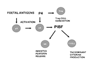
The foetus expressing paternal antigens ought to be “rejected” by the maternal immune system. However, the immunological relationship of the mother and the foetus does not follow the rules of transplantation immunology. Maternal immune functions are re-adjusted during pregnancy, to create a tolerant environment for the developing foetus. Progesterone and its downstream mediator; the progesterone induced blocking factor (PIBF) are important in this process. The mRNA transcribed from the PIBF1 gene contains 18 exons, and codes for a 90 kDa protein. The 90 kDa form is associated with the centrosome and plays a role in cell cycle regulation, while smaller isoforms produced by alternative spicing are secreted, and bind to the glycosylphosphatidylinositol (GPI) anchored PIBF receptor. Upon ligation, the former forms a heterodimer with the alpha chain of the interleukin-4 (IL-4) receptor and activates the Janus kinase/signal transducers and activators of transcription (Jak/STAT) pathway, via which, PIBF induces increased production of T helper2 (Th2) cytokines. PIBF regulates natural killer (NK) cytotoxicity, by inhibiting perforin release from the cytoplasmic granules of NK cells. During normal human pregnancy, the serum concentrations of PIBF increase with gestational age, and lower than normal serum levels predict spontaneous pregnancy termination. Depletion of PIBF during the peri-implantation period in mice, results in lower implantation and increased resorption rates, together with increased decidual and peripheral NK activity, downregulation of the genes implicated in T cell activation in CD4+ cells, and Th1 differentiation of the T cells. PIBF is expressed in rapidly proliferating immature cells as well as several tumours, and regulates invasion. The PIBF gene has been identified in the chromosomal region 13q21-q22—which is a common site for somatic deletions in a variety of malignant tumours. These data suggest that PIBF might be involved in tumorigenesis.
The foetus expressing paternal antigens ought to be “rejected” by the maternal immune system. However, the immunological relationship of the mother and the foetus does not follow the rules of transplantation immunology. Maternal immune functions are re-adjusted during pregnancy, to create a tolerant environment for the developing foetus. Progesterone and its downstream mediator; the progesterone induced blocking factor (PIBF) are important in this process. The mRNA transcribed from the PIBF1 gene contains 18 exons, and codes for a 90 kDa protein. The 90 kDa form is associated with the centrosome and plays a role in cell cycle regulation, while smaller isoforms produced by alternative spicing are secreted, and bind to the glycosylphosphatidylinositol (GPI) anchored PIBF receptor. Upon ligation, the former forms a heterodimer with the alpha chain of the interleukin-4 (IL-4) receptor and activates the Janus kinase/signal transducers and activators of transcription (Jak/STAT) pathway, via which, PIBF induces increased production of T helper2 (Th2) cytokines. PIBF regulates natural killer (NK) cytotoxicity, by inhibiting perforin release from the cytoplasmic granules of NK cells. During normal human pregnancy, the serum concentrations of PIBF increase with gestational age, and lower than normal serum levels predict spontaneous pregnancy termination. Depletion of PIBF during the peri-implantation period in mice, results in lower implantation and increased resorption rates, together with increased decidual and peripheral NK activity, downregulation of the genes implicated in T cell activation in CD4+ cells, and Th1 differentiation of the T cells. PIBF is expressed in rapidly proliferating immature cells as well as several tumours, and regulates invasion. The PIBF gene has been identified in the chromosomal region 13q21-q22—which is a common site for somatic deletions in a variety of malignant tumours. These data suggest that PIBF might be involved in tumorigenesis.
DOI: https://doi.org/10.37349/ei.2021.00027
This article belongs to the special issue Human Reproduction: Involvement of the Immune System
Coronavirus disease 2019 (COVID-19) emerges as an expeditiously growing pandemic, in the human population caused by the highly transmissible RNA virus severe acute respiratory syndrome of coronavirus 2 (SARS-CoV-2). Prognosis of SARS-CoV-2 infection predominantly occurs at the angiotensin-converting enzyme 2 receptor and transmembrane protease serine type 2 positive (ACE2 + TMPRSS2)+ epithelial cells of the mucosal surface like nasal, oral mucosae, and/or the conjunctival surface of the eye where it has interacted along with the immune system. The primary host response towards the pathogen starts from an immune microenvironment of nasopharynx-associated lymphoid tissue (NALT) and mucosa-associated lymphoid tissue (MALT). The presence of exhausted lymphocytes, lymphopenia, pneumonia and cytokine storm is the hallmark of COVID-19. The multifaceted nature of co-morbidity factors like obesity and type 2 diabetes and its effects on immunity can alter the pathogenesis of SARS-CoV-2 infection. Adipose tissue is a crucial endocrine organ that secretes a plethora of factors like adipokines, cytokines, and chemokines that have a profound impact on metabolism and augments the expression of mucosal pro-inflammatory cytokines, like tumor necrosis factor-alpha (TNF-α), interferon-gamma (IFN-γ), and the interleukin-12 (IL-12)/IL-23. Mucosal immunization could be a superior approach to activate mucosal and systemic immune responses against pathogenic invasion at mucosal surface entry ports. Mucosal vaccines are also able to generate strong systemic humoral immunity—required to neutralize any virus particle that dodges the primary immune response. To develop an efficient vaccine against mucosal pathogens, considering the designing of the delivery route, immunomodulatory features, and adjuvants are very important. In this article, we further provide evidence to understand the significant role of mucosal immunity, along with secretory and circulating immunoglobulin A (IgA) antibodies in generating a novel mucosal vaccine against COVID-19. Moreover, along with mucosal vaccines, we should look for combination treatment strategies with plant bioactive molecules. Glycan-binding lectins against viral proteins for targeted activation of mucosal immune response are one of such examples. This might play a promising role to halt this emerging virus.
Coronavirus disease 2019 (COVID-19) emerges as an expeditiously growing pandemic, in the human population caused by the highly transmissible RNA virus severe acute respiratory syndrome of coronavirus 2 (SARS-CoV-2). Prognosis of SARS-CoV-2 infection predominantly occurs at the angiotensin-converting enzyme 2 receptor and transmembrane protease serine type 2 positive (ACE2 + TMPRSS2)+ epithelial cells of the mucosal surface like nasal, oral mucosae, and/or the conjunctival surface of the eye where it has interacted along with the immune system. The primary host response towards the pathogen starts from an immune microenvironment of nasopharynx-associated lymphoid tissue (NALT) and mucosa-associated lymphoid tissue (MALT). The presence of exhausted lymphocytes, lymphopenia, pneumonia and cytokine storm is the hallmark of COVID-19. The multifaceted nature of co-morbidity factors like obesity and type 2 diabetes and its effects on immunity can alter the pathogenesis of SARS-CoV-2 infection. Adipose tissue is a crucial endocrine organ that secretes a plethora of factors like adipokines, cytokines, and chemokines that have a profound impact on metabolism and augments the expression of mucosal pro-inflammatory cytokines, like tumor necrosis factor-alpha (TNF-α), interferon-gamma (IFN-γ), and the interleukin-12 (IL-12)/IL-23. Mucosal immunization could be a superior approach to activate mucosal and systemic immune responses against pathogenic invasion at mucosal surface entry ports. Mucosal vaccines are also able to generate strong systemic humoral immunity—required to neutralize any virus particle that dodges the primary immune response. To develop an efficient vaccine against mucosal pathogens, considering the designing of the delivery route, immunomodulatory features, and adjuvants are very important. In this article, we further provide evidence to understand the significant role of mucosal immunity, along with secretory and circulating immunoglobulin A (IgA) antibodies in generating a novel mucosal vaccine against COVID-19. Moreover, along with mucosal vaccines, we should look for combination treatment strategies with plant bioactive molecules. Glycan-binding lectins against viral proteins for targeted activation of mucosal immune response are one of such examples. This might play a promising role to halt this emerging virus.
DOI: https://doi.org/10.37349/ei.2021.00025
This article belongs to the special issue Vaccine-induced Immune Responses Against SARS-CoV-2 Infections
This article is a tribute and homage to Gerard Chaouat who invited me to contribute this article. My years in France have remained very memorable to me. Reviewed briefly is the vaccine that was made against human chorionic gonadotropin (hCG) to prevent unwanted pregnancy in sexually active women. It has now been developed as a genetically engineered recombinant vaccine and passed onto industry for its production under good manufacturing practices (GMP) conditions for confirmatory trials. The trials have received the approval of the Drugs Controller General of India. The trials have started but have been interrupted by the coronavirus disease 2019 (COVID-19) pandemic. This vaccine is likely to have another highly beneficial application in the treatment of cancers expressing ectopically hCG.
This article is a tribute and homage to Gerard Chaouat who invited me to contribute this article. My years in France have remained very memorable to me. Reviewed briefly is the vaccine that was made against human chorionic gonadotropin (hCG) to prevent unwanted pregnancy in sexually active women. It has now been developed as a genetically engineered recombinant vaccine and passed onto industry for its production under good manufacturing practices (GMP) conditions for confirmatory trials. The trials have received the approval of the Drugs Controller General of India. The trials have started but have been interrupted by the coronavirus disease 2019 (COVID-19) pandemic. This vaccine is likely to have another highly beneficial application in the treatment of cancers expressing ectopically hCG.
DOI: https://doi.org/10.37349/ei.2021.00026
This article belongs to the special issue Human Reproduction: Involvement of the Immune System
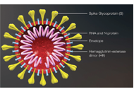
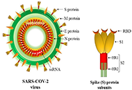
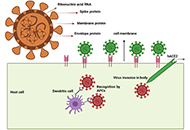
Severe acute respiratory syndrome coronavirus 2 (SARS-CoV-2; βCoV), the causative agent of coronavirus disease 2019 (COVID-19), causes severe lower respiratory tract infections and acute respiratory failure syndrome (ARDS). Deaths due to the ongoing COVID-19 pandemic for more than a year are still seen worldwide. Therefore, vaccine trials have gained importance. The discovery of the genome and protein structure of SARS-CoV-2 in a short time allowed the development of nucleic acid-based vaccines (mRNA and DNA vaccines), vector vaccines, inactivated virus vaccines, protein-based vaccines, virus-like particle vaccines, and live attenuated virus vaccines. Many companies, universities, and institutes around the world continue to develop effective vaccines against SARS-CoV-2. In this review, the structural features, classification, genome, and intracellular entry of SARS-CoV-2 coronaviruses, stimulation of the immune system and immunity, COVID-19 vaccine types, and the latest status of clinical trials of these vaccines have been reviewed.
Severe acute respiratory syndrome coronavirus 2 (SARS-CoV-2; βCoV), the causative agent of coronavirus disease 2019 (COVID-19), causes severe lower respiratory tract infections and acute respiratory failure syndrome (ARDS). Deaths due to the ongoing COVID-19 pandemic for more than a year are still seen worldwide. Therefore, vaccine trials have gained importance. The discovery of the genome and protein structure of SARS-CoV-2 in a short time allowed the development of nucleic acid-based vaccines (mRNA and DNA vaccines), vector vaccines, inactivated virus vaccines, protein-based vaccines, virus-like particle vaccines, and live attenuated virus vaccines. Many companies, universities, and institutes around the world continue to develop effective vaccines against SARS-CoV-2. In this review, the structural features, classification, genome, and intracellular entry of SARS-CoV-2 coronaviruses, stimulation of the immune system and immunity, COVID-19 vaccine types, and the latest status of clinical trials of these vaccines have been reviewed.
DOI: https://doi.org/10.37349/ei.2021.00023
This article belongs to the special issue Vaccine-induced Immune Responses Against SARS-CoV-2 Infections
The maternal syndrome preeclampsia is triggered by syncytiotrophoblast (STB) stress; the heterogeneity of the syndrome is caused by the different pathways leading to this STB stress. Inflammation plays a pivotal role in the pathogenesis of preeclampsia. While, the immune system at large is therefore intimately involved in the causation of this heterogeneous syndrome, the role of the adaptive immune system is more controversial. The classic paradigm placed preeclampsia as the disease of the nulliparous pregnant women. Up to the later part of the 20th century, human reproduction, particularly in Western societies, was characterised by a low rate of pre-marital sex, and the great majority of children being born within one stable sexual relationship. More prolonged periods of regular sexual intercourse within a stable relationship have been demonstrated to reduce the risk of preeclampsia and fetal growth restriction. Primarily animal studies have indeed shown that repetitive sperm exposure leads to partner specific mucosal tolerance. Societal changes made partner change over the reproductive period of individual women extremely common. For the adaptive immune system of multiparous women, being pregnant in a new sexual relationship (primipaternity) would represent being faced with a new “hemi-allograft”. In these pregnancies, potential couple-specific immune “maladaptation” could lead to the superficial cytotrophoblast invasion of the spiral arteries, known to be associated with early-onset preeclampsia. Having a new pregnancy in a different relationship does indeed increase the risk for this type of preeclampsia. Large epidemiologic population studies identified prolonged birth interval but not “primipaternity” as a risk factor for preeclampsia in multiparous women. This apparent contradiction is explained by the fact that the great majority of preeclampsia cases in these population studies involve term preeclampsia. In late-onset preeclampsia, the far more common phenotype of the syndrome, STB stress is not caused by lack of proper spiral artery modification, but involves maternal genetic predisposition to cardiovascular and metabolic disease, with in particular obesity/metabolic syndrome representing major players. Partner or couple specific issues are not detectable in this disease phenotype.
The maternal syndrome preeclampsia is triggered by syncytiotrophoblast (STB) stress; the heterogeneity of the syndrome is caused by the different pathways leading to this STB stress. Inflammation plays a pivotal role in the pathogenesis of preeclampsia. While, the immune system at large is therefore intimately involved in the causation of this heterogeneous syndrome, the role of the adaptive immune system is more controversial. The classic paradigm placed preeclampsia as the disease of the nulliparous pregnant women. Up to the later part of the 20th century, human reproduction, particularly in Western societies, was characterised by a low rate of pre-marital sex, and the great majority of children being born within one stable sexual relationship. More prolonged periods of regular sexual intercourse within a stable relationship have been demonstrated to reduce the risk of preeclampsia and fetal growth restriction. Primarily animal studies have indeed shown that repetitive sperm exposure leads to partner specific mucosal tolerance. Societal changes made partner change over the reproductive period of individual women extremely common. For the adaptive immune system of multiparous women, being pregnant in a new sexual relationship (primipaternity) would represent being faced with a new “hemi-allograft”. In these pregnancies, potential couple-specific immune “maladaptation” could lead to the superficial cytotrophoblast invasion of the spiral arteries, known to be associated with early-onset preeclampsia. Having a new pregnancy in a different relationship does indeed increase the risk for this type of preeclampsia. Large epidemiologic population studies identified prolonged birth interval but not “primipaternity” as a risk factor for preeclampsia in multiparous women. This apparent contradiction is explained by the fact that the great majority of preeclampsia cases in these population studies involve term preeclampsia. In late-onset preeclampsia, the far more common phenotype of the syndrome, STB stress is not caused by lack of proper spiral artery modification, but involves maternal genetic predisposition to cardiovascular and metabolic disease, with in particular obesity/metabolic syndrome representing major players. Partner or couple specific issues are not detectable in this disease phenotype.
DOI: https://doi.org/10.37349/ei.2021.00022
This article belongs to the special issue Human Reproduction: Involvement of the Immune System
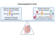
The testis is designated as one of the immune previleged sites in the body and harbours a unique immunoregulatory environment, which is important for preventing an immune response against sperm antigens which otherwise are recognized as “foreign” by the immune system. The blood-testis barrier along with the unique immune cells repertoire and various immunoregulatory & immunosuppressive factors secreted by the Leydig cells, Sertoli cells and peritubular cells act in concert to maintain the tolerogenic environment in the testis. Abberations in immunotolerant mechanisms in the testis can lead to generation of anti-sperm antibodies that have an association with male infertility. It can also lead to inflammatory conditions of the male reproductive tract manifested as epididymitis and orchitis, generally due to bacterial or viral infections. In addition, non-infectious epididymitis and orchitis, having autoimmune origin have also been reported in males. While the immune privilege status of human testis protects the germ cells from an immune attack, it can also make the testis a succeptible reservoir for viruses such as human immunodeficiency virus-1, Zika virus and severe acute respiratory syndrome coronavirus-2, all of which have adverse consequences on male reproduction.
The testis is designated as one of the immune previleged sites in the body and harbours a unique immunoregulatory environment, which is important for preventing an immune response against sperm antigens which otherwise are recognized as “foreign” by the immune system. The blood-testis barrier along with the unique immune cells repertoire and various immunoregulatory & immunosuppressive factors secreted by the Leydig cells, Sertoli cells and peritubular cells act in concert to maintain the tolerogenic environment in the testis. Abberations in immunotolerant mechanisms in the testis can lead to generation of anti-sperm antibodies that have an association with male infertility. It can also lead to inflammatory conditions of the male reproductive tract manifested as epididymitis and orchitis, generally due to bacterial or viral infections. In addition, non-infectious epididymitis and orchitis, having autoimmune origin have also been reported in males. While the immune privilege status of human testis protects the germ cells from an immune attack, it can also make the testis a succeptible reservoir for viruses such as human immunodeficiency virus-1, Zika virus and severe acute respiratory syndrome coronavirus-2, all of which have adverse consequences on male reproduction.
DOI: https://doi.org/10.37349/ei.2021.00021
This article belongs to the special issue Human Reproduction: Involvement of the Immune System
Vaccination against coronavirus disease 2019 (COVID-19) is one of the most effective tools to curb the pandemic. Multiple vaccine candidates based on different platforms are available for emergency use presently. However, in common all the vaccines target spike protein, which is a dominant immunogen of severe acute respiratory syndrome corona virus 2 (SARS-CoV-2). Adequate immunogenicity and efficacy are demonstrated by many of the vaccines in clinical phase III trials. The emergence of the new variant of concern is believed to be associated with less susceptibility to the post-infection or post-vaccination mounted immunity. It is a global concern currently threatening the progression of the vaccination drive. Nevertheless, the results of the presently available phase III clinical trials promote COVID-19 vaccination to prevent disease severity and COVID-19 related deaths. Cross-immunity towards the new variants of concern especially against the South African variant is yet to be explored and managed adequately.
Vaccination against coronavirus disease 2019 (COVID-19) is one of the most effective tools to curb the pandemic. Multiple vaccine candidates based on different platforms are available for emergency use presently. However, in common all the vaccines target spike protein, which is a dominant immunogen of severe acute respiratory syndrome corona virus 2 (SARS-CoV-2). Adequate immunogenicity and efficacy are demonstrated by many of the vaccines in clinical phase III trials. The emergence of the new variant of concern is believed to be associated with less susceptibility to the post-infection or post-vaccination mounted immunity. It is a global concern currently threatening the progression of the vaccination drive. Nevertheless, the results of the presently available phase III clinical trials promote COVID-19 vaccination to prevent disease severity and COVID-19 related deaths. Cross-immunity towards the new variants of concern especially against the South African variant is yet to be explored and managed adequately.
DOI: https://doi.org/10.37349/ei.2021.00024
This article belongs to the special issue Vaccine-induced Immune Responses Against SARS-CoV-2 Infections
Viral vectors have been frequently applied for vaccine development. It has also been the case for vaccines against severe acute respiratory syndrome coronavirus 2 (SARS-CoV-2) to tackle the coronavirus disease 2019 (COVID-19) pandemic. A multitude of different viral vectors have been mainly targeting the SARS-CoV-2 spike (S) protein as antigen. Intramuscular injection has been most commonly used, but also intranasal administration has been tested. Adenovirus vector-based vaccines are the most advanced with several vaccines receiving Emergency Use Authorization (EUA). The simian ChAdOx1 nCoV-19 vaccine applied as a prime-boost regimen has provided 62.1–90% vaccine efficacy in clinical trials. The Ad26.COV2.S vaccine requires only one immunization to provide protection against SARS-CoV-2. The rAd26-S/rAd5-S vaccine utilizes the Ad26 serotype for the prime immunization followed by a boost with the Ad5 serotype resulting in 91.2% vaccine efficacy. All adenovirus-based vaccines are used for mass vaccinations. Moreover, vaccine candidates based on vaccinia virus and lentivirus vectors have been subjected to clinical evaluation. Among self-replicating RNA viruses, vaccine vectors based on measles virus, rhabdoviruses, and alphaviruses have been engineered and tested in clinical trials. In addition to the intramuscular route of administration vaccine candidates based on influenza viruses and adenoviruses have been subjected to intranasal delivery showing antibody responses and protection against SARS-CoV-2 challenges in animal models. The detection of novel more transmissible and pathogenic SARS-CoV-2 variants added concerns about the vaccine efficacy and needs to be monitored. Moreover, the cause of recently documented rare cases of vaccine-induced immune thrombotic thrombocytopenia (VITT) must be investigated.
Viral vectors have been frequently applied for vaccine development. It has also been the case for vaccines against severe acute respiratory syndrome coronavirus 2 (SARS-CoV-2) to tackle the coronavirus disease 2019 (COVID-19) pandemic. A multitude of different viral vectors have been mainly targeting the SARS-CoV-2 spike (S) protein as antigen. Intramuscular injection has been most commonly used, but also intranasal administration has been tested. Adenovirus vector-based vaccines are the most advanced with several vaccines receiving Emergency Use Authorization (EUA). The simian ChAdOx1 nCoV-19 vaccine applied as a prime-boost regimen has provided 62.1–90% vaccine efficacy in clinical trials. The Ad26.COV2.S vaccine requires only one immunization to provide protection against SARS-CoV-2. The rAd26-S/rAd5-S vaccine utilizes the Ad26 serotype for the prime immunization followed by a boost with the Ad5 serotype resulting in 91.2% vaccine efficacy. All adenovirus-based vaccines are used for mass vaccinations. Moreover, vaccine candidates based on vaccinia virus and lentivirus vectors have been subjected to clinical evaluation. Among self-replicating RNA viruses, vaccine vectors based on measles virus, rhabdoviruses, and alphaviruses have been engineered and tested in clinical trials. In addition to the intramuscular route of administration vaccine candidates based on influenza viruses and adenoviruses have been subjected to intranasal delivery showing antibody responses and protection against SARS-CoV-2 challenges in animal models. The detection of novel more transmissible and pathogenic SARS-CoV-2 variants added concerns about the vaccine efficacy and needs to be monitored. Moreover, the cause of recently documented rare cases of vaccine-induced immune thrombotic thrombocytopenia (VITT) must be investigated.
DOI: https://doi.org/10.37349/ei.2021.00020
This article belongs to the special issue Vaccine-induced Immune Responses Against SARS-CoV-2 Infections
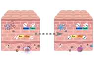
Both innate and adaptive immune cells exist in the skin, predominantly in the dermis layer. Recent studies have focused on how and which circadian rhythms contribute to maintain good health. Over recent years, we have gained a better understanding of the molecular mechanisms that control biological clocks and circadian rhythms. Circadian rhythms maintain homeostasis by providing day and night information to various physiological functions of our body. However, excessively high immune system activity can lead to a risk of developing autoimmune or allergic diseases. Recently, increasing numbers of studies with human and mouse models have been conducted to investigate the mechanisms underlying circadian regulation of the skin homeostasis. In this review, circadian regulation in the skin will be discussed from different points of view. Skin is referred as the largest organ of the body and is directly exposed to the external environment, including large changes in diurnal temperature, light, and pathogens. Immune cells as well as skin cells are the ones protecting us from these stimulants. Associations of the circadian system and these cells have been revealed in many ways, however, the specific roles of the peripheral clocks in these cells remain unknown. Circadian regulation in the skin diseases is discussed specifically in atopic dermatitis and other skin allergic symptoms as well as psoriasis.
Both innate and adaptive immune cells exist in the skin, predominantly in the dermis layer. Recent studies have focused on how and which circadian rhythms contribute to maintain good health. Over recent years, we have gained a better understanding of the molecular mechanisms that control biological clocks and circadian rhythms. Circadian rhythms maintain homeostasis by providing day and night information to various physiological functions of our body. However, excessively high immune system activity can lead to a risk of developing autoimmune or allergic diseases. Recently, increasing numbers of studies with human and mouse models have been conducted to investigate the mechanisms underlying circadian regulation of the skin homeostasis. In this review, circadian regulation in the skin will be discussed from different points of view. Skin is referred as the largest organ of the body and is directly exposed to the external environment, including large changes in diurnal temperature, light, and pathogens. Immune cells as well as skin cells are the ones protecting us from these stimulants. Associations of the circadian system and these cells have been revealed in many ways, however, the specific roles of the peripheral clocks in these cells remain unknown. Circadian regulation in the skin diseases is discussed specifically in atopic dermatitis and other skin allergic symptoms as well as psoriasis.
DOI: https://doi.org/10.37349/ei.2021.00019
This article belongs to the special issue Cross Talk Among Skin Cells and Immune Cells
Aim:
Previously, we reported increased number of T helper 17 (Th17) cells in vitiligo. However, in our recent study, tryptase and interleukin (IL)17 double positive cells which identified by polyclonal anti-IL17 antibody with specificity for IL17A, B, D, F was observed, but these mast cells cannot be stained by monoclonal anti-IL17 antibody with specificity for IL17A. Therefore, this study was aimed to clarify the role of mast cells in induction and progression of vitiligo.
Methods:
Mast cells were stained with two antibodies against IL17 and one antibody against tryptase by immunofluorescent staining. Furthermore, immunoelectron microscopy (IEM) analyses were conducted using anti-tryptase. In vitro, cultured epidermal keratinocytes were treated with agents which released by mast cells. Expression levels of mRNA were analyzed by real-time polymerase chain reaction (PCR), expression of protein levels was analyzed by western blotting.
Results:
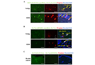
An increased number of tryptase positive mast cells was observed at the lesional skin of upper dermis in vitiligo and rhododendrol-induced leukoderma (RDIL). These mast cells showed prominent degranulation in vitiligo. Interestingly, the melanosome forming glycoprotein non-metastatic melanoma protein B (GPNMB) is downregulated in the lesional basal keratinocytes in vitiligo and mast cell tryptase contributes to this phenomenon. In addition, small interfering GPNMB RNA (siGPNMB RNA)-introduced keratinocytes increased melanocyte survival through stem cell factor (SCF) production in the melanocyte/keratinocyte co-culture system.
Conclusions:
Mast cells might be two-faced in vitiligo induction, progression, and recovery through the differential function of histamine and tryptase.
Aim:
Previously, we reported increased number of T helper 17 (Th17) cells in vitiligo. However, in our recent study, tryptase and interleukin (IL)17 double positive cells which identified by polyclonal anti-IL17 antibody with specificity for IL17A, B, D, F was observed, but these mast cells cannot be stained by monoclonal anti-IL17 antibody with specificity for IL17A. Therefore, this study was aimed to clarify the role of mast cells in induction and progression of vitiligo.
Methods:
Mast cells were stained with two antibodies against IL17 and one antibody against tryptase by immunofluorescent staining. Furthermore, immunoelectron microscopy (IEM) analyses were conducted using anti-tryptase. In vitro, cultured epidermal keratinocytes were treated with agents which released by mast cells. Expression levels of mRNA were analyzed by real-time polymerase chain reaction (PCR), expression of protein levels was analyzed by western blotting.
Results:
An increased number of tryptase positive mast cells was observed at the lesional skin of upper dermis in vitiligo and rhododendrol-induced leukoderma (RDIL). These mast cells showed prominent degranulation in vitiligo. Interestingly, the melanosome forming glycoprotein non-metastatic melanoma protein B (GPNMB) is downregulated in the lesional basal keratinocytes in vitiligo and mast cell tryptase contributes to this phenomenon. In addition, small interfering GPNMB RNA (siGPNMB RNA)-introduced keratinocytes increased melanocyte survival through stem cell factor (SCF) production in the melanocyte/keratinocyte co-culture system.
Conclusions:
Mast cells might be two-faced in vitiligo induction, progression, and recovery through the differential function of histamine and tryptase.
DOI: https://doi.org/10.37349/ei.2021.00018
This article belongs to the special issue Cross Talk Among Skin Cells and Immune Cells
Aim:
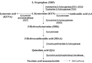
Chronic inflammation is closely associated with tryptophan (TRP)-kynurenine (KYN) metabolic pathway. However, TRP-KYN pathway has not been fully elucidated in psoriasis, a systemic inflammatory disease with skin lesions and extracutaneous manifestations. Herein, we studied comprehensively serum profiles of TRP-KYN pathway metabolites in psoriatic patients (PSOs) to clarify the involvement of this pathway in the pathophysiology of psoriasis and to evaluate serum biomarkers reflecting systemic inflammation in PSOs.
Methods:
The concentrations of main TRP metabolites, TRP, KYN, 3-hydroxykynurenine (3HK), kynurenic acid (KYNA), 3-hydroxyanthranilic acid (3HAA), and anthranilic acid (AA), were determined by high-performance liquid chromatography in the sera from 65 PSOs and 35 healthy controls (HCs). The levels of these metabolites and the ratios of metabolites were compared between these subjects. The correlations between these values and the psoriasis area severity index (PASI) scores were analyzed. Skin samples from PSOs and HCs were subjected to immunohistochemical staining for kynureninase, catabolic enzyme from KYN or 3HK to downstream. Cytokine concentrations were comprehensively measured in the same samples and the correlations between the cytokine levels and TRP-KYN pathway metabolite levels were examined.
Results:
Serum TRP, KYN, and KYNA concentrations were lower and the 3HAA concentrations were higher in PSOs than in HCs. The ratios of 3HK/KYN, 3HAA/3HK, and 3HK/AA were higher in PSOs than in HCs. The AA levels and the ratio of AA/KYN were weakly positively correlated, and TRP, KYNA, and 3HK levels and the ratios of KYNA/KYN and 3HAA/AA were weakly negatively correlated with the PASI scores. The AA, KYN, and KYNA levels were positively correlated with the interferon gamma-induced protein 10 (IP-10) concentrations. Kynureninase expression was enhanced in the epidermis, both involved and uninvolved skin.
Conclusions:
Serum profiles of TRP-KYN pathway metabolites differed between PSOs and HCs. TRP-KYN pathway-associated processes, including kynureninase activation, may be involved in the pathogenesis of psoriasis, and thus serve as targets for psoriasis therapy.
Aim:
Chronic inflammation is closely associated with tryptophan (TRP)-kynurenine (KYN) metabolic pathway. However, TRP-KYN pathway has not been fully elucidated in psoriasis, a systemic inflammatory disease with skin lesions and extracutaneous manifestations. Herein, we studied comprehensively serum profiles of TRP-KYN pathway metabolites in psoriatic patients (PSOs) to clarify the involvement of this pathway in the pathophysiology of psoriasis and to evaluate serum biomarkers reflecting systemic inflammation in PSOs.
Methods:
The concentrations of main TRP metabolites, TRP, KYN, 3-hydroxykynurenine (3HK), kynurenic acid (KYNA), 3-hydroxyanthranilic acid (3HAA), and anthranilic acid (AA), were determined by high-performance liquid chromatography in the sera from 65 PSOs and 35 healthy controls (HCs). The levels of these metabolites and the ratios of metabolites were compared between these subjects. The correlations between these values and the psoriasis area severity index (PASI) scores were analyzed. Skin samples from PSOs and HCs were subjected to immunohistochemical staining for kynureninase, catabolic enzyme from KYN or 3HK to downstream. Cytokine concentrations were comprehensively measured in the same samples and the correlations between the cytokine levels and TRP-KYN pathway metabolite levels were examined.
Results:
Serum TRP, KYN, and KYNA concentrations were lower and the 3HAA concentrations were higher in PSOs than in HCs. The ratios of 3HK/KYN, 3HAA/3HK, and 3HK/AA were higher in PSOs than in HCs. The AA levels and the ratio of AA/KYN were weakly positively correlated, and TRP, KYNA, and 3HK levels and the ratios of KYNA/KYN and 3HAA/AA were weakly negatively correlated with the PASI scores. The AA, KYN, and KYNA levels were positively correlated with the interferon gamma-induced protein 10 (IP-10) concentrations. Kynureninase expression was enhanced in the epidermis, both involved and uninvolved skin.
Conclusions:
Serum profiles of TRP-KYN pathway metabolites differed between PSOs and HCs. TRP-KYN pathway-associated processes, including kynureninase activation, may be involved in the pathogenesis of psoriasis, and thus serve as targets for psoriasis therapy.
DOI: https://doi.org/10.37349/ei.2021.00017
This article belongs to the special issue Cross Talk Among Skin Cells and Immune Cells
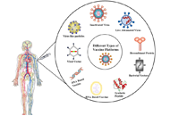
There have been significant developments in the design of nanostructured scaffolds for eliciting robust immune responses named vaccine. The technique is to produce strong immune responses is to manipulate the appearance of a pathogen. Subsequently pathogens such as viruses and bacteria often demonstrate of multiple copies of ligands on their surfaces, the immune system is predominantly sensitive towards multivalent presentations of antigens. Consequently, when designing a vaccine, it is beneficial to garnish a nanostructured surface with multiple copies of an antigen so it can effectively act as an immune booster. Different methods are there for the development of the vaccine, from them most of the techniques are well developed and reported and some of in the developing state. This review focuses primarily on cellular and non-cellular vaccines, the whole cells or cellular proteins either as the source of antigens or the platform in which to deliver the antigens. Purpose of this review, understand and discussion on the various vaccine platforms which will contribute noteworthy information to vaccine research and development (R and D).
There have been significant developments in the design of nanostructured scaffolds for eliciting robust immune responses named vaccine. The technique is to produce strong immune responses is to manipulate the appearance of a pathogen. Subsequently pathogens such as viruses and bacteria often demonstrate of multiple copies of ligands on their surfaces, the immune system is predominantly sensitive towards multivalent presentations of antigens. Consequently, when designing a vaccine, it is beneficial to garnish a nanostructured surface with multiple copies of an antigen so it can effectively act as an immune booster. Different methods are there for the development of the vaccine, from them most of the techniques are well developed and reported and some of in the developing state. This review focuses primarily on cellular and non-cellular vaccines, the whole cells or cellular proteins either as the source of antigens or the platform in which to deliver the antigens. Purpose of this review, understand and discussion on the various vaccine platforms which will contribute noteworthy information to vaccine research and development (R and D).
DOI: https://doi.org/10.37349/ei.2021.00016
This article belongs to the special issue Vaccine-induced Immune Responses Against SARS-CoV-2 Infections
Coronavirus disease-2019 (COVID-19) caused by severe acute respiratory syndrome coronavirus (SARS-CoV)-2 spread globally and creates an alarming situation. Following the SARS-CoV-2 paradigm, therapeutic efficacy is achieved via repurposing several antiviral, antibacterial, and antimalarial drugs. Innate and adaptive immune cells work close to combat infection through the intricate production of antibodies (Abs) and inflammatory cytokines. As an essential component of the immune system, Abs play an important role in eliminating viruses and maintaining homeostasis. B lymphocytes (B cells) are effector cells, stringent to produce neutralizing Abs to combat infection. After recognizing SARS-CoV-2 antigens by a surface receptor called B cell receptors (BCRs) on the plasma membrane, the BCRs transmembrane signal transduction and immune activation results in Ab production and development of immune memory. Thus, it ensures that plasma B cells can quickly start an intricate immune response to generate efficient protective Abs to clear the pathogen. Nevertheless, considering therapeutic challenges in the context of the new coronavirus pandemic, this review addresses the molecular mechanism of the immune activation and function of novel SARS-CoV-2 specific B cells in the production of SARS-CoV-2 specific Abs. Additionally, these studies highlighted the Ab-mediated pathogenesis, the intriguing role of nano-scale signaling subunits, non-structural proteins during COVID-19 infection, and structural insights of SARS-CoV-2 specific Abs.
Coronavirus disease-2019 (COVID-19) caused by severe acute respiratory syndrome coronavirus (SARS-CoV)-2 spread globally and creates an alarming situation. Following the SARS-CoV-2 paradigm, therapeutic efficacy is achieved via repurposing several antiviral, antibacterial, and antimalarial drugs. Innate and adaptive immune cells work close to combat infection through the intricate production of antibodies (Abs) and inflammatory cytokines. As an essential component of the immune system, Abs play an important role in eliminating viruses and maintaining homeostasis. B lymphocytes (B cells) are effector cells, stringent to produce neutralizing Abs to combat infection. After recognizing SARS-CoV-2 antigens by a surface receptor called B cell receptors (BCRs) on the plasma membrane, the BCRs transmembrane signal transduction and immune activation results in Ab production and development of immune memory. Thus, it ensures that plasma B cells can quickly start an intricate immune response to generate efficient protective Abs to clear the pathogen. Nevertheless, considering therapeutic challenges in the context of the new coronavirus pandemic, this review addresses the molecular mechanism of the immune activation and function of novel SARS-CoV-2 specific B cells in the production of SARS-CoV-2 specific Abs. Additionally, these studies highlighted the Ab-mediated pathogenesis, the intriguing role of nano-scale signaling subunits, non-structural proteins during COVID-19 infection, and structural insights of SARS-CoV-2 specific Abs.
DOI: https://doi.org/10.37349/ei.2021.00011
This article belongs to the special issue Vaccine-induced Immune Responses Against SARS-CoV-2 Infections
Head and neck squamous cell carcinoma (HNSCC) is a relatively widespread cancer with high mortality rates. Many patients with locally advanced disease are treated with combinations of surgery, radiation, and chemotherapy, while others are considered incurable and develop recurrent/metastatic (R/M) disease. Despite these treatment modalities, the 5-year survival rate of HNSCC has remained at 50% due to limited treatment options in patients with recurrent disease. Immunotherapy has been shown to induce durable responses in R/M patients, but only a minority of patients currently respond. A major hurdle in tumor immunotherapy is identifying the non-responders and markers to predict resistance in patients who at first responded to the therapy. In HNSCC patients, the tumor microenvironment (TME) assumes a vital role to either diminish or augment immune responses. There is an urgent need for extensive studies to be undertaken to better understand how tumor cells escape immune surveillance and resist immune attack. In this review, the impact of TME on the efficiency of immunotherapy, addressing the factors that mediate therapy resistance are highlighted. The composition of the TME encompassing the immunosuppressive cells including myeloid-derived suppressor cell (MDSC), regulatory T cells (Treg), mesenchymal stem cell (MSC), cancer-associated fibroblast (CAF), and tumor-associated macrophages (TAMs) and intrinsic factors like hypoxia, reactive oxygen species (ROS), extracellular matrix (ECM), angiogenesis, and epithelial-mesenchymal transition (EMT), how this debilitates immunosurveillance, and also discuss existing and potential strategies aimed at targeting these cellular and molecular TME components are reviewed. Understanding the interactions between the TME and immunotherapy is not only important in dissevering the mechanisms of action of immunosuppression but also offers scope for developing newer strategies to improve the competence of current immunotherapies.
Head and neck squamous cell carcinoma (HNSCC) is a relatively widespread cancer with high mortality rates. Many patients with locally advanced disease are treated with combinations of surgery, radiation, and chemotherapy, while others are considered incurable and develop recurrent/metastatic (R/M) disease. Despite these treatment modalities, the 5-year survival rate of HNSCC has remained at 50% due to limited treatment options in patients with recurrent disease. Immunotherapy has been shown to induce durable responses in R/M patients, but only a minority of patients currently respond. A major hurdle in tumor immunotherapy is identifying the non-responders and markers to predict resistance in patients who at first responded to the therapy. In HNSCC patients, the tumor microenvironment (TME) assumes a vital role to either diminish or augment immune responses. There is an urgent need for extensive studies to be undertaken to better understand how tumor cells escape immune surveillance and resist immune attack. In this review, the impact of TME on the efficiency of immunotherapy, addressing the factors that mediate therapy resistance are highlighted. The composition of the TME encompassing the immunosuppressive cells including myeloid-derived suppressor cell (MDSC), regulatory T cells (Treg), mesenchymal stem cell (MSC), cancer-associated fibroblast (CAF), and tumor-associated macrophages (TAMs) and intrinsic factors like hypoxia, reactive oxygen species (ROS), extracellular matrix (ECM), angiogenesis, and epithelial-mesenchymal transition (EMT), how this debilitates immunosurveillance, and also discuss existing and potential strategies aimed at targeting these cellular and molecular TME components are reviewed. Understanding the interactions between the TME and immunotherapy is not only important in dissevering the mechanisms of action of immunosuppression but also offers scope for developing newer strategies to improve the competence of current immunotherapies.
DOI: https://doi.org/10.37349/ei.2021.00013
